Category: Implants and Prostheses

Implant Enables Man With Severed Spinal Cord to Walk

Michel Rocatti walking using the spinal stimulation system. ©NeuroRestore-Jimmy Ravier

In a world first, Michel Roccati, a man with a completely severed spinal cord was able to walk again outside the lab with the help of a portable electrical stimulation system that causes his legs to take a step in conjunction with his intention to move and a walker to steady him.

This was a further development of a technology that in 2018 helped David M’zee, who had been left paralysed by a partial spinal cord injury suffered in a sports accident, to walk again. A research team led by Professors Grégoire Courtine, at École Polytechnique Fédérale de Lausanne (EPFL) and Jocelyne Bloch, at Lausanne University Hospital (CHUV) had developed an electrical stimulation system to help people with spinal cord injuries walk again.

They had wanted to see if electrodes could stimulate movement in the parts of the spine damaged so badly that signals no longer reach the nervous system from the brain. That pioneering study was detailed in Nature and Nature NeuroscienceThanks to the electrodes making up for the weakness of the signals in his damaged spinal cord, M’zee was able to voluntarily move his legs and could walk several hundred metres at a time, sometimes without the aid of the rails on the treadmill.

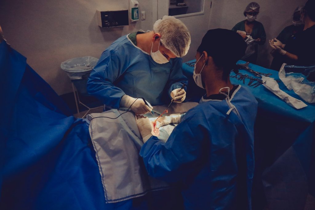
Now a new milestone has just been reached with the technology, and the research team enhanced their system with more sophisticated implants controlled by advanced software. These implants can stimulate the region of the spinal cord that activates the trunk and leg muscles. Thanks to this new technology, three patients with complete spinal cord injury were able to walk again outside the lab. “Our stimulation algorithms are still based on imitating nature,” said Prof Courtine. “And our new, soft implanted leads are designed to be placed underneath the vertebrae, directly on the spinal cord. They can modulate the neurons regulating specific muscle groups. By controlling these implants, we can activate the spinal cord like the brain would do naturally to have the patient stand, walk, swim or ride a bike, for example.”

On a cold, snowy day last December, Michel Roccati – an Italian man who became paralysed after a motorcycle accident four years earlier – braved the icy wind to try out the system outdoors, in central Lausanne. He had recently undergone the surgical procedure in which Prof Bloch placed the new, implanted lead on his spinal cord.

Scientists attached two small remote controls to Michel’s walker and connected them wirelessly to a tablet that forwards the signals to a pacemaker in Michel’s abdomen. The pacemaker in turn relays the signals to the implanted spinal lead that stimulates specific neurons, causing Michel to move. Grasping the walker, Michel pressed a button corresponding to either the left or right leg with the firm intention of taking a step forward, and his feet rose and fell in short steps.

“The first few steps were incredible – a dream come true!” he says. “I’ve been through some pretty intense training in the past few months, and I’ve set myself a series of goals. For instance, I can now go up and down stairs, and I hope to be able to walk one kilometre by this spring.”

Two other patients have also successfully tested the new system, which is described in Nature Medicine. “Our breakthrough here is the longer, wider implanted leads with electrodes arranged in a way that corresponds exactly to the spinal nerve roots,” said Bloch. “That gives us precise control over the neurons regulating specific muscles.” Ultimately, it allows for greater selectivity and accuracy in controlling the motor sequences for a given activity.

While extensive training is necessary for patients to get comfortable using the device, the speed and scope of rehabilitation is amazing. “All three patients were able to stand, walk, pedal, swim and control their torso movements in just one day, after their implants were activated!” said Prof Courtine. “That’s thanks to the specific stimulation programs we wrote for each type of activity. Patients can select the desired activity on the tablet, and the corresponding protocols are relayed to the pacemaker in the abdomen.”

While the progress achievable in a single day is astonishing, the gains after several months are even more impressive. The three patients followed a training regimen based on the stimulation programs and were able to regain muscle mass, move around more independently, and take part in social activities like having a drink standing at a bar. What’s more, because the technology is miniaturized, the patients can perform their training exercises outdoors and not only inside a lab.

Presently there is one key limitation, Prof Bloch said: “We need at least six centimetres of healthy spinal cord under the lesion. That’s where we implant our electrodes.”

As for Roccati, after nine months of Lausanne-based rehab, he now lives independently in Italy. “I continued rehab at home, working alone, with all the devices,” he said. “And I see improvements every day.”

Source: École Polytechnique Fédérale de Lausanne

Bionic Eye Demonstration Paves the Way to Human Trials

The Phoenix99 Bionic Eye. Credit: University of Sydney

A bionic eye under development has shown to be safe and stable for long-term implantation in a three-month animal study, paving the way towards human trials.

The Phoenix99 Bionic Eye, being developed by University of Sydney and UNSW, is an implantable system, designed to restore a form of vision to patients living with severe vision impairment and blindness caused by degenerative diseases, such as retinitis pigmentosa. The device consists of two main implants: a stimulator attached to the eye and a communication module positioned under the skin behind the ear.

Published in Biomaterials, the researchers used a sheep model to observe how the body responds and heals when implanted with the device, with the results allowing for further refinement of the surgical procedure. The biomedical research team is now confident the device could be trialled in human patients and have applied for ethical approval.

The Phoenix99 Bionic Eye works by stimulating the retina which, in healthy eyes, the cells in one of the layers turn incoming light into electrical messages. In some retinal diseases, the cells responsible for this crucial conversion degenerate, causing vision impairment. The system bypasses these malfunctioning cells by stimulating the remaining cells directly, effectively tricking the brain into believing that light was sensed.

“Importantly, we found the device has a very low impact on the neurons required to ‘trick’ the brain. There were no unexpected reactions from the tissue around the device and we expect it could safely remain in place for many years,” said Mr Samuel Eggenberger, a biomedical engineer who is completing his doctorate with Head of School of Biomedical Engineering Professor Gregg Suaning.

“Our team is thrilled by this extraordinary result, which gives us confidence to push on towards human trials of the device. We hope that through this technology, people living with profound vision loss from degenerative retinal disorders may be able to regain a useful sense of vision,” said Mr Eggenberger.

Professor Gregg Suaning said the positive results are a significant milestone for the Phoenix99 Bionic Eye.

“This breakthrough comes from combining decades of experience and technological breakthroughs in the field of implantable electronics,” said Prof Suaning.

A patient is implanted with the Phoenix99, and a stimulator is positioned on the eye and a communication module implanted behind the ear. A tiny camera attached to glasses captures the visual scene in front of the wearer, and the images are processed into a set of stimulation instructions which are sent wirelessly through to the communication module of the prosthesis.

The implant then transfers the instructions to the stimulation module, which delivers electrical impulses to the neurons of the retina. The electrical impulses, delivered in patterns matching the images recorded by the camera, trigger neurons which forward the messages to the brain, which interprets the signals as seeing the scene.

Source: University of Sydney

UK Man to Receive World’s First 3D-printed Eye

Photo by Victor Freita on Pexels

Moorfields Eye Hospital patient in the UK will be the first to benefit solely from a fully digital 3D printed prosthetic eye. Steve Verze, an engineer, will go home from the Old Street hospital with only a printed eye fitted that day. He first tried his eye on November 11 alongside a traditional acrylic prosthetic.

This new 3D printing process avoids the invasive process of making a mould of the eye socket: a procedure so difficult that in children it can require putting them under general anaesthetic.

Steve said: “I’ve needed a prosthetic since I was 20, and I’ve always felt self-conscious about it. When I leave my home I often take a second glance in the mirror, and I’ve not liked what I’ve seen. This new eye looks fantastic and, being based on 3D digital printing technology, it’s only going to be better and better.”

Professor Mandeep Sagoo, consultant ophthalmologist at Moorfields and professor of ophthalmology at the NIHR Biomedical Research Centre at Moorfields Eye Hospital UCL and Institute of Ophthalmology, said: “We are excited about the potential for this fully digital prosthetic eye.

“We hope the forthcoming clinical trial will provide us with robust evidence about the value of this new technology, showing what a difference it makes for patients. It clearly has the potential to reduce waiting lists.”

The printed eye is more realistic, with clearer definition and giving real depth to the pupil. The way light travels through the full depth of the printed eye is more natural than current prosthetics, which simply have the iris hand-painted onto a black disc embedded in the eye, with no light passage through the eye.

The current process can take six weeks but 3D printing halves that time, and the scanning ensures a precise fit. 

Source: Islington Gazette

A Novel Brain Implant Relieves Treatment-resistant Depression

Image by Falkurian Design on Unsplash

A proof-of-principle trial has shown that an electrical implant wired into the brain can detect and treat depression, thanks to positive results for the first patient to be fitted with the device.

The patient, Sarah (36), says the matchbox-sized implant in her skull has turned her life around since it was fitted a year ago. Her depression persisted despite anti-depressants and electroconvulsive therapy.

Sarah said that “any kind of relief” was better than her suffering. “My daily life had become so restricted. I felt tortured each day. I barely moved or did anything.”

The device, including its battery, was inserted into her skull beneath the scalp and holes were drilled for wires into her brain.

 Recalling how the implant changed her life, she said: “When the implant was first turned on, my life took an immediate upward turn. My life was pleasant again.

“Within a few weeks, the suicidal thoughts disappeared. When I was in the depths of depression all I saw is what was ugly.”

After 15 months, she has so far experienced no side effects from the device.

“In the early few months, the lessening of the depression was so abrupt, and I wasn’t sure if it would last,” she said. “But it has lasted. And I’ve come to realise that the device really augments the therapy and self-care I’ve learned while being a patient here at UCSF.”

The treatment however has to be personalised to the individual and their unique brain circuitry. Researcher Dr Katherine Scangos, a psychiatrist at University of California, San Francisco, said locating the ‘depression circuits’ in Sarah’s brain was what made the innovation possible.
“We found one location, which is an area called the ventral striatum, where stimulation consistently eliminated her feelings of depression.

“And we also found a brain activity area in the amygdala that could predict when her symptoms were most severe.”

Dr Scangos, who has enrolled two other patients in the trial and hopes to recruit nine more, said they need to repeat the work, looking for any changes in biomarkers or brain circuits. 
She said, “We didn’t know if we were going to be able to treat her depression at all because it was so severe. So in that sense we are really excited about this. It’s so needed in the field right now.”

However, the researchers stress that much more research is needed to see if this novel treatment is effective in other patients, and if it can be applied to other disorders.

The study is reported in Nature Medicine.

Source: BBC News

Deaths From Medical Devices Are Underreported in the US

Photo by Vidal Balielo Jr. from Pexels

Researchers have found that a number of deaths related to medical device adverse events were improperly categorised in the FDA’s Manufacturer and User Facility Device Experience (MAUDE) database, according to a new study.

Flagging terms commonly associated with death, the study investigators used a natural language processing algorithm to identify 290 141 reports where serious injury or death was reported; 52.1% of these events were reported as deaths, and 47.9% were classified as either malfunction, injury, or missing (report was uncategorised), reported Christina Lalani, MD, of the University of California San Francisco, and colleagues, in JAMA Internal Medicine.

Overall, 23% of reports with a death were not placed in the death category, amounting to some 31 552 reports filed from December 31, 1991, to April 30, 2020.

Whether to classify the event as a malfunction, injury, death, or ‘other’ is up to the physician or manufacturer. According to the FDA, the reporter is required to categorise an adverse event as an official death if the cause of death is unknown, or if the device “may have caused or contributed to a death.”

The three most common product codes among the adverse event reports were for ventricular assist bypass devices (38 708 reports), dialysate concentrate for haemodialysis (25 261 reports), and transcervical contraceptive tubal occlusion devices (14 387 reports).

The natural language processing algorithm scanned through reports, identifying terms such as “patient died,” “patient expired,” “could not be resuscitated,” and “time of death.” Of the 70 terms that were associated with a death, 62 (88.6%) were found among miscategorised adverse event reports involving a patient death. And, out of all 62, there were 17 terms that had an estimated percentage of 100%, meaning that “every time that term was used, the patient had died, even though the reporter had not classified the report as death,” the team wrote.

Only 18 terms had sample sizes large enough for researchers to calculate confidence intervals; among them, the words “death” or “deaths” were linked to 12% of adverse event reports in which a patient died, but were classified as malfunction, other, or missing — the highest rate of all the analysed terms.

The researchers acknowledged a major limitation in that only reports with at least one death-associated term were included, in contrast to all the reports from the MAUDE database. Improperly categorised deaths likely contribute to an underestimate.

“The classification chosen by the reporter is vital, as the FDA must review all adverse events reported as deaths, which is not the case for other reporting categories,” the authors wrote. Improving the reports’ accuracy is crucial, since patient death frequency can prompt the FDA to pursue investigations into the device’s safety, they added.

The researchers pointed out an inherent conflict of interest as 95.9% of the reports evaluated in the study were submitted by manufacturers.

“It may not be in their interest to facilitate identification of serious problems with their own devices in a timely manner,” they wrote. “There have been multiple instances of delays by manufacturers in reporting serious malfunctions and deaths that were associated with medical devices, as well as complete failures to report.”

Therefore, it’s likely that a significant number of patients have been unknowingly treated with devices that were later revealed to be dangerous, Dr Lalani and colleagues noted. For example, they referenced the reporting failures that occurred from 2002 to 2013, when 32 000 women reported adverse events associated with the permanent birth control device Essure while the FDA only received 1023 adverse event reports from the manufacturer.

They concluded that patients and care providers should submit reports directly to the FDA as well as or instead of the manufacturer.

Source: MedPage Today

Journal information: Lalani C, et al “Reporting of death in US Food and Drug Administration medical device adverse event reports in categories other than death” JAMA Intern Med 2021; DOI: 10.1001/jamainternmed.2021.3942.

Liquid Metal Sensors Recreate a Sense of Touch

Photo by ThisisEngineering RAEng on Unsplash

To recreate a sense of ‘touch’, researchers have incorporated stretchable tactile sensors using liquid metal on the fingertips of a prosthetic hand. 

When manipulating an object, humans are heavily reliant on sensation in their fingertips, each of which has over 3000 pressure-sensitive touch receptors. While there are many high-tech, dexterous prosthetics available today, they all lack the sensation of ‘touch‘, resulting in objects inadvertently being dropped or crushed by a prosthetic hand.
To make a prosthetic hand interface that feels more natural and intuitive, researchers from Florida Atlantic University’s College of Engineering and Computer Science and collaborators incorporated stretchable tactile sensors using liquid metal on a prosthetic hand’s fingertips. Encapsulated within silicone-based elastomers, this technology provides key advantages over traditional sensors, including high conductivity, compliance, flexibility and stretchability.

For the study, published in the journal Sensors, researchers used individual fingertips on the prosthesis to distinguish between different speeds of a sliding motion along different textured surfaces. The four different textures had one variation: the distance between the ridges. To detect the textures and speeds, researchers trained four machine learning algorithms. For each of the ten surfaces, 20 trials were performed to test the ability of the machine learning algorithms to distinguish between the different textured surfaces.

Results showed that integrating tactile information from the fingertip sensors simultaneously distinguished between complex, multi-textured surfaces – demonstrating a new form of hierarchical intelligence. The algorithms could accurately distinguish between the fingertip speeds. This new technology could improve prosthetic hand control and provide haptic feedback for amputees to restore a sense of touch.

“Significant research has been done on tactile sensors for artificial hands, but there is still a need for advances in lightweight, low-cost, robust multimodal tactile sensors,” said senior author Erik Engeberg, PhD, an associate professor in the Department of Ocean and Mechanical Engineering. “The tactile information from all the individual fingertips in our study provided the foundation for a higher hand-level of perception enabling the distinction between ten complex, multi-textured surfaces that would not have been possible using purely local information from an individual fingertip. We believe that these tactile details could be useful in the future to afford a more realistic experience for prosthetic hand users through an advanced haptic display, which could enrich the amputee-prosthesis interface and prevent amputees from abandoning their prosthetic hand.”

Researchers compared four different machine learning algorithms for their successful classification capabilities. The time-frequency features of the liquid metal sensors were extracted to train and test the machine learning algorithms. Of these, a neural network algorithm generally performed the best at the speed and texture detection with a single finger and had a 99.2 percent accuracy to distinguish between ten different multi-textured surfaces using four liquid metal sensors from four fingers simultaneously.

“The loss of an upper limb can be a daunting challenge for an individual who is trying to seamlessly engage in regular activities,” said Stella Batalama, Ph.D., dean, College of Engineering and Computer Science. “Although advances in prosthetic limbs have been beneficial and allow amputees to better perform their daily duties, they do not provide them with sensory information such as touch. They also don’t enable them to control the prosthetic limb naturally with their minds. With this latest technology from our research team, we are one step closer to providing people all over the world with a more natural prosthetic device that can ‘feel’ and respond to its environment.”

Source: Florida Atlantic University

Journal information: Abd, M.A., et al. (2021) Hierarchical Tactile Sensation Integration from Prosthetic Fingertips Enables Multi-Texture Surface Recognition. Sensors. doi.org/10.3390/s21134324.

Carbon Fibre Electrodes Allow Unprecedented Neural Recording

Image by Robina Weemeijer on Unsplash

A tiny, implantable carbon fibre electrode has the potential to provide a long-term brain-computer interface which can record electrical signals over lengthy periods of time.

The carbon fibre electrodes were developed at the University of Michigan and demonstrated in rats. The new research shows the promise of carbon fibre electrodes in recording electrical signals from the brain without damaging brain tissue. Directly implanting carbon fiber electrodes into the brain allows the capturing of bigger and more specific signals than current technologies.

This technology could lead to advances that could give amputees and those with spinal injuries control of advanced prosthetics, stimulate the sacral nerve to restore bladder control, stimulate the cervical vagus nerve to treat epilepsy and provide deep brain stimulation as a possible treatment for Parkinson’s.  

“There are interfaces out there that can be implanted directly into the brain but, for a variety of reasons, they only last from months to a few years,” said Elissa Welle, a recent PhD graduate from the U-M Department of Biomedical Engineering. “Any time you’re opening up the skull for a procedure involving the brain, it’s a big deal.”

Brain implants are typically made from silicon due to its ability to conduct electricity and its historic use in cleanroom technology. But silicon is not very biocompatible and leads to the formulation of scar tissue over long periods. Such electodes will eventually degrade and no longer capture brain signals, requiring removal.

Carbon fibres may be the answer to getting high-quality signals with an interface that lasts years, not months. And by laser cutting and sharpening carbon fibers into tiny, subcellular electrodes in the lab with the help of a small blowtorch, U-M engineers have harnessed the potential for excellent signal capture in a form the body is more likely to accept.

“After implantation, it sits inside the brain in a way that does not interfere with the surrounding blood vessels, because it’s smaller than those blood vessels,” Welle said. “They’ll move around and adjust to an object that small, rather than get torn as they would when encountering larger implants.”

Part of the electrode’s compatibility in brain tissue is down to smaller size, but its needle-like shape may also minimise compacting of any surrounding tissue. Larger carbon-based electrodes have been shown to actually encourage neural tissue to grow instead of degrading. The team is hopeful that similar potential for their carbon fibre electrodes will be revealed by further testing.

Carbon fibre electrodes in a previous study dramatically outperformed conventional silicon electrodes with 34% of electrodes recording a neuron signal compared to 3%. Laser cutting then improved this number to 71% at 9 weeks after implantation. Flame sharpening has now enabled these high performance probes to be implanted directly into the cerebral cortex, negating the need for a temporary insertion aid, or shuttle, as well as into the rat’s cervical vagus nerve.

It is relatively easy to insert electrodes into the brain. But the researchers have also taken on the more difficult task of inserting the sharpened carbon fibre electrodes into nerves, with micrometre diameters.

Those findings show that potential for these electrodes goes beyond prosthetic manipulation, according to Cindy Chestek, a U-M associate professor of biomedical engineering, and principal investigator of the The Cortical Neural Prosthetics Lab.

“Someone who is paralysed may have no control over things like their bladder, for example,” Prof Chestek said. “We may be able to utilise these smaller electrodes to stimulate and record signals from areas that can’t be reached by larger ones, maybe the neck or spinal cord, to help give patients some level of control.”

Source: University of Michigan

Innovative 3D Printing Makes Stronger and More Flexible Implants

Photo by Tom Claes on Unsplash

A new 3D printing process developed by University of Nottingham researchers, allows customised production of artificial body parts and other medical devices with built-in functionality offering shape and durability, while also cutting bacterial infection risk.

“Most mass-produced medical devices fail to completely meet the unique and complex needs of their users,” explained lead researcher Dr Yinfeng He, Centre for Additive Manufacturing. “Similarly, single-material 3D printing methods have design limitations that cannot produce a bespoke device with multiple biological or mechanical functions.”

“But for the first time, using a computer-aided, multi-material 3D-print technique, we demonstrate it is possible to combine complex functions within one customised healthcare device to enhance patient wellbeing.”

The team’s hope is that their new design process can be applied to 3D-print any highly customised medical device.

For example, the method could be adapted to create a single-part prosthetic limb or joint with greater comfort and functionality; or printing customised pills containing multiple drugs – known as polypills – optimised to release their contents in a planned sequence.

What it can do

For this study, the researchers applied a computer algorithm to design and manufacture 3D-printed objects made up of two polymer materials with differing stiffness that also prevent bacterial biofilm build-up. Combining these two materials, they produced an implant with the required strength and flexibility.

Artificial finger joint replacements currently use both silicone and metal parts, offering the wearer a standardised level of dexterity but must be rigid enough to implant into bone. The team 3D-printed a finger joint as a demonstration, which offered these dual requirements in one device, while also being able to customise its size and strength to meet individual patient requirements. They can even make use of intrinsically bacteria-resistant and bio-functional multi-materials, combating infection without extra antibiotics.

A new high-resolution characterisation technique (3D orbitSIMS) was used to 3D-map the chemistry of the print structures and to test the bonding between them throughout the part. This showed that the two materials were intermingling at their interfaces; a sign of good bonding and therefore a stronger device.

The study was carried out by the Centre for Additive Manufacturing (CfAM) and funded by the Engineering and Physical Sciences Research Council. The complete findings are published in Advanced Science, in a paper entitled: ‘Exploiting generative design for 3D printing of bacterial biofilm resistant composite devices’.

Prior to making the technique commercialised, the researchers plan to try out more advanced materials with extra functionalities such as controlling immune responses and promoting stem cell attachment.

Source: University of Nottingham

Journal reference: He, Y., et al. (2021) Exploiting Generative Design for 3D Printing of Bacterial Biofilm Resistant Composite Devices. Advanced Science. doi.org/10.1002/advs.202100249.

Ventricular Assist Device Pulled from Market due to Failures

Photo from Olivier Collett on Unsplash

The HeartWare system, a left ventricular assist device (LVAD) for advanced heart failure patients, is being discontinued immediately, according to the Food and Drug Administration.

The manufacturer, Medtronic, is halting global distribution and sale of its HeartWare system in the wake of observational evidence of increased neurological adverse events and mortality for its LVAD compared with similar mechanical circulatory support (MCS) devices.

Last December, some HeartWare LVADs were recalled because of complaints that the pump may delay or fail to start. So far 100 of these complaints have been received, including 14 patient deaths and 13 cases where an explant was necessary, the FDA noted.

“We have been carefully monitoring the adverse events associated with this device and support its removal from the marketplace,” said Bram Zuckerman, MD, director of the Office of Cardiovascular Devices at the FDA’s Center for Devices and Radiological Health, in a statement.

Medtronic now advises physicians to immediately stop new implants of the HeartWare device, but does not recommend explants.

The company is working on a plan for ongoing support of the some 4000 patients around the world who currently have this LVAD. It received commercial approval for use in the US in November 2012.

The FDA named Abbott’s HeartMate 3 as one alternative LVAD for patients with end-stage heart failure. This device features a magnetic levitation system that keeps the rotor separate without mechanical contact.

“The FDA is working closely with both Medtronic and Abbott to ensure patient care is optimised during this transition period and that there is an adequate supply of devices available to provide this patient population with options for end-stage heart failure treatment,” said Dr Zuckerman.

In a separate press release, Abbott reassured the public that it has the ability to meet increased demand for MCS devices as a result of HeartWare withdrawal from clinical use.

Source: MedPage Today

Tiny Implant Shelters Diabetes-curing Cells

Photo by Photomix Company from Pexels

A team of researchers have developed a miniscule device that allows them to implant insulin-secreting cells into diabetic mice, which secrete insulin in response to blood sugar without being destroyed by the immune system.

The findings are published in the journal Science Translational Medicine.

“We can take a person’s skin or fat cells, make them into stem cells and then grow those stem cells into insulin-secreting cells,” said co-senior investigator Jeffrey R Millman, PhD, an associate professor of medicine at Washington University. “The problem is that in people with Type 1 diabetes, the immune system attacks those insulin-secreting cells and destroys them. To deliver those cells as a therapy, we need devices to house cells that secrete insulin in response to blood sugar, while also protecting those cells from the immune response.”

Prof Millman, also an associate professor of biomedical engineering, had previously developed and honed a method to make stem cells and then grow them into insulin-secreting beta cells. Prof Millman previously used those beta cells to reverse diabetes in mice, but it was not clear how the insulin-secreting cells might safely be implanted into people with diabetes.

Prof Millman explained why the new device’s structure was so important.

“The device, which is about the width of a few strands of hair, is micro-porous—with openings too small for other cells to squeeze into—so the insulin-secreting cells consequently can’t be destroyed by immune cells, which are larger than the openings,” he said. “One of challenges in this scenario is to protect the cells inside of the implant without starving them. They still need nutrients and oxygen from the blood to stay alive. With this device, we seem to have made something in what you might call a Goldilocks zone, where the cells could feel just right inside the device and remain healthy and functional, releasing insulin in response to blood sugar levels.”

Millman’s laboratory collaborated with researchers from the laboratory of Minglin Ma, PhD, an associate professor of biomedical engineering at Cornell and the study’s other co-senior investigator. Prof Ma has been working to develop biomaterials that can help implant beta cells safely into animals and, eventually, people with Type 1 diabetes.

In recent years a number of implants have been tried to varying degrees of success. For this study, the team led by Prof Ma developed a nanofibre-integrated cell encapsulation (NICE) device. They filled those implants with insulin-secreting beta cells grown from stem cells and then implanted the devices into the abdomens of diabetic mice.

“The combined structural, mechanical and chemical properties of the device we used kept other cells in the mice from completely isolating the implant and, essentially, choking it off and making it ineffective,” Prof Ma explained. “The implants floated freely inside the animals, and when we removed them after about six months, the insulin-secreting cells inside the implants still were functioning. And importantly, it is a very robust and safe device.”

The cells in the implants continued to secrete insulin and control blood sugar in the mice for up to 200 days — even without any immunosuppressive drugs being administered.
“We’d rather not have to suppress someone’s immune system with drugs, because that would make the patient vulnerable to infections,” Prof Millman said. “The device we used in these experiments protected the implanted cells from the mice’s immune systems, and we believe similar devices could work the same way in people with insulin-dependent diabetes.”

Profs Millman and Ma stress that a considerable amount of work is needed before the device can be trialled in a clinical setting.

Source: Washington University School of Medicine in St Louis

Journal information: X. Wang et al., “A nanofibrous encapsulation device for safe delivery of insulin-producing cells to treat type 1 diabetes,” Science Translational Medicine (2021)