Category: Immune System

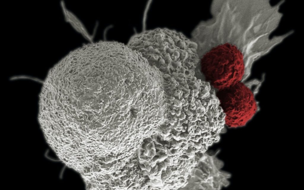
Scientists Strengthen Evidence Linking Autoimmunity and Schizophrenia

Image by Pikisuperstar On Freepix

Links have been reported between schizophrenia and autoimmunity. In a study published in Brain Behavior and Immunity, Japanese researchers identified autoantibodies that target a ‘synaptic adhesion protein’, neurexin 1α, in a subset of patients with schizophrenia. When injected into mice, the autoantibodies caused many schizophrenia-related changes.

What is a synaptic protein, and why might it be linked to schizophrenia? Synaptic adhesion proteins are specialised proteins that bind to create physical connections between brain cells. These connections, called synapses, allow the cells to communicate by passing molecules back and forth. Both synapses and autoimmunity are known to be associated with schizophrenia, so the research team from Tokyo Medical and Dental University (TMDU) decided to investigate autoantibodies that target synaptic proteins in patients with schizophrenia.

“In around 2% of our patient population, we identified autoantibodies against the synaptic protein neurexin 1α, which is expressed by one cell in the synapse and binds to proteins known as neuroligins on the other cell in the synapse,” says lead author of the study Hiroki Shiwaku. “Once we had identified these autoantibodies, we wanted to see if they were able to cause schizophrenia-related changes.”

To do this, the researchers isolated autoantibodies from some of the patients with schizophrenia and injected them into the cerebrospinal fluid of mice, so that the autoantibodies would travel into the brain. In these mice, the autoantibodies blocked neurexin 1α and neuroligin binding and altered some related synaptic properties. The administration of these autoantibodies also resulted in fewer synapses in the brains of mice and schizophrenia-related behaviours, such as reduced social behaviour toward unfamiliar mice and reduced cognitive function.

“Together, our results strongly suggest that autoantibodies against neurexin 1α can cause schizophrenia-related changes, at least in mice,” explains Hiroki Shiwaku. “These autoantibodies may therefore represent a therapeutic target for a subset of patients with schizophrenia.”

Schizophrenia has a wide variety of both symptoms and treatment responses, and many patients have symptoms that are resistant to currently available treatment options. Therefore, the identification of possible disease-causing autoantibodies is important for improving symptom control in patients with schizophrenia. It is hoped that the results of this investigation will allow patients with autoantibodies that target neurexin 1α – all of whom were resistant to antipsychotic treatment in the present study — to better control their symptoms in the future.

Source: Tokyo Medical and Dental University

Autoimmune Disorders Now Affect Roughly One in Ten Individuals

Photo by Jacek Dylag on Unsplash

A population-based study of 22 million people in the UK estimates that around one in ten individuals in the UK now live with an autoimmune disorder. The findings, published in The Lancet, also highlight important socioeconomic, seasonal and regional differences for several autoimmune disorders, providing new clues as to what factors may be involved in these conditions.

There are more than 80 known autoimmune diseases, including conditions like rheumatoid arthritis, type 1 diabetes and multiple sclerosis, some of which have been increasing in the last few decades.

This has raised the question whether overall incidence of autoimmune disorders is on the rise and what factors are involved, such as environmental factors or behavioural changes in society. The exact causes of autoimmune diseases remain largely unknown, including how much can be attributed to a genetic predisposition to disease and how much is down to exposure to environmental factors.

The study used anonymised electronic health data from 22 million individuals in the UK to investigate 19 of the most common autoimmune diseases. The authors examined whether incidence of autoimmune diseases is rising over time, who is most affected by these conditions and how different autoimmune diseases may co-exist with each other.

They found that the 19 autoimmune diseases studied affect around 10% of the population. This is higher than previous estimates, which ranged from 3–9% and often relied on smaller sample sizes and included fewer autoimmune conditions. The analysis also highlighted a higher incidence in women (13%) than men (7%).

The research discovered evidence of socioeconomic, seasonal and regional disparities for several autoimmune disorders, suggesting that these conditions are unlikely to be caused by genetic differences alone. This observation may point to the involvement of potentially modifiable risk factors such as smoking, obesity or stress. It was also found that in some cases a person with one autoimmune disease is more likely to develop a second, compared to someone without an autoimmune disease.

Dr Nathalie Conrad at the University of Oxford said: “We observed that some autoimmune diseases tended to co-occur with one another more commonly than would be expected by chance or increased surveillance alone. This could mean that some autoimmune diseases share common risk factors, such as genetic predispositions or environmental triggers. This was particularly visible among rheumatic diseases and among endocrine diseases. But this phenomenon was not generalised across all autoimmune diseases. Multiple sclerosis, for example, stood out as having low rates of co-occurrence with other autoimmune diseases, suggesting a distinct pathophysiology.”

These findings reveal novel patterns that will inform the design of further research into the possible common causes of different autoimmune diseases.

Professor Geraldine Cambridge at UCL Medicine said: “Our study highlights the considerable burden that autoimmune diseases place upon individuals and the wider population. Disentangling the commonalities and differences within this large and varied set of conditions is a complex task. There is a crucial need, therefore, to increase research efforts aimed at understanding the underlying causes of these conditions, which will support the development of targeted interventions to reduce the contribution of environmental and social risk factors.”

Source: University College London

Vitamin D may be Necessary for Effective Immunotherapy in Skin Cancer

3D structure of a melanoma cell derived by ion abrasion scanning electron microscopy. Credit: Sriram Subramaniam/ National Cancer Institute

Vitamin D has many effects on the body, including regulation of the immune system. New research indicates that for patients with advanced skin cancer, it may be important to maintain normal vitamin D levels when receiving immunotherapy in the form of immune checkpoint inhibitors. The findings are published in CANCER.

To see whether levels of vitamin D might impact the effectiveness of immune checkpoint inhibitors, investigators analysed the blood of 200 patients with advanced melanoma both before and every 12 weeks during immunotherapy treatment.

A favourable response rate to immune checkpoint inhibitors was observed in 56.0% of patients in the group with normal baseline vitamin D levels or normal levels obtained with vitamin D supplementation, compared with 36.2% in the group with low vitamin D levels without supplementation. Progression‐free survival in these groups was 11.25 and 5.75 months, respectively.

“Of course, vitamin D is not itself an anti-cancer drug, but its normal serum level is needed for the proper functioning of the immune system, including the response that anti-cancer drugs like immune checkpoint inhibitors affect,” said lead author Łukasz Galus, MD, of Poznan University of Medical Sciences, in Poland. “In our opinion, after appropriately randomised confirmation of our results, the assessment of vitamin D levels and its supplementation could be considered in the management of melanoma.”

Source: Wiley

Monocytes are Still Capable of Proliferation, Study Shows

Scanning electron micrograph of a macrophage. Credit: NIAID

Cell proliferation from stem cells is vital for organisms to grow and form vital organs. In cancer, however, cell proliferation is no longer controlled and becomes chaotic. Research published in Nature Immunology revealed that monocytes, which are blood immune cells previously thought to be already differentiated, can in fact still proliferate. The study’s researchers at the University of Liège have discovered that, in a healthy individual, monocytes also have this ability to proliferate, in order to replace tissue macrophages.

Monocytes are white blood cells that derive from the bone marrow. A monocyte is part of the innate immune response and functions to regulate cellular homeostasis, especially in the setting of infection and inflammation. They account for approximately 5% of circulating nucleated cells in normal adult blood.

The formation of complex multicellular organisms necessitates billions of cells to be produced from progenitor cells, proliferating and taking on particular morphologies and functions while assembling into tissues and organs. Most of the cells that constitute a living organism are understood to arise from stem cells. When they stop proliferating, they specialise, differentiate and form muscles, brain, bones, immune cells, etc. When proliferation is no longer properly regulated, this can lead to the development of various diseases, chief if which is cancer.

Professor Thomas Marichal (Professor at ULiège, Welbio investigator at the WEL Research Institute) and his team from the GIGA Institute at ULiège discovered that this ability to proliferate is not merely restricted to stem cells, but is also an as-yet-unknown function of blood immune cells, the monocytes.

Indeed, blood monocytes, previously considered as differentiated cells, are capable of proliferating and generating a pool of monocytes in the tissues in order to give rise to macrophages, which are important immune cells that protect us against microbes and support the proper functioning of our organs.

“This is a major fundamental discovery, which changes our conception of the involvement of cell proliferation in the constitution and maintenance of our immune system,” explains Thomas Marichal, director of the study. “Our finding also suggests that the information that can be drawn from an enumeration of blood monocytes, classically carried out during a blood test, would reflect only little of what is happening at the level of the tissues, during ‘infection or inflammation, for example, since monocytes can proliferate when they enter tissues.”

He also adds, “Fortunately, this proliferation is extremely well controlled and does not lead to a humoral process. It has only one goal: to allow, as effectively as possible, the replacement of immune cells that populate our tissues: the macrophages.”

This discovery was possible thanks to the development of new tools and the use of innovative technologies. “This study is a great example of how technological advances can drive breakthrough scientific discoveries. It would have been extremely difficult, if not impossible, to study with such a resolution this population of proliferating monocytes only 10 years ago. This required the use of state-of-the-art equipment recently acquired at the GIGA Institute, the generation of complex genomic data and very sophisticated bioinformatics analyses,” explains Domien Vanneste, first author of the study.

This study paves the way for future investigations that will evaluate the possibility of manipulating or controlling monocyte proliferation for therapeutic purposes, at the benefit of an enhanced health.

Source: University de Liege

Research Supports Letting A Moderate Fever Run its Course

Photo by Mockup Graphics on Unsplash

It may be better to let a mild fever run its course instead of automatically reaching for medication, new University of Alberta research suggests. Researchers found that, in fish models, untreated moderate fever helped them to quickly their infections, keep inflammation in check and repair damaged tissue. “We let nature do what nature does, and in this case it was very much a positive thing,” says Professor Daniel Barreda, immunologist and lead author on the study which is published in eLife.

Moderate fever is self-resolving, meaning that the body can both induce it and shut it down naturally without medication, Barreda explains. The health advantages of natural fever to humans still have to be confirmed through research, but the researchers say because the mechanisms driving and sustaining fever are shared among animals, it is reasonable to expect similar benefits are going to happen in humans.

That suggests the need to resist taking non-steroidal anti-inflammatory drugs at the first signs of a mild temperature, he says. “They take away the discomfort felt with fever, but you’re also likely giving away some of the benefits of this natural response.”

The study also sheds light on some benefits of moderate fever, which Barreda notes has been evolutionarily conserved across the animal kingdom for 550 million years. “Every animal examined has this biological response to infection.”

For the study, fish were given a bacterial infection and their behaviour was then tracked and evaluated using machine learning. Outward symptoms were similar to those seen in humans with fever, including immobility, fatigue and malaise. These were then matched to important immune mechanisms inside the animals.

The research showed that natural fever offers an integrative response that not only activates defences against infection, but also helps control it. The researchers found that fever helped to clear the fish of infection in about seven days – half the time it took for those animals not allowed to exert fever. Fever also helped to shut down inflammation and repair injured tissue.

“Our goal is to determine how to best take advantage of our medical advances while continuing to harness the benefits from natural mechanisms of immunity,” says Barreda.

Source: University of Alberta

Towards Treating Dangerous Immunotherapy Side Effects

Photo by Tima Miroshnichenko on Pexels

While immunotherapy has been shown to greatly improve survival rates for certain types of cancer, in some cases, it can lead to a dangerous over-activation of the immune system. In a recent review published in Journal for ImmunoTherapy of Cancer, potential therapies have been identified, which might make it possible to continue with immunotherapy even when facing severe side effects.

The rare immunotherapy side effect of over-activation was only clinically recognised during regular clinical use rather than in clinical trials or animal experiments. To better understand this over-activation, Lisa LiuMarco Gerling, and colleagues analysed data from all published international reports on this issue after cancer immunotherapy. Their findings indicate that potentially life-threatening inflammation may occur more frequently than previously thought, and might be treatable with existing drugs such as steroids or anti-inflammatory therapies commonly used for rheumatoid arthritis.

“It will be exciting to follow up on the main findings of our systematic review, says Marco Gerling at the Department of Biosciences and Nutrition, Karolinska Institutet and lead author.

“We believe that inhibition of a specific inflammatory molecule, interleukin-6, could allow patients to continue immunotherapy despite strong, systemic activation of the immune system”, he continues. “But we need more data to support the regular use of interleukin-6 inhibitors. We also want to thank Narcisa Hannerz and Sabine Gillsund from Karolinska University Library for their invaluable help with finding articles for this review.“

Source: Karolinska Institutet

Cytotoxic T Cells Become ‘Marathon Runners’ to Wage Long Immune Battles

Shown here is a pseudo-colored scanning electron micrograph of an oral squamous cancer cell (white) being attacked by two cytotoxic T cells (red), part of a natural immune response. Photo by National Cancer Institute on Unsplash

When it comes to chronic infections and cancer, cytotoxic T cells play a central role in our defences. Research published in the journal Immunity has revealed that these cells can specialise into “sprinters” to fight a strong, short-term infection or into “marathon runners” for the long battle against chronic infections and cancer.

Professor Daniel Pinschewer at the Department of Biomedicine of the University of Basel led a study into understanding how cytotoxic T cells adapt to infection and cancer.

“These T cells can become specialised in two different ways: either as a kind of sprinter or as marathon runners,” explains Pinschewer. “However, the latter can also convert into sprinters at any time, in order to stamp out an infection.”

Chronic infections are a special case: the T cells are activated and a strong inflammatory response occurs at the same time. “This tends to ‘shock’ the T cells into developing into sprinters, which can only intervene effectively in the short term to remove infected cells,” says the virologist. “If all T cells behaved like that, our immune defences would break down pretty soon.”

Biological messenger counteracts the “shock”

The researchers examined how, in spite of this, the immune system is still able to provide enough T cells for the endurance race against chronic infections. According to their results, a biological messenger called interleukin-33 (IL-33) plays a key role. It allows the T cells to remain in their “marathon runner” state. “IL-33 takes away the shock of the inflammation, so to speak,” explains Dr Anna-Friederike Marx, lead author of the study.

In addition, the biological messenger causes the marathon T cells to proliferate, so that more endurance runners are available to combat the infection. “Thanks to IL-33, there are enough cytotoxic T cells around for the long haul that can still pull off a final sprint after their marathon,” says Marx.

The findings could help improve the treatment of chronic infections such as hepatitis C. It is conceivable that IL-33 could be administered to support an effective immune response. Thinking along the same lines, IL-33 could be one key to improving cancer immunotherapy, to enable T cells to wage an efficient and long-lasting offensive against tumour cells.

Source: University of Basel

Pseudomonas Aeruginosa Locks out Immune Cells

Pseudomonas
Scanning Electron Micrograph of Pseudomonas aeruginosa. Credit: CDC/Janice Carr

Pseudomonas aeruginosa bacteria are a common menace in hospital wards, causing life-threatening infections, and are often resistant to antibiotics. Researchers have discovered a mechanism that likely contributes to the severity of P. aeruginosa infections, which could also be a target for future treatments. The results were recently appeared in the journal EMBO Reports.

Many bacterial species use sugar-binding molecules called lectins to attach to and invade host cells. Lectins can also influence the immune response to bacterial infections. However, these functions have hardly been researched so far. A research consortium led by Prof Dr Winfried Römer at the University of Freiburg and Prof Dr Christopher G. Mueller at the CNRS/University of Strasbourg has investigated the effect of the lectin LecB from P. aeruginosa on the immune system. It found that isolated LecB can render immune cells ineffective: The cells are then no longer able to migrate through the body and trigger an immune response. The administration of a substance directed against LecB prevented this effect and led to the immune cells being able to move unhindered again.

LecB blockades immune cells

As soon as they perceive an infection, cells of the innate immune system migrate to a nearby lymph node, where they activate T and B cells, triggering a targeted immune response. LecB, according to the current study, prevents this migration. “We assume that LecB not only acts on the immune cells themselves in this process, but also has an unexpected effect on the cells lining the inside of the blood and lymph vessels,” Römer explains. “When LecB binds to these cells, it triggers extensive changes in them.” Indeed, the researchers observed that important structural molecules were relocated to the interior of the cells and degraded. At the same time, the cell skeleton became more rigid. “The cell layer thus becomes an impenetrable barrier for the immune cells,” Römer said.

An effective agent against LecB

Can this effect be prevented? To find out, the researchers tested a specific LecB inhibitor that resembles the sugar building blocks to which LecB otherwise binds. “The inhibitor prevented the changes in the cells, and T-cell activation was possible again,” Mueller said. The inhibitor was developed by Prof Dr Alexander Titz, who conducts research at the Helmholtz Institute for Pharmaceutical Research Saarland and Saarland University.

Further studies are needed to determine how clinically relevant the inhibition of the immune system by LecB is to the spread of P. aeruginosa infection and whether the LecB inhibitor has potential for therapeutic application. “The current results provide further evidence that lectins are a useful target for the development of new therapies, especially for antibiotic-resistant pathogens such as P. aeruginosa,” the authors conclude.

Source: University of Freiburg

Skipping Breakfast may Weaken Immune Cells

Photo by Malvestida on Unsplash

Fasting may be detrimental to fighting off infection, and could lead to an increased risk of heart disease, suggests a new study published in Immunity. The research in mouse models, shows that skipping meals triggers a response in the brain that negatively affects immune cells. The results could lead to a better understanding of how chronic fasting may affect the body long term.

“There is a growing awareness that fasting is healthy, and there is indeed abundant evidence for the benefits of fasting. Our study provides a word of caution as it suggests that there may also be a cost to fasting that carries a health risk,” says lead author Filip Swirski, PhD, Director of the Cardiovascular Research Institute at Icahn Mount Sinai. “This is a mechanistic study delving into some of the fundamental biology relevant to fasting. The study shows that there is a conversation between the nervous and immune systems.”

As part of a larger study on different fasting times affect the immune systems, researchers focused on the role of breakfast. They fed one group of mice breakfast right after waking up (breakfast is their largest meal of the day), and the other group had no breakfast. Researchers collected blood samples in both groups when mice woke up (baseline), then four hours later, and eight hours later.

Analysing the blood work, researchers saw a difference in the number of monocytes, which are white blood cells that are made in the bone marrow and travel through the body, where they play many critical roles, from fighting infections, to heart disease, to cancer.

At baseline, all mice had the same amount of monocytes. But after four hours, monocytes in mice from the fasting group were dramatically affected. Researchers found that 90% of these cells disappeared from the bloodstream, and the number further declined at eight hours. Meanwhile monocytes in the non-fasting group were unaffected.

In fasting mice, researchers discovered the monocytes travelled back to the bone marrow to hibernate. At the same time, production of new cells in the bone marrow diminished. The normally short-lived monocytes in the bone marrow survived longer as a consequence of staying in the bone marrow, and aged differently than the monocytes that stayed in the blood.

The researchers continued to fast mice for up to 24 hours, and then reintroduced food. The cells hiding in the bone marrow surged back into the bloodstream within a few hours, provoking heightened level of inflammation. Instead of protecting against infection, these altered monocytes were more inflammatory, making the body less resistant to fighting infection.

Researchers found that specific regions in the brain controlled the monocyte response during fasting. This study demonstrated that fasting elicits a stress response in the brain (feeling ‘hangry’) and this instantly triggers a large-scale migration of these white blood cells from the blood to the bone marrow, and then back to the bloodstream shortly after food is reintroduced.

Dr Swirski emphasised that while there is also evidence of the metabolic benefits of fasting, this new study is a useful advance in the full understanding of the body’s mechanisms.

“The study shows that, on the one hand, fasting reduces the number of circulating monocytes, which one might think is a good thing, as these cells are important components of inflammation. On the other hand, reintroduction of food creates a surge of monocytes flooding back to the blood, which can be problematic. Fasting, therefore regulates this pool in ways that are not always beneficial to the body’s capacity to respond to a challenge such as an infection,” explains Dr Swirski. “Because these cells are so important to other diseases like heart disease or cancer, understanding how their function is controlled is critical.”

Source: The Mount Sinai Hospital / Mount Sinai School of Medicine

How the Body Responds to Life-threatening Disease from HSV-1

Source: National Cancer Institute on Unsplash

Analysing an infant’s genome has allowed scientists to find a new way genetics influences the body’s antiviral response by studying a life-threatening disease caused by a common virus: herpes simplex virus 1 (HSV-1). The findings, published in Science Immunologyhold potential as a genetic marker doctors could use to gauge a child’s risk of herpes encephalitis, although such mutations are generally very rare in the population.

The researchers analysed genetic data from a patient with immunodeficiency and hospitalised at nine months old with herpes encephalitis, a rare but life-threatening brain inflammation after HSV-1 infection. They identified novel mutations in the gene GTF3A, and found that these mutations impair the innate immune response.

Many people are infected in childhood with the HSV-1 virus but the vast majority don’t suffer from encephalitis. The most common symptom of HSV-1 is oral cold sores, but many people show no signs at all. HSV-1 is more threatening to children and adults who are immunodeficient, whose immune system cannot control the virus well.

“Genetic and mechanistic analyses of uncommon viral diseases like herpes encephalitis are quite rare. In fact, the causes underlying severe herpes encephalitis are often unknown,” says Michaela Gack, PhD, FRIC’s scientific director. “This information provides us with invaluable insight into the fundamental molecular processes that govern our immune response and opens up opportunities for future research on severe disease outcomes.”

The Ghent research team led by Filomeen Haerynck, MD, PhD, reached out to Dr Gack’s team after finding the mutations in the gene. Dr Gack’s lab studies interactions between the human immune system and viruses on a molecular level.

The GTF3A mutations shape how cells respond to viral activity through the genetic makeup of a protein called TFIIIA. TFIIIA plays a role in helping a human enzyme produce certain types of RNA that can determine specific functions inside cells. Some RNAs can elicit an anti-herpes viral immune response.

Dr Gack’s team tested cells that have the mutations, and found that because of defects in certain immunostimulatory RNAs, the cells were more susceptible to HSV-1 infection and lost the ability to control the HSV-1 virus.

The affected gene is part of the body’s defence system that produces interferons to combat viruses. Interferons are crucial to the human immune response and for suppressing virus infection and spread.

This new genetic pathway could be helpful in understanding the immune response to other viruses, like Epstein-Barr virus, a common virus linked to mononucleosis and associated with certain types of cancer and multiple sclerosis.

“Understanding the molecular processes underlying antiviral responses is key to treating or possibly preventing severe viral infections that change patients’ and families’ lives,” Dr Gack said. “Our findings on critical immune defence proteins may translate into new therapies in the future.”

Source: Cleveland Clinic