COVID patients placed on ventilators can take a long time to regain consciousness. New research published the Proceedings of the National Academy of Sciences now shows that these delays may serve a purpose: protecting the brain from oxygen deprivation.
The existence of such a brain-preserving state could explain why some patients wake up days or even weeks after they stop receiving ventilation, and it suggests that physicians should take these lengthy recovery times into account when determining a patient’s prognosis.
In their study, investigators connect the pattern seen among those who have survived severe COVID with similar delays known to occur in a small fraction of cardiac arrest patients.
“The delayed recoveries in COVID patients are very much like the rare cases we’ve documented in previous research. In this new paper, we describe a mechanism to explain what we’re seeing in both types of patients,” said study co-senior author Dr Nicholas D. Schiff, a neurology professor at Weill Cornell Medicine.
He suggests that this mechanism is the brain protecting itself, pointing to animals, most notably painted turtles, that can tolerate extended periods without oxygen.
More than a decade ago, Dr Schiff and his colleagues first observed these delays among comatose cardiac arrest patients who received cooling therapy to reduce brain damage caused by a loss of blood flow. In one such case, a 71-year-old patient took 37 days to awaken, before ultimately making a near-complete recovery.
During the pandemic, Dr Schiff performed neurology consultations for COVID patients, and he soon began seeing similar, delayed awakenings occurring when patients were taken off ventilators and stopped receiving movement-limiting sedatives.
In a separate analysis of a large cohort of COVID patients from Weill Cornell Medicine and two other major U.S. medical centres, Dr Schiff and his colleagues, including co-author of the current paper, Dr Emery N. Brown, professor of anaesthesia at Harvard Medical School, found that a quarter of patients who survived ventilation took 10 days or longer to recover consciousness. The more oxygen deprivation they suffered while on the ventilator, the longer that delay.
In the prior study of cardiac patients, the researchers recorded a distinctive pattern in brain activity, one also seen in patients under deep anaesthesia. (Recordings from COVID patients are extremely limited.) Dr Schiff read that a similar pattern had been seen in the brains of painted turtles, which can withstand up to five months without oxygen under ice in the winter. To do so, they activate the same inhibitory system within the brain targeted by anaesthetics given to human cardiac and COVID patients but in novel ways developed by evolutionary specialisations.
Drs Schiff and Brown propose that, by chance, the same protective response emerges in the patients.
“It is our theory that oxygen deprivation as well as practices in the ICU, including commonly used anaesthetics, expose elements of strategies that animals use to survive in extreme conditions,” Dr Schiff said.
“These observations may offer new insights into the mechanisms of how certain anaesthetics produce unconsciousness and new approaches for ICU sedation and for fostering recovery from disorders of consciousness,” Dr Brown added.
When patients fail to regain consciousness for an extended time, physicians may recommend withdrawing life-supporting care. This threshold is typically set at 14 days or less for cardiac patients, while no such guidelines exist for COVID.
In light of this new research, however, so long as they lack brain injuries, physicians should avoid making negative projections about these patients’ potential to recover, note the researchers.
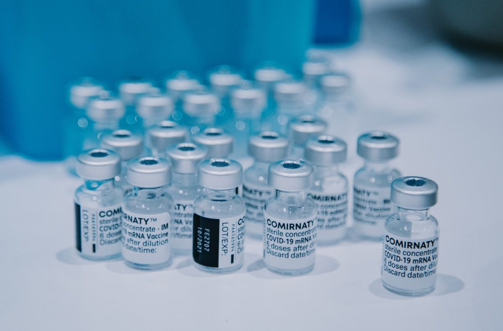
The European Medicines Agency (EMA) recommended that heavy menstrual bleeding should be added to the product information as a side effect of unknown frequency of the mRNA COVID vaccines Comirnaty (Pfizer/BioNtech) and Spikevax (Moderna).
Heavy menstrual bleeding may be defined as bleeding characterised by an increased volume and/or duration which interferes with the person’s physical, social, emotional and material quality of life. Cases of heavy menstrual bleeding have been reported after the first, second and booster doses of Comirnaty and Spikevax.
The EMA’s Pharmacovigilance Risk Assessment Committee (PRAC) assessed this safety signal after reviewing the available data, including cases reported during clinical trials, cases spontaneously reported in Eudravigilance and findings from the medical literature.
After a review of the available data, the PRAC concluded that there is at least a reasonable possibility that the occurrence of heavy menstrual bleeding is causally associated with these vaccines and therefore recommended the update of the product information.
The available data reviewed involved mostly cases which appeared to be non-serious and temporary in nature.
There is no evidence to suggest the menstrual disorders experienced by some people have any impact on reproduction and fertility. Available data provides reassurance about the use of mRNA COVID vaccines before and during pregnancy. A review carried out by EMA’s Emergency Task Force showed that mRNA COVID vaccines do not cause pregnancy complications for expectant mothers and their babies, and they are as effective at reducing the risk of hospitalisation and deaths in pregnant people as they are in non-pregnant people.
Red mitochondria in airway cells become coated with green SARS-COV-2 proteins after viral infection: Researchers discovered that the virus that causes COVID-19 damages lungs by attacking mitochondria. Credit: Stephen Archer
Viruses and bacteria have a very long history. Because viruses can’t reproduce without a host, they’ve been attacking bacteria for millions of years. Some of those bacteria eventually became mitochondria, synergistically adapting to life within eukaryotic cells (cells that have a nucleus containing chromosomes).
Ultimately, mitochondria became the powerhouses within all human cells.
This is the story of how a team, assembled during the pandemic, recognized the mechanism by which these viruses were causing lung injury and lowering oxygen levels in patients: It is a throwback to the primitive war between viruses and bacteria – more specifically, between this novel virus and the evolutionary offspring of bacteria, our mitochondria.
SARS-CoV-2 is the third novel coronavirus to cause human outbreaks in the 21st century, following SARS-CoV in 2003 and MERS-CoV in 2012. We need to better understand how coronaviruses cause lung injury to prepare for the next pandemic.
How COVID-19 affects lungs
People with severe COVID-19 pneumonia often arrive at the hospital with unusually low oxygen levels. They have two unusual features distinct from patients with other types of pneumonia:
First, they suffer widespread injury to their lower airway (the alveoli, which is where oxygen is taken up).
Second, they shunt blood to unventilated areas of the lung, which is called ventilation-perfusion mismatch. This means blood is going to parts of the lung where it won’t get sufficiently oxygenated.
We already knew that mitochondria are not just the powerhouse of the cell, but also its main consumers and sensors of oxygen. Mitochondria control the process of programmed cell death (called apoptosis), and they regulate the distribution of blood flow in the lung by a mechanism called hypoxic pulmonary vasoconstriction.
This mechanism has an important function. It directs blood away from areas of pneumonia to better ventilated lobes of the lung, which optimizes oxygen-uptake. By damaging the mitochondria in the smooth muscle cells of the pulmonary artery, the virus allows blood flow to continue into areas of pneumonia, which also lowers oxygen levels.
It appeared plausible that SARS-CoV-2 was damaging mitochondria. The results of this damage – an increase in apoptosis in airway epithelial cells, and loss of hypoxic pulmonary vasoconstriction – were making lung injury and hypoxaemia (low blood oxygen) worse.
Our discovery, published in Redox Biology, explains how SARS-CoV-2, the coronavirus that causes COVID-19 pneumonia, reduces blood oxygen levels.
We show that SARS-CoV-2 kills airway epithelial cells by damaging their mitochondria. This results in fluid accumulation in the lower airways, interfering with oxygen uptake. We also show that SARS-CoV-2 damages mitochondria in the pulmonary artery smooth muscle cells, which inhibits hypoxic pulmonary vasoconstriction and lowers oxygen levels.
Attacking mitochondria
Coronaviruses damage mitochondria in two ways: by regulating mitochondria-related gene expression, and by direct protein-protein interactions. When SARS-CoV-2 infects a cell, it hijacks the host’s protein synthesis machinery to make new virus copies. However, these viral proteins also target host proteins, causing them to malfunction. We soon learned that many of the host cellular proteins targeted by SARS-CoV-2 were in the mitochondria.
How SARS-CoV-2 targets mitochondria to kill lung cells and prevent oxygen sensing. Credit: Brooke Ring, provided by Stephen Archer
Viral proteins fragment the mitochondria, depriving cells of energy and interfering with their oxygen-sensing capability. The viral attack on mitochondria starts within hours of infection, turning on genes that break the mitochondria into pieces (called mitochondrial fission) and make their membranes leaky (an early step in apoptosis called mitochondrial depolarization).
In our experiments, we didn’t need to use a replicating virus to damage the mitochondria – simply introducing single SARS-CoV-2 proteins was enough to cause these adverse effects. This mitochondrial damage also occurred with other coronaviruses that we studied.
We are now developing drugs that may one day counteract COVID-19 by blocking mitochondrial fission and apoptosis, or by preserving hypoxic pulmonary vasoconstriction. Our drug discovery efforts have already enabled us to identify a promising mitochondrial fission inhibitor, called Drpitor1a.
Our team’s infectious diseases expert, Gerald Evans, notes that this discovery also has the potential to help us understand Long COVID. “The predominant features of that condition – fatigue and neurologic dysfunction – could be due to the lingering effects of mitochondrial damage caused by SARS-CoV-2 infection,” he explains.
Bacteria are regularly attacked by viruses, called bacteriophages, that need a host to replicate in. The bacteria in turn fight back, using an ancient form of immune system called the CRISPR-cas system, that chops up the viruses’ genetic material. Humans have recently exploited this CRISPR-cas system for a Nobel Prize-winning gene editing discovery.
The ongoing competition between bacteria and viruses is a very old one; and recall that our mitochondria were once bacteria. So perhaps it’s not surprising at all that SARS-CoV-2 attacks our mitochondria as part of the COVID-19 syndrome.
Pandemic pivot
The original team members on this project are heart and lung researchers with expertise in mitochondrial biology. In early 2020 we pivoted to apply that in another field – virology – in an effort to make a small contribution to the COVID-19 puzzle.
Our discovery owes a lot to our virology collaborators. Early in the pandemic, University of Toronto virologist Gary Levy offered us a mouse coronavirus (MHV-1) to work with, which we used to make a model of COVID-19 pneumonia. Che Colpitts, a virologist at Queen’s University, helped us study the mitochondrial injury caused by another human beta coronavirus, HCoV-OC43.
Finally, Arinjay Banerjee and his expert SARS-CoV-2 virology team at Vaccine and Infectious Disease Organization (VIDO) in Saskatoon performed key studies of human SARS-CoV-2 in airway epithelial cells. VIDO is one of the few Canadian centres equipped to handle the highly infectious SARS-CoV-2 virus.
Our team’s super-resolution microscopy expert, Jeff Mewburn, notes the specific challenges the team had to contend with.
“Having to follow numerous and extensive COVID-19 protocols, they were still able to exhibit incredible flexibility to retool and refocus our laboratory specifically on the study of coronavirus infection and its effects on cellular/mitochondrial functions, so very relevant to our global situation,” he said.
Our discovery will hopefully be translated into new medicines to counter future pandemics.
According to a recent study published in Frontiers in Cardiovascular Medicine, the risk of developing myocarditis is seven times higher with a COVID infection than with the COVID vaccine, by scientists. Patients with myocarditis can experience chest pains, shortness of breath or an irregular heartbeat. In severe cases, the inflammation can lead to heart failure and death.
“Our findings show that the risk of myocarditis from being infected by COVID is far greater than from getting the vaccine,” said Dr Navya Voleti, a resident physician at Penn State College of Medicine. “Moving forward, it will be important to monitor the potential long-term effects in those who develop myocarditis.”
Typically resulting from viral infection, myocarditis is a known complication of SARS-CoV-2 infection, and heart complications have been associated with mRNA COVID vaccination – particularly myocarditis in teenage boys. However, the relative risk of myocarditis due to vaccines and infections had not been clearly established.
The researchers conducted the largest study to date on the risk of developing myocarditis as a result of SARS-CoV-2 infection vs experiencing inflammation after COVID vaccination. The researchers compared vaccinated and unvaccinated COVIP patients to uninfected controls. The myocarditis risk was 15 times higher in COVID patients, regardless of vaccination status, compared to individuals who did not contract the virus.
Next, the researchers separately compared the rates of myocarditis in those who received the vaccines to those in unvaccinated individuals. According to the findings, the rates of myocarditis in people who were vaccinated against COVID were only twofold higher than in unvaccinated people.
Based on all the findings, the researchers concluded that the risk of myocarditis due to COVID was seven times higher than the risk related to the vaccines.
Investigators conducted a systematic review and meta-analysis of 22 studies published worldwide from December 2019 through May 2022. The studies included nearly 58 million patients who reported cardiac complications and belonged to one of two groups: the 55.5 million who were vaccinated against COVID compared to those who were not vaccinated (vaccination group), and the 2.5 million who contracted the virus compared to those who did not contract the virus (COVID group).
In the vaccination group, the researchers separately compared the risk of myocarditis for various COVID vaccines. The median age of the study population was 49 years; 49% were men; and the median follow-up time after infection or COVID vaccination was 28 days.
The researchers found that among those diagnosed with myocarditis after receiving the vaccine or having COVID, the majority (61%) were men. Of patients diagnosed with myocarditis in both vaccination and COVID groups, 1.07% were hospitalised and 0.015% died.
“COVID infection and the related vaccines both pose a risk for myocarditis. However, the relative risk of heart inflammation induced by COVID infection is substantially greater than the risk posed by the vaccines,” said Dr. Paddy Ssentongo, a resident physician in the Department of Medicine at Penn State Health Milton S. Hershey Medical Center and the lead author of the study. “We hope our findings will help mitigate vaccine hesitancy and increase vaccine uptake.”
The pharmaceutical industry is facing a serious challenge as it struggles to source enough non-human primates (NHPs) such as macaques for research and testing. Alongside demand created by HIV/AIDS research, the pandemic has tightened supplies of the animals further as China, a major supplier, has clamped down on exports.
Since NHPs have great genetic and physiological similarity to humans, scientists use these animals, most commonly rhesus macaques, to study medical conditions and conduct trials which are not yet possible in humans. In 2019, US scientists used 68 257 NHPs in research, according to US government data.
As a result of this shortage, many projects may not be able to be completed, according to industry insiders, with implications for medical research. Pre-pandemic prices of $11 000 per macaque have risen to $35 000.
In July last year, Nature reported that the US government pledged to increase funding to make primates available for clinical research. However, this would not do anything to address the current shortage.
To make room for more NHPs, the US National Institutes of Health (NIH) has invested about US$29 million to refurbish housing, build outdoor enclosures and making other infrastructure improvements at the US National Primate Research Centers (NPRCs), which it funds.
“A couple of years ago, we were feeling the pinch,” Nancy Haigwood, director of the Oregon NPRC in Beaverton, which houses about 5 000 non-human primates. But because of the pandemic, “we are truly out of animals”, she told Nature. “We’re turning away everyone.”
China had been a cheap source of cynomolgus macaques (Macaca fascicularis) since 1985, but in 2013 began to prioritise local research, restricting exports. Adding to this was soaring demand was sparked by multiple NIH grants awarded in 2016 to study HIV/AIDS, according to a 2018 report. Housing and feeding NHPs is costly, and NPRCs could not expand due to budget caps. The report warned of a coming shortage of various primates in coming years.
The situation has drawn the public’s attention – and opposition. Complaints made to airlines has resulted in many no longer carrying the animals, making transportation a major challenge. Air France was one of the last holdouts, and last year said it would stop carrying NHPs for research purposes.
With the arrival of the pandemic and the need for NHP research and testing, vaccine research was naturally prioritised, while trying to supply other projects as well.
When COVID hit, China completely suspended exports of macaques, hitting pharmaceutical companies hardest, which prefer that species for drug trials. Even if the export ban were to be lifted, the Chinese demand for macaques in research is so high that there would be few available for export: of 30 000 macaques that became suitable for use in research last year, 28 000 were used.
Other restrictions constrain the supply, such as a European Union requirement that all non-human primates for research come from self-sustaining colonies by November this year. The UK also carried through this directive following its exit from the EU.
The emerging Omicron variant BA.2.75.2 largely evades neutralising antibodies in the blood and resists several monoclonal antibody antiviral treatments, according to a study published in the journal The Lancet Infectious Diseases. The findings suggest a risk of increased COVID infections in the northern hemisphere’s winter, unless the new updated bivalent vaccines help to boost immunity in the population.
“While antibody immunity is not completely gone, BA.2.75.2 exhibited far more dramatic resistance than variants we’ve previously studied, largely driven by two mutations in the receptor binding domain of the spike protein,” said the study’s corresponding author Ben Murrell, assistant professor at the Department of Microbiology, Tumor and Cell Biology, Karolinska Institutet.
The study shows that antibodies in random serum samples from 75 blood donors in Stockholm were approximately only one-sixth as effective at neutralising BA.2.75.2 compared with the now-dominant variant BA.5. The serum samples were collected at three time points: in November 2021 before the emergence of Omicron, in April 2022 after a large wave of infections in the country, and at the end of August to early September after the BA.5 variant became dominant.
The researchers found that only one of the clinically available monoclonal antibody treatments that were tested, bebtelovimab, managed to effectively neutralise the new variant.
BA.2.75.2 is a mutated version of another Omicron variant, BA.2.75. Since it was first discovered earlier this fall, it has spread to several countries but so far represents only a minority of registered cases.
A possible increase in infections
“We now know that this is just one of a constellation of emerging variants with similar mutations that will likely come to dominate in the near future,” Ben Murrell says, adding “we should expect infections to increase this winter.”
Some questions remain. It is unclear whether these new variants will drive an increase in hospitalisation rates. Also, while current vaccines have, in general, had a protective effect against severe disease for Omicron infections, there is not yet data showing the degree to which the updated COVID vaccines provide protection from these new variants. “We expect them to be beneficial, but we don’t yet know by how much,” Ben Murrell says.
Nirmatrelvir-ritonavir (Paxlovid) is often given to heart disease patients with symptomatic COVID to prevent progression to severe disease – but it can interact with some previously prescribed medications. A review paper published in the Journal of the American College of Cardiology examines the potential drug-drug interactions (DDIs) between Paxlovid and commonly used cardiovascular medications, as well as potential options to mitigate severe adverse effects.
“Awareness of the presence of drug-drug interactions of Paxlovid with common cardiovascular drugs is key. System-level interventions by integrating drug-drug interactions into electronic medical records could help avoid related adverse events,” said Sarju Ganatra, MD, senior author of the review.
He continued: “The prescription of Paxlovid could be incorporated into an order set, which allows physicians, whether it be primary care physicians or cardiology providers, to consciously rule out any contraindications to the co-administration of Paxlovid. Consultation with other members of the health care team, particularly pharmacists, can prove to be extremely valuable. However, a health care provider’s fundamental understanding of the drug-drug interactions with cardiovascular medications is key.”
In December 2021, Paxlovid received emergency use authorisation from the US Food and Drug Administration as an oral antiviral agent for the treatment of symptomatic, non-hospitalised adults with mild to moderate COVID infection who are at high risk for progression to severe disease. Patients with heart disease and other risk factors, including diabetes, high blood pressure, chronic kidney disease and smoking make up a large portion of the high-risk population for whom Paxlovid is beneficial.
According to the authors, Paxlovid has been shown to be very effective in patients with existing heart disease, but it has significant DDIs with commonly used cardiovascular medications, highlighting the importance for all clinicians to be familiar with these DDIs. As there is limited clinical information regarding DDI-related adverse events, the authors used existing knowledge and data regarding how therapies like Paxlovid typically react with other medications to provide guidance regarding potential interactions and the associated likely consequences based on the degree of interaction.
The review provides an in-depth overview of a variety of cardiovascular medications used to treat many forms of heart disease. Five of the most important cardiovascular drug interactions with Paxlovid to be aware of include:
Anti-arrhythmic agents
Many anti-arrhythmic agents are metabolised in a way that increases plasma levels when co-administered with Paxlovid. While it may be possible to start Paxlovid after 2–2.5-day temporary discontinuation of the anti-arrhythmic agents, this may not be feasible from a practical standpoint. Clinicians are advised to consider alternative COVID therapies and avoid co-administration of these agents with Paxlovid. Sotalol, another anti-arrhythmic agent, is renally cleared and does not interact with Paxlovid.
Antiplatelet agents and anticoagulants
Antiplatelet agents are used for the treatment of coronary artery disease, particularly if a patient has received a stent. Aspirin and prasugrel are safe to co-administer with Paxlovid. There is an increased risk of blood clots when Paxlovid is given alongside clopidogrel and an increased risk of bleeding when given with ticagrelor. When possible, these agents should be switched to prasugrel. If patients have contraindication to taking prasugrel, then co-administration of Paxlovid should be avoided and alternative COVID therapies should be considered.
Anticoagulants such as warfarin may be co-administered with Paxlovid but require close monitoring of clotting factors in bloodwork. The plasma levels of all direct oral anticoagulants increase when co-administered with Paxlovid, therefore dose adjustment or temporary discontinuation and use of alternative anticoagulants may be required.
Certain statins
Co-administration of simvastatin or lovastatin with Paxlovid can lead to increased plasma levels and subsequent myopathy and rhabdomyolysis, a condition in which the breakdown of muscle tissue releases a damaging protein into the bloodstream. These agents should be stopped prior to initiation of Paxlovid. A dose reduction of atorvastatin and rosuvastatin is reasonable when co-administered with Paxlovid. The other statins are considered safe when given along with Paxlovid.
Ranolazine
Plasma concentration of ranolazine, used to treat angina and other heart-related chest pain, is exponentially increased in the presence of CPY450 inhibitors like Paxlovid, thereby increasing the risk of clinically significant QT prolongation and torsade de pointes (a type of arrhythmia). Co-administration of Paxlovid is therefore contraindicated. Temporary discontinuation of ranolazine is advised if prescribing Paxlovid.
Immunosuppressive agents
The plasma levels of immunosuppressive agents prescribed for patients who have undergone heart transplantation exponentially rise to toxic levels when co-administered with Paxlovid. Temporary reduction of dosing of immunosuppressive agents would require frequent monitoring and be logistically difficult. Therefore, alternative COVID therapies should be considered in these patients.
The authors conclude awareness and availability of other COVID therapies enable clinicians to offer alternative treatment options to patients who are unable to take Paxlovid due to DDIs.
A team of Japanese researchers have shown that low concentrations of cetylpyridinium chloride, used in certain mouthwashes, has an antiviral effect on SARS-CoV-2. Their findings were published in the journal Scientific Reports.
SARS-CoV-2 is transmitted via aerosols, which are spread from the oral and nasal cavities. In addition to infecting the cells of the respiratory tract, SARS-CoV-2 is also known to infect the cells of the lining of the mouth and the salivary glands.
Store-bought mouthwashes contain a number of antibiotic and antiviral components, such as cetylpyridinium chloride (CPC), which has been shown to reduce the oral viral load of SARS-CoV-2, chiefly by disrupting the virus’s lipid envelope. While there are other chemicals with similar effects, CPC has the advantage of being tasteless and odourless.
The researchers, led by Professor Kyoko Hida at Hokkaido University, were interested in studying the effects of CPC in Japanese mouthwashes, which typically contain a fraction of the CPC compared to previously tested mouthwashes. They tested the effects of CPC on cell cultures that express trans-membrane protease serine 2 (TMPRSS2), which is required for SARS-CoV-2 entry into the cell.
They found that, within 10 minutes of application, 30–50 µg/mL of CPC inhibited the infectivity and capability for cell entry of SARS-CoV-2. Interestingly, commercially available mouthwashes that contain CPC performed better than CPC alone. They also showed that saliva did not alter the effects of CPC. Testing with the original, alpha, beta and gamma variants of SARS-CoV-2 showed similar effetcts of CPC.
This study shows that low concentrations of CPC in commercial mouthwash suppress the infectivity of four variants of SARS-CoV-2. The authors have already begun assessing the effect on CPC-containing mouthwashes on viral loads in saliva of COVID patients. Future work will also focus on fully understanding the mechanism of effect, as lower concentrations of CPC do not disrupt lipid membranes.
Intravenous treatment with omega-3 fatty acids in elderly hospitalised patients in intensive care due to COVID seems to help the immune system’s ability to cope with the virus, according to a study published in the journal Clinical and Translational Medicine. These findings could lead to a complementary, cost-effective treatment for COVID.
In COVID patients, the immune system and the body’s activation of white blood cells are over-activated. It can lead to a so-called systemic inflammatory storm, which worsens the disease state and can cause complications such as sepsis and heart failure.
Researchers at Karolinska Institutet, among others, have now shown that omega-3 fatty acids can stimulate active healing of inflammation, without inhibiting the immune response. By accelerating the healing of the inflammation without compromising the body’s immune system, it could be possible to counteract the most serious complications of COVID, researchers believe.
Stimulated inflammation-healing molecules
The treatment effect was found by mapping inflammatory biomarkers and immunological reactions.
“First, we showed that fatty acid metabolism to inflammation-healing molecules was stimulated in those patients treated with omega-3 fatty acids. By isolating immune cells before, during, and after treatment, we were able to show that immune function improved,” said corresponding author Magnus Bäck, senior consultant in cardiology and professor at Karolinska Institutet.
Planning further studies
Researchers are now planning for larger clinical studies, which will be needed to show whether the course of the disease in severe COVID is improved through treatment with omega-3 fatty acids.
“It is important that even our weakest and frailest patients have the opportunity to participate in studies when the enemy, in this case, COVID, is on the attack and that they can fight the disease with the help of the medicine,” said Dorota Religa, senior consultant and professor in geriatrics at Karolinska Institutet.
“Stimulating the healing of inflammation with omega-3 fatty acids has the potential to lead to a new, cost-effective low-risk treatment for COVID-19, as a complement to existing treatment,” said Prof Magnus Bäck.
Although SARS-CoV-2 infections mainly attack the lungs, in many cases they can also damage other organs, such as the colon: around 60% of patients experienced digestive tract impacts. A study published in the International Journal of Molecular Sciences analysed the manifestations of COVID in the lungs and colon, identifying the differences at a molecular level.
Their findings serve as the basis for the identification of novel biomarkers and the development of new treatment strategies.
The University of Vienna scientific team, led by Diana Mechtcheriakova, studied the singularities and commonalities in the impact of COVID on the lungs and other organs. Using complex dataset analyses, the researchers recognised that a different molecular mechanism is at work in pulmonary and gastrointestinal manifestations. While SARS-CoV-2 infections of the lungs evoke classic immune system responses, in the gastrointestinal tract they evoke responses related to liver and lipid metabolism.
The fact that SARS-CoV-2 infections not only manifest in the lungs but frequently also manifest in other organs, such as the heart, kidneys, skin or gut, can be attributed to the particular structure of the virus. During the course of COVID, up to 60% of patients experience gastrointestinal symptoms, which may be associated with a longer duration of disease and/or a worse outcome. The results of this study will add to our understanding of the organ- and tissue-specific molecular processes triggered by SARS-CoV-2.
“Our findings can advance the identification of new biomarkers and treatment strategies for COVID, taking account of the specific responses in manifestations outside the lung,” said Diana Mechtcheriakova, Head of the Molecular Systems Biology and Pathophysiology Research Group at MedUni Vienna, holding out the prospect of promising follow-up studies.