The emerging Omicron variant BA.2.75.2 largely evades neutralising antibodies in the blood and resists several monoclonal antibody antiviral treatments, according to a study published in the journal The Lancet Infectious Diseases. The findings suggest a risk of increased COVID infections in the northern hemisphere’s winter, unless the new updated bivalent vaccines help to boost immunity in the population.
“While antibody immunity is not completely gone, BA.2.75.2 exhibited far more dramatic resistance than variants we’ve previously studied, largely driven by two mutations in the receptor binding domain of the spike protein,” said the study’s corresponding author Ben Murrell, assistant professor at the Department of Microbiology, Tumor and Cell Biology, Karolinska Institutet.
The study shows that antibodies in random serum samples from 75 blood donors in Stockholm were approximately only one-sixth as effective at neutralising BA.2.75.2 compared with the now-dominant variant BA.5. The serum samples were collected at three time points: in November 2021 before the emergence of Omicron, in April 2022 after a large wave of infections in the country, and at the end of August to early September after the BA.5 variant became dominant.
The researchers found that only one of the clinically available monoclonal antibody treatments that were tested, bebtelovimab, managed to effectively neutralise the new variant.
BA.2.75.2 is a mutated version of another Omicron variant, BA.2.75. Since it was first discovered earlier this fall, it has spread to several countries but so far represents only a minority of registered cases.
A possible increase in infections
“We now know that this is just one of a constellation of emerging variants with similar mutations that will likely come to dominate in the near future,” Ben Murrell says, adding “we should expect infections to increase this winter.”
Some questions remain. It is unclear whether these new variants will drive an increase in hospitalisation rates. Also, while current vaccines have, in general, had a protective effect against severe disease for Omicron infections, there is not yet data showing the degree to which the updated COVID vaccines provide protection from these new variants. “We expect them to be beneficial, but we don’t yet know by how much,” Ben Murrell says.
Nirmatrelvir-ritonavir (Paxlovid) is often given to heart disease patients with symptomatic COVID to prevent progression to severe disease – but it can interact with some previously prescribed medications. A review paper published in the Journal of the American College of Cardiology examines the potential drug-drug interactions (DDIs) between Paxlovid and commonly used cardiovascular medications, as well as potential options to mitigate severe adverse effects.
“Awareness of the presence of drug-drug interactions of Paxlovid with common cardiovascular drugs is key. System-level interventions by integrating drug-drug interactions into electronic medical records could help avoid related adverse events,” said Sarju Ganatra, MD, senior author of the review.
He continued: “The prescription of Paxlovid could be incorporated into an order set, which allows physicians, whether it be primary care physicians or cardiology providers, to consciously rule out any contraindications to the co-administration of Paxlovid. Consultation with other members of the health care team, particularly pharmacists, can prove to be extremely valuable. However, a health care provider’s fundamental understanding of the drug-drug interactions with cardiovascular medications is key.”
In December 2021, Paxlovid received emergency use authorisation from the US Food and Drug Administration as an oral antiviral agent for the treatment of symptomatic, non-hospitalised adults with mild to moderate COVID infection who are at high risk for progression to severe disease. Patients with heart disease and other risk factors, including diabetes, high blood pressure, chronic kidney disease and smoking make up a large portion of the high-risk population for whom Paxlovid is beneficial.
According to the authors, Paxlovid has been shown to be very effective in patients with existing heart disease, but it has significant DDIs with commonly used cardiovascular medications, highlighting the importance for all clinicians to be familiar with these DDIs. As there is limited clinical information regarding DDI-related adverse events, the authors used existing knowledge and data regarding how therapies like Paxlovid typically react with other medications to provide guidance regarding potential interactions and the associated likely consequences based on the degree of interaction.
The review provides an in-depth overview of a variety of cardiovascular medications used to treat many forms of heart disease. Five of the most important cardiovascular drug interactions with Paxlovid to be aware of include:
Anti-arrhythmic agents
Many anti-arrhythmic agents are metabolised in a way that increases plasma levels when co-administered with Paxlovid. While it may be possible to start Paxlovid after 2–2.5-day temporary discontinuation of the anti-arrhythmic agents, this may not be feasible from a practical standpoint. Clinicians are advised to consider alternative COVID therapies and avoid co-administration of these agents with Paxlovid. Sotalol, another anti-arrhythmic agent, is renally cleared and does not interact with Paxlovid.
Antiplatelet agents and anticoagulants
Antiplatelet agents are used for the treatment of coronary artery disease, particularly if a patient has received a stent. Aspirin and prasugrel are safe to co-administer with Paxlovid. There is an increased risk of blood clots when Paxlovid is given alongside clopidogrel and an increased risk of bleeding when given with ticagrelor. When possible, these agents should be switched to prasugrel. If patients have contraindication to taking prasugrel, then co-administration of Paxlovid should be avoided and alternative COVID therapies should be considered.
Anticoagulants such as warfarin may be co-administered with Paxlovid but require close monitoring of clotting factors in bloodwork. The plasma levels of all direct oral anticoagulants increase when co-administered with Paxlovid, therefore dose adjustment or temporary discontinuation and use of alternative anticoagulants may be required.
Certain statins
Co-administration of simvastatin or lovastatin with Paxlovid can lead to increased plasma levels and subsequent myopathy and rhabdomyolysis, a condition in which the breakdown of muscle tissue releases a damaging protein into the bloodstream. These agents should be stopped prior to initiation of Paxlovid. A dose reduction of atorvastatin and rosuvastatin is reasonable when co-administered with Paxlovid. The other statins are considered safe when given along with Paxlovid.
Ranolazine
Plasma concentration of ranolazine, used to treat angina and other heart-related chest pain, is exponentially increased in the presence of CPY450 inhibitors like Paxlovid, thereby increasing the risk of clinically significant QT prolongation and torsade de pointes (a type of arrhythmia). Co-administration of Paxlovid is therefore contraindicated. Temporary discontinuation of ranolazine is advised if prescribing Paxlovid.
Immunosuppressive agents
The plasma levels of immunosuppressive agents prescribed for patients who have undergone heart transplantation exponentially rise to toxic levels when co-administered with Paxlovid. Temporary reduction of dosing of immunosuppressive agents would require frequent monitoring and be logistically difficult. Therefore, alternative COVID therapies should be considered in these patients.
The authors conclude awareness and availability of other COVID therapies enable clinicians to offer alternative treatment options to patients who are unable to take Paxlovid due to DDIs.
A team of Japanese researchers have shown that low concentrations of cetylpyridinium chloride, used in certain mouthwashes, has an antiviral effect on SARS-CoV-2. Their findings were published in the journal Scientific Reports.
SARS-CoV-2 is transmitted via aerosols, which are spread from the oral and nasal cavities. In addition to infecting the cells of the respiratory tract, SARS-CoV-2 is also known to infect the cells of the lining of the mouth and the salivary glands.
Store-bought mouthwashes contain a number of antibiotic and antiviral components, such as cetylpyridinium chloride (CPC), which has been shown to reduce the oral viral load of SARS-CoV-2, chiefly by disrupting the virus’s lipid envelope. While there are other chemicals with similar effects, CPC has the advantage of being tasteless and odourless.
The researchers, led by Professor Kyoko Hida at Hokkaido University, were interested in studying the effects of CPC in Japanese mouthwashes, which typically contain a fraction of the CPC compared to previously tested mouthwashes. They tested the effects of CPC on cell cultures that express trans-membrane protease serine 2 (TMPRSS2), which is required for SARS-CoV-2 entry into the cell.
They found that, within 10 minutes of application, 30–50 µg/mL of CPC inhibited the infectivity and capability for cell entry of SARS-CoV-2. Interestingly, commercially available mouthwashes that contain CPC performed better than CPC alone. They also showed that saliva did not alter the effects of CPC. Testing with the original, alpha, beta and gamma variants of SARS-CoV-2 showed similar effetcts of CPC.
This study shows that low concentrations of CPC in commercial mouthwash suppress the infectivity of four variants of SARS-CoV-2. The authors have already begun assessing the effect on CPC-containing mouthwashes on viral loads in saliva of COVID patients. Future work will also focus on fully understanding the mechanism of effect, as lower concentrations of CPC do not disrupt lipid membranes.
Intravenous treatment with omega-3 fatty acids in elderly hospitalised patients in intensive care due to COVID seems to help the immune system’s ability to cope with the virus, according to a study published in the journal Clinical and Translational Medicine. These findings could lead to a complementary, cost-effective treatment for COVID.
In COVID patients, the immune system and the body’s activation of white blood cells are over-activated. It can lead to a so-called systemic inflammatory storm, which worsens the disease state and can cause complications such as sepsis and heart failure.
Researchers at Karolinska Institutet, among others, have now shown that omega-3 fatty acids can stimulate active healing of inflammation, without inhibiting the immune response. By accelerating the healing of the inflammation without compromising the body’s immune system, it could be possible to counteract the most serious complications of COVID, researchers believe.
Stimulated inflammation-healing molecules
The treatment effect was found by mapping inflammatory biomarkers and immunological reactions.
“First, we showed that fatty acid metabolism to inflammation-healing molecules was stimulated in those patients treated with omega-3 fatty acids. By isolating immune cells before, during, and after treatment, we were able to show that immune function improved,” said corresponding author Magnus Bäck, senior consultant in cardiology and professor at Karolinska Institutet.
Planning further studies
Researchers are now planning for larger clinical studies, which will be needed to show whether the course of the disease in severe COVID is improved through treatment with omega-3 fatty acids.
“It is important that even our weakest and frailest patients have the opportunity to participate in studies when the enemy, in this case, COVID, is on the attack and that they can fight the disease with the help of the medicine,” said Dorota Religa, senior consultant and professor in geriatrics at Karolinska Institutet.
“Stimulating the healing of inflammation with omega-3 fatty acids has the potential to lead to a new, cost-effective low-risk treatment for COVID-19, as a complement to existing treatment,” said Prof Magnus Bäck.
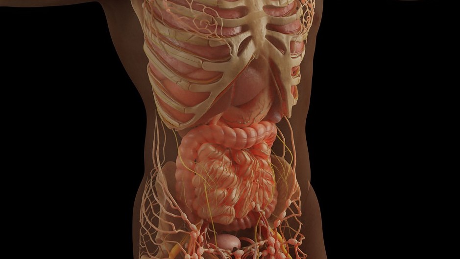
Although SARS-CoV-2 infections mainly attack the lungs, in many cases they can also damage other organs, such as the colon: around 60% of patients experienced digestive tract impacts. A study published in the International Journal of Molecular Sciences analysed the manifestations of COVID in the lungs and colon, identifying the differences at a molecular level.
Their findings serve as the basis for the identification of novel biomarkers and the development of new treatment strategies.
The University of Vienna scientific team, led by Diana Mechtcheriakova, studied the singularities and commonalities in the impact of COVID on the lungs and other organs. Using complex dataset analyses, the researchers recognised that a different molecular mechanism is at work in pulmonary and gastrointestinal manifestations. While SARS-CoV-2 infections of the lungs evoke classic immune system responses, in the gastrointestinal tract they evoke responses related to liver and lipid metabolism.
The fact that SARS-CoV-2 infections not only manifest in the lungs but frequently also manifest in other organs, such as the heart, kidneys, skin or gut, can be attributed to the particular structure of the virus. During the course of COVID, up to 60% of patients experience gastrointestinal symptoms, which may be associated with a longer duration of disease and/or a worse outcome. The results of this study will add to our understanding of the organ- and tissue-specific molecular processes triggered by SARS-CoV-2.
“Our findings can advance the identification of new biomarkers and treatment strategies for COVID, taking account of the specific responses in manifestations outside the lung,” said Diana Mechtcheriakova, Head of the Molecular Systems Biology and Pathophysiology Research Group at MedUni Vienna, holding out the prospect of promising follow-up studies.
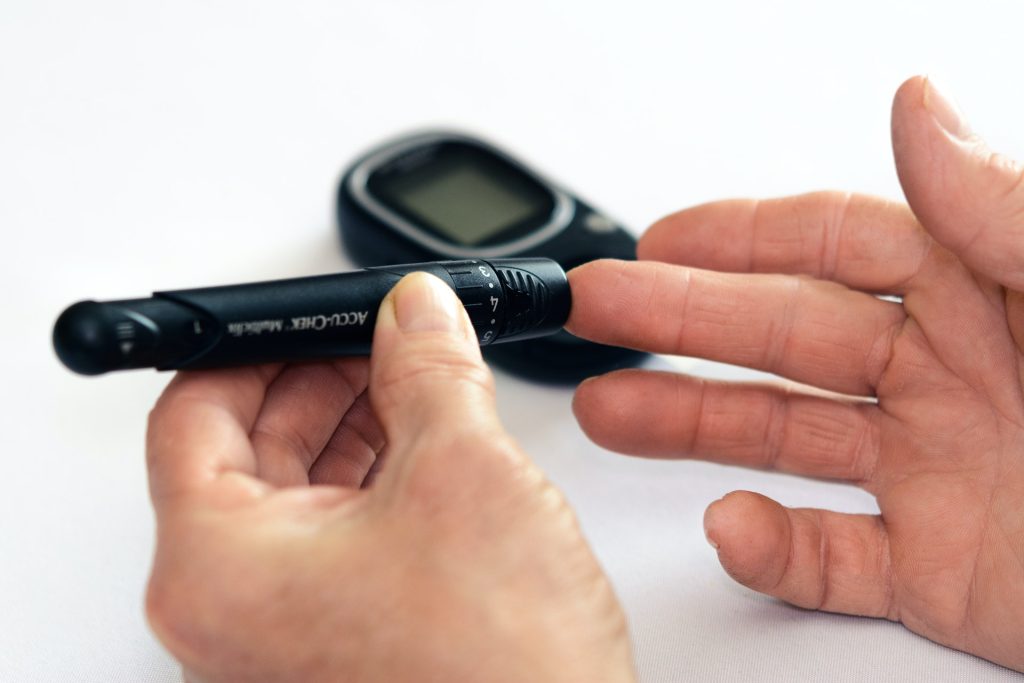
New research on Scottish data has found SARS-CoV-2 infection is linked to an increased incidence of new-onset type 1 diabetes in under-35s – but only in the first month after infection, likely due to increased testing as well as COVID bringing forward the progression of diabetes.
The study, presented at this year’s European Association for the Study of Diabetes (EASD) Annual Meeting, linked data on COVID tests results to the Scottish diabetes register for the period between March 2020 and November 2021, and tested whether this period of COVID infection was associated with an increased risk of diabetes.
“Our findings call into question whether a direct association between COVID and new-onset type 1 diabetes in adults and children exists”, says co-lead author Professor Helen Colhoun from Public Health Scotland and the University of Edinburgh, Scotland. “One recent report by researchers at the US Centers for Disease Control and Prevention (CDC), analysing two large insurance-claim databases of those under age 18, found that children with COVID were 2.5 times as likely to be diagnosed with diabetes over a month after infection than those who were never infected. If replicated, this is going to create a large number of people with newly diagnosed diabetes and might also alter the risk–benefit balance for COVID vaccination in young children. Importantly, we did not confirm that finding.”
In type 1 diabetes, which usually appears during childhood or adolescence, the immune system attacks insulin-producing cells, but it is not known why. One theory is that the immune system may be triggered by a viral infection and then accidentally also attacks insulin-producing cells. It has also been suggested that viral infections may increase the rate of progression of type 1 diabetes in people who still have normal blood sugar levels.
In this study, Prof Colhoun and colleagues linked individual-level data on PCR-confirmed SARS-CoV-2 infections from the Electronic Communication of Surveillance Database, which captures all PCR tests for COVID-19 nationally, with precise dates of all new type 1 diabetes diagnoses from the national register in Scotland (that is updated daily).
During the study period a confirmatory PCR test was mandatory for all those with a positive lateral flow test.
The important aspect of this study is the exact dates of diabetes diagnosis were available, unlike in some earlier studies, ensuring that the time sequence of COVID and type 1 diabetes could be established.
Between March 2020 and November 2021, a total of 365 080 children and adults had at least one detected SARS-CoV-2 infection, and 1074 were diagnosed with type 1 diabetes.
The analysis found no association between SARS-CoV-2 infection and new-onset type 1 diabetes 30 days or more after infection, or in those aged younger than 16 years, contrary to several previously reported studies.
However, the researchers did find that children and adults with a first positive SARS-CoV-2 test were 2.5 times as likely to be diagnosed with diabetes within 30 days of infection compared to those who did not have a previous registered infection; this risk was more than three times higher in those younger than 16 years.
But the authors stress strong arguments against a causal effect of COVID underlying this association.
Further analyses investigating the pattern of COVID- testing in relation to type 1 diabetes diagnosis found an increased frequency of SARS-CoV-2 testing in the days before and after diabetes presentation, for both negative and positive results. This suggests, says the authors, that the association may partly be explained by higher detection of infection at this time.
The authors also note that the average time from the onset of type 1 diabetes symptoms to diagnosis under 16s in England is around 25 days. So, it is likely that many of those who tested positive for COVID-19 within 30 days of a diabetes diagnosis already had type 1 diabetes at the time of infection.
No link was found between between COVID vaccination status and new-onset type 1 diabetes in adults (few children were vaccinated during the study period), providing further evidence against a causal effect of SARS-CoV-2 infection on the development of diabetes.
The researchers also looked at trends in type 1 diabetes incidence in Scottish children aged 0–14 years before and during the pandemic, finding that the incidence in 2020–2021 was around 20% higher than the 7-year average for 2015–2021.
However, they point out that based on estimates from England, the time course of the increase in diabetes incidence in those aged 0–14 years predated most of the cumulative incidence of SARS-CoV-2 infection in this age group (June 2021 onwards), suggesting no causal link between COVID and rates of diabetes.
“Our findings show that causes other than COVID infection itself need to be considered in relation to the increased incidence of type 1 diabetes”, said co-lead author Professor Paul McKeigue from Public Health Scotland and the University of Edinburgh, Scotland. “We need to consider what has happened regarding the spread of viruses such as enteroviruses during the pandemic, and whether there are any other environmental factors, such as sunlight exposure and vitamin D levels, that might have altered during lockdown that might also be relevant.”
The authors note that although their study was large, further analyses with more recent data is needed, and that the delay in mass testing meant many COVID cases in the young went undetected.
A recent study in the journal Frontiers in Immunology may explain why some cases of COVID are asymptomatic: common foods, vaccines, bacteria and viruses may all prime the immune system to attack SARS-CoV-2 due to bearing similar antigens to the virus. The study paves the way for new immunotherapies or vaccines that lead to stronger immunity against COVID.
Proteins present in bacteria, human cells, vaccines, and even foods may all share similarities with those in SARS-CoV-2. The researchers behind this latest study hypothesised that similarities between SARS-CoV-2 and other common proteins may affect our susceptibility to the virus.
After the initial infection by a pathogen has passed, T and B cells retain a memory of it, ready to rapidly produce more antibodies if needed. Food allergies are a result of the immune system targeting the proteins in what are otherwise harmless substances.
Testing antibody cross-reactions
Could such an ‘immune memory’ to proteins we have encountered in our past underlie immune resistance and reduced susceptibility to Covid-19? To begin to test this hypothesis, these researchers investigated whether antibodies that target proteins in the SARS-CoV-2 virus could also bind to proteins in other agents, such as foods or common bacteria.
The researchers tested the ability of these antibodies to bind to 180 different proteins from common foods, two different vaccines, and 15 bacterial and viral proteins. The antibodies reacted most strongly with a common gut bacterium called E. faecalis and a vaccine against diphtheria, tetanus, and pertussis. Interestingly, they also reacted very strongly against proteins found in common foods, including broccoli, roasted almonds, pork, cashews, milk, soy, and pineapple.
Eat for immunity?
Unfortunately, you will likely not be able to eat your way to COVID immunity. ‘Immunity’ against a food type, for instance, is typically characterised by a food allergy. “Usually only people with leaky gut can make antibodies against food, so I wouldn’t actually recommend eating foods that give you leaky gut, because this would give you a whole new set of problems,” said Dr Aristo Vodjani of Cyrex Laboratories in Arizona, lead author on the study.
Indeed, the researchers caution that although these agents could potentially provide some protection from SARS-CoV-2, they are no replacement for current vaccines. Further studies are also needed to confirm that these proteins do indeed confer some protection, and if so, whether it is mediated through a short-lived antibody response or a longer-term memory cell response.
The findings may shed some light on our variable responses to COVID infection. With more research, these results could lead to more effective treatments or better vaccines against the virus. Another application may lie in assessing an individual’s susceptibility to the virus before they have even been infected.
In a study published in the American Journal of Obstetrics & Gynecology, researchers found that, if a woman is infected by SARS-CoV-2 during her pregnancy, the infection damages the placenta’s immune response to further infections – even if the infection was mild.
“This is the largest study to date of placentas from women who had COVID during their pregnancies,” said Professor Kristina Adams Waldorf, at the University of Washington School of Medicine and senior author of the study. “We were surprised to find that women who had COVID during their pregnancies had placentas with an impaired immune response to new infection.”
This finding, Prof Adams Waldorf added, “was the tip of the iceberg” in how COVID might affect foetal or placental development.
During the early stages of the pandemic, many thought that COVID did not appear to harm the developing foetus because there were so few babies born with COVID infection, she noted.
“But what we’re seeing now is that the placenta is vulnerable to COVID-19, and the infection changes the way the placenta works, and that in turn is likely to impact the development of the foetus,” Prof Adams Waldorf said.
“To date, the studies about how COVID might affect foetal or child development are very limited as the children are still very young,” noted co-author Dr Helen Feltovich, professor at Intermountain Healthcare.
“Our study suggests that babies born to mothers infected with COVID at any point during their pregnancy will need to be monitored as they grow up,” she said.
Studies led by Adams Waldorf have shown that pregnant women who contract COVID have a significantly higher mortality rate than those who do not contract COVID. Other studies have found that pregnant women are more likely to risk hospitalisations or preterm birth, according to the Center for Disease Control and Prevention.
It’s unknown how different COVID variants may affect the mother or foetus, Profs Adams Waldorf and Feltovich agree.
“Studying each of the variants in real time is really challenging because they just keep coming so fast, we can’t keep up,” Prof Adams Waldorf said. “We do know that the COVID Delta variant was worse for pregnant individuals, because there was a spike in stillbirths, maternal deaths and hospitalisations at that time.”
Regardless of the variant, Prof Adams Waldorf stressed taking precautions, such as vaccination and booster shots, limiting social contact a bubble of vaccinated individuals even if it means isolating for the duration of the pregnancy.
“The disease may be mild, or it may be severe, but we’re still seeing these abnormal effects on the placenta,” she said. “It seems that after contracting COVID in pregnancy, the placenta is exhausted by the infection, and can’t recover its immune function.”
In this study, a total of 164 pregnant individuals were studied, consisting of 24 uninfected healthy patients as a control group and 140 individuals who contracted COVID. Both groups delivered at about the same time, 37 to 38 weeks. Preterm birth occurred at nearly three times the rate with the patients with COVID when compared with those without. About 75% of the COVID patients had either asymptomatic COVID or mild symptoms.
Findings from a prospective seroprevalence study in Spain reveal falling SARS-CoV-2 antibody levels in unvaccinated but infected individuals a year after their infection. This reinforces the necessity of vaccination even after infection, and confirms that hybrid immunity – gained from both infection and vaccination – is the most robust. The study findings appears in BMC Medicine.
Both infection and vaccination against SARS-CoV-2 contribute to population immunity, an important factor for deciding when and to whom booster shots should be offered. Although immunity against a pathogen is more than antibodies, the easiest strategy for assessing population immunity is to perform seroepidemiological studies.
“Most of the serological studies performed after COVID vaccination focused on specific groups such as healthcare workers, did not distinguish between people with or without previous infection, or did not have clinical and immunological data of the infection,” explained senior co-author Manolis Kogevinas, ISGlobal researcher.
In this study, the research team performed a second measurement in a population-based cohort from Catalonia six months after the start of the vaccination campaign (the first one was just after the first confinement), to monitor the level and type of antibodies against five viral antigens (the whole Spike (S) protein, the RBD receptor binding domain, the S2 fragment, the full nucleocaspid (N) protein, or the N-terminal fragment). They also used information from a questionnaire and health records to identify potential factors that determine the magnitude and duration of the antibody response in unvaccinated, vaccinated, or vaccinated and infected persons. A total of 1076 people, aged 43 to 72 years, were included in the analysis.
The results yielded three main conclusions: First, that in 36% of infected but unvaccinated persons, antibodies were no longer detectable almost a year after the infection, particularly in those older than 60 years and smokers.
The second conclusion was that vaccination induced significantly higher antibody levels in people who had a prior infection, as compared to those without prior infection; and that these levels were strongly associated with the magnitude of the response during the infection. “Our data underscore the importance of vaccinating people even if they have been previously infected, and confirm that hybrid immunity is superior and more durable. This means that people who have been vaccinated but have not been infected would need a booster earlier than those who have,” pointed out Marianna Karachaliou, first author of the study together with Gemma Moncunill.
The third was that the factor most strongly associated with the level of antibodies is the type of vaccine, with Moderna’s Spikevax generating the highest levels of antibodies. Other factors also appear to play a role: people older than 60 or with mental illness had lower antibody levels post-vaccination. “The association between mental health and antibody responses requires further investigation, but it is known that people with disorders such as depression, chronic stress or schizophrenia have a lower response to vaccination in general,” explained co-author Carlota Dobaño.
Among those vaccinated, only 2.1% had no antibodies at the time of testing and approximately 1% had a breakthrough infection. “However, it should be noted that this study was done before the Omicron variant became dominant,” warned Kogevinas.
A major scientific report from Africa is featured in the journal Science today. This scientific report shows how the rapid expansion of genomics surveillance in Africa allowed the continent to describe the introduction and spread of the SARS-CoV-2 variants in African countries in real time during the COVID pandemic.
The scientific report includes over 300 authors from Africa and abroad who worked together to describe and analyse over 100 000 genomes and characterise SARS-CoV-2 variants in real time. This was the largest consortium of African scientists and public health institutions ever to work together to support data-driven COVID response in Africa.
This report shows how the large investment, collaboration and capacity building in genomic surveillance on the African continent enabled real-time public health response. Particularly it describes the setting up of the Africa Centres for Disease Control (CDC) – Africa Pathogen Genomics Initiative (Africa PGI) and the continental network by the Africa CDC and World Health Organisation (WHO) Regional Office for Africa (WHO AFRO) to expand access to sequencing and cover surveillance blind spots, in parallel with the growth of the number of countries that are able to sequence SARS-CoV-2 within their own country.
“The publication highlights that sustained investment for diagnostics and genomic surveillance inAfrica was needed to not only combatSARS-CoV-2 on the continent, but establish a platform to address the emerging, re-emerging, endemic infectious disease threats, such as Ebola, HIV/AIDS, TB and Malaria. These investments are crucialfor pandemic preparedness and response and will serve the health of the continent well intothe 21st century,” said Dr. Yenew Kebede, Head Division of Laboratory Systems and Acting Head: Surveillance and Disease Intelligence at the Africa CDC.
African Scientists receiving training in genomics surveillance at the KwaZulu-Natal Research Innovation and Sequencing Platform (KRISP), South Africa.
“The enormous leap Africa made in genomic surveillance during the past two years is the silver lining in the COVID pandemic,” said Dr Matshidiso Moeti, WHO Regional Director for Africa. “The continent is now better prepared to face down both old and emerging pathogens. This is a model of how when Africans are in the driving seat we can come up with lasting change and stay a step ahead of dangerous diseases.”
“It has been an inspiring experience to continuously share knowledge, support and learn from colleagues all over the continent during the pandemic. We witnessed small countries with no previous genomics experience become empowered in sequencing and bioinformatics methods, and how they started to actively participate in regular pathogen genomic surveillance for SARS-CoV-2. I think it will be a real model of how scientists and public health officials across countries can form a unified front against infectious diseases in the future,” says Houriiyah Tegally, Bioinformatician at KRISP and CERI and first author on this report.
The results also highlight the early warning capacity that genomic surveillance in Africa has had for the rest of the world with the detection of new lineages and variants, the most relevant being the detection of the Beta and various Omicron subvariants. The report highlights that most SARS-CoV-2 variants, which caused an epidemic in Africa, were introduced from abroad.
The scientists proceeded carefully in analysing genomic and epidemiological data collected in over 50 countries that experienced quite heterogenous epidemics in order to reconstruct transmission dynamics of the virus in the most accurate way. “The phylogeographic methods that we employ to investigate the movement of the SARS-CoV-2 virus and its variants into, out of, and within the African continent account for uneven testing and sampling proportions across countries, arising from the realities of doing genomic sequencing in the middle of a pandemic, often in low resourced settings,” explains Dr Eduan Wilkinson, head of bioinformatics at CERI at Stellenbosch University and senior author on this report.
The initial waves of infections in Africa were primarily seeded by multiple introductions of viral lineages from abroad (mainly Europe). The Alpha variant that emerged in Europe at the end of 2020, was responsible for infections in 43 countries with evidence of community transmission in Ghana, Nigeria, Kenya, Gabon and Angola. For the Delta variant, the bulk of introductions were attributed to India (~72%), mainland Europe (~8%), the UK (~5%), and the US (~2.5%). Viral introductions of Delta also occurred between African countries in 7% of inferred introduction. For Omicron, the scientific results indicate more reintroductions of the variant back into Africa, with at least 69 (95% CI: 60 – 78) from Europe and 102 (95% CI: 92 – 112) from North America than from other African countries. This was amplified for Omicron BA.2; the results suggest at least 99 separate introduction or reintroduction events of BA.2 into African countries, ~65% of which are from Europe and ~30% from Asia.
“The ironical part of these results is that most of the introductions of variants in Africa were from abroad, but Africa was the most discriminated and penalized continent in the world with travel bans imposed. Instead of unscientific and inappropriate reactions, we should be building on the infrastructure established in Africa so that the continent can rapidly pivot to other epidemics without the fear of being punished,” says Prof Tulio de Oliveira, director of CERI and KRISP, which lead the consortium analysis with the Africa CDC and WHO AFRO.
“This study is a testament of the Africa CDC – Africa PGI efforts to expand access to sequencing to member states and create a platform of coordination and collaboration among institutions within and outside of the continent,” said Dr. Ahmed Ogwell, Acting Director of the Africa CDC.