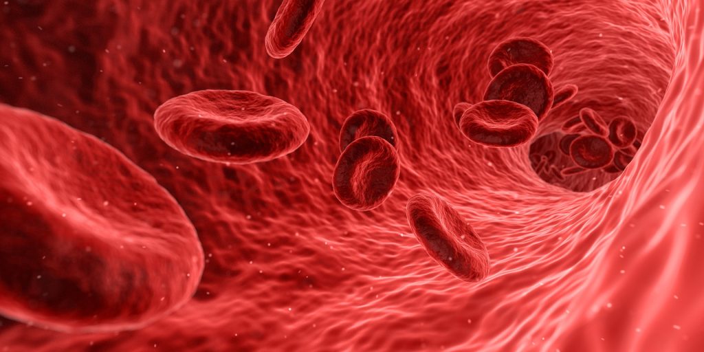
New research is raising concerns that long-term use of renin-angiotensin system (RAS) inhibitor drugs such as ACE inhibitors could be contributing to kidney damage.
The researchers stress that patients should continue taking the medications. But the scientists are urging studies to better understand the drugs’ long-term effects.
“Our studies show that renin-producing cells are responsible for the damage. We are now focusing on understanding how these cells, which are so important to defend us from drops in blood pressure and maintain our well-being, undergo such transformation and induce kidney damage,” said UVA’s Dr Maria Luisa Sequeira Lopez. “What is needed is to identify what substances these cells make that lead to uncontrolled vessel growth.”
A billion people around the world are affected by chronic hypertension. In a study published in JCI Insight, University of Virginia (UVA) researchers were seeking to better understand why severe forms of the condition are often accompanied by atherosclerosis in the kidney, leading to organ damage.
They found that renin cells, which help regulate blood pressure through renin production, play an important role. Harmful changes in the renin cells can cause the cells to invade the walls of the kidney’s blood vessels. The renin cells then trigger a buildup of another cell type, smooth muscle cells, that cause the vessels to thicken and stiffen, resulting in impeded kidney blood flow.
Long-term use of RAS inhibitor drugs, such as ACE inhibitors, or angiotensin receptor blockers, have a similar effect. But the study found that long-term use of the drugs was associated with hardened kidney vessels in both lab mice and humans
The researchers note that the medications can be lifesaving for patients, so they stress the importance of continuing to take them. But they say additional studies are needed to better understand the drugs’ long-term effects on the kidneys.
“It would be important to conduct prospective, randomised controlled studies to determine the extent of functional and tissue damage in patients taking medications for blood pressure control,” said UVA’s Dr Ariel Gomez. “It is imperative to find out what molecules these cells make so that we can counteract them to prevent the damage while the hypertension is treated with the current drugs available today.”
David Bennett, a 57 year old US man, is doing well after being the world’s first human transplant of a pig heart, according to the man’s son, David Bennett Jr.
When his father first told him of the pig heart option, his son was incredulous, telling the BBC: “I didn’t believe him, I thought he was suffering from delirium at first.”
However, when he did some research on the work done, he realised it was a reality and that they were “walking into the unknown”.
He added that according to Dr Bartley Griffith, who performed the surgery, his father has a good prognosis of 6–9 months. The experimental seven-hour procedure at the University of Maryland Medical Center in Baltimore was considered the last hope of saving Mr Bennett’s life, though it is currently unclear what his long-term chances of survival are.
“It was either die or do this transplant,” Mr Bennett explained a day before the surgery, adding that it was his “last choice”.
Dr Griffith said heart failure and an irregular heartbeat made him ineligible for a human heart transplant or a heart pump.
Xenotransplantation, as these inter-species transplants are called, have failed, largely because patients’ bodies quickly rejected the animal organ. Notably, in 1984, Baby Fae, a dying infant, lived 21 days with a baboon heart.
What makes this attempt different is that the Maryland surgeons used a heart from a pig that had been genetically modified to remove a sugar in its cells that’s responsible for that hyper-fast organ rejection. Many biotech companies are working on adapting pig organs for xenotransplantation.
“I think you can characterise it as a watershed event,” Dr David Klassen, chief medical officer at the United Network for Organ Sharing (UNOS), which oversees the US transplant system.
Dr Klassen nevertheless cautioned that it’s only a first tentative step into exploring whether xenotransplantation might finally work this time.
The Food and Drug Administration, which oversees such experiments, allowed the surgery under what’s called a “compassionate use” emergency authorisation, available when a patient with a life-threatening condition has no other options.
Surgeon Bartley Griffith said the surgery would bring the world “one step closer to solving the organ shortage crisis”. At present, 17 people die every day in the US waiting for a transplant, with more than 100 000 reportedly on the waiting list. A record 3800 heart transplants were done last year, according to the UNOS.
Physicians have recommended that children with autism spectrum disorder (ASD) receive screening for abnormally high or low cholesterol levels at least once during their childhood, since ASD is a risk factor for cardiovascular disease in both children and adults.
The recommendation stemmed from a recent study, published in Translational Psychiatry, that found reduced levels of high density lipoprotein cholesterol (HDL-C) in individuals from families with two or more children with ASD. Additionally, they found reduced or elevated levels of other lipids, apolipoprotein A1 (ApoA1) and apolipoprotein B (ApoB). Individuals with low HDL-C levels or ApoA1 levels had lower adaptive functioning than other individuals with ASD.
“This latest research is part of our ongoing work to understand some of the co-occurring conditions with ASD,” said Elaine Tierney, MD, a child and adolescent psychiatrist with Kennedy Krieger Institute. “Our work indicates that lipids are abnormal in many individuals with ASD. Our findings, in addition to studies that show an increase in heart disease in individuals with ASD, lead us to recommend that children with ASD be screened for abnormal total and HDL cholesterol levels. We hope our work underscores the importance of cholesterol screening and raises awareness for families in the ASD community.”
Previously, Dr Tierney and colleagues identified that Smith-Lemli-Opitz Syndrome (SLOS), a genetic condition of impaired cholesterol biosynthesis, is associated with autism. This led to a recommendation that all children with ASD be screened for SLOS if they exhibit some of its characteristics, such as slow growth, microcephaly, mental retardation and other birth defects, although the severity of this rare disease can vary.
A novel study has isolated powerful anticoagulants from the saliva of ticks, which may have reduced potential for bleeding.
Blood-feeding animals rely on specific molecules in their saliva to overcome defence mechanisms of their mammalian hosts for successful survival. The saliva of ticks, for example, contains molecules that can prevent blood from clotting, and which can also suppress inflammation or immune response to enable continuous feeding on the same bite site for days, sometimes undetected by the host. The harmful effects of these parasites can actually be harnessed for medical treatments.
In their paper, published in Nature Communications, the authors explain how the cardiovascular team developed a series of thrombin inhibitors to be potent anticoagulants based on sequences of inhibitors of blood coagulation enzyme thrombin found in the tropical bont tick Amblyomma variegatum.
The team developed a series of thrombin inhibitors to be powerful anticoagulants.
Anticoagulants are used in conditions where there is an increased propensity to form blood clots in our body depriving blood supplies to important tissues and organs, otherwise known as thrombosis. These drugs are needed in many diseases caused by blood clots including heart attacks, strokes, deep vein thrombosis, pulmonary embolism and even some severe complications caused by SARS-CoV-2 infection.
These next-generation anticoagulants will now need to be tested in human trials to determine if they can effectively counteract clotting without the bleeding side effects of currently available anticoagulants.
Chronic exposure to air pollution in the form of particulate matter contributes to the risk of cardiovascular and respiratory diseases, and in particular has been linked to hypertension, according to a study published in Scientific Reports.
Air pollution, accounting for more than 4.2 million deaths annually, is a significant health risk. The study assessed the impact of particulate pollution on the long-term incidence of hypertension in Spain, supporting the need to improve air quality to the extent possible in order to reduce the risk of cardiometabolic diseases among the population.
To this end, researchers have carried out a study, di@bet.es, which recruited 1103 participants aged 18–83. None of the participants presented with hypertension at the start of the study (2008–2010), and they were monitored until 2016–17. Participants were assigned air pollution concentrations for particulate matter, obtained through modeling and air quality readings. During this period, 282 cases of incident hypertension were recorded.
The study was carried out in collaboration with the air pollution department of the Research Centre for Energy, Environment and Technology (CIEMAT).
As explained by endocrinologist Sergio Valdés, “Several previous studies have described the short- and long-term association of ambient air pollutants with hypertension and blood pressure levels, but few studies have addressed the association between long-term exposure to these particles and the incidence of hypertension in a prospective manner. Therefore, the di@bet.es study has offered us the opportunity to do so in the Spanish population.”
Participants underwent a medical examination and had blood samples taken. They also answered questionnaires to obtain demographic information and variables such as smoking, exercise and diet.
Gemma Rojo, last study author, stated that “our data is consistent with a large body of evidence suggesting that air pollution may contribute to the pathogenesis of hypertension. It also supports the idea that the particulate component of air pollution is the greatest threat to the cardiovascular system.”
In this regard, she noted, “Although previous associations between exposure to gaseous pollutants and hypertension have shown some discrepancies, most studies reporting long-term exposure to particulate matter and incident high blood pressure have reported positive associations consistent with our findings.”
As Sergio Valdés explained, “our results support the need to improve air quality to the extent possible in order to reduce the risk of high blood pressure among our population, as even moderate levels such as those we report here increase the risk significantly.”
Human heart muscle cells stop multiplying after birth, making any heart injury later in life a permanent one, reducing function and leading to heart failure. Now, however, researchers have new evidence that manipulating certain nerve cells or their controlling genes might trigger the formation of new heart muscle cells and restore heart function after heart attacks and other cardiac disorders.
The study, published in Science Advances, sheds new light on how some neurons regulate the number of heart muscle cells. While nerve cells have long been known to regulate heart function, their role and impact during heart development and their effect on muscle cell growth has been unclear.
“Our study sought to examine the role of so-called sympathetic neurons on heart development after birth, and what we found is that by manipulating them, there could be tremendous potential for regulating the total number of muscle cells in the heart even after birth,” said lead author Emmanouil Tampakakis, MD, assistant professor of medicine at the Johns Hopkins University School of Medicine.
The nerve cells that make up the sympathetic nervous system (SNS) control automatic processes in the body such as digestion, heart rate and respiration. The SNS is typically associated with ‘fight-or-flight’ responses.
Researchers in this study made a genetically modified mouse model by blocking sympathetic heart neurons in developing mouse embryos, and analysed the drivers of heart muscle cell proliferation through the first two weeks of life after birth.
What they found was a significant decrease in the activity of a pair of genes – the period 1 and period 2 genes – already known to control the circadian cycle. Remarkably, removing those two circadian genes in mouse embryos, the researchers saw increased neonatal heart size and an increase in the number of cardiomyocytes, or heart muscle cells, by up to 10%. Thus, sympathetic nerves on heart muscle cells could likely be mediated through these two circadian genes.
Circadian, or ‘clock’, genes are components of the circadian rhythm pattern that in mammals regulates bodily functions on a roughly 24-hour cycle aligned with hours of daylight and darkness.
“Shortly after birth, mammals, including people and mice, stop producing heart muscle cells. And unlike other organs, like the liver, the heart can’t regenerate after it’s damaged,” said Prof Tampakakis. “We’ve shown that it may be possible to manipulate nerves and/or circadian genes, either through drugs or gene therapies, to increase the number of heart cells after birth.”
Up to a billion heart muscle cells can be lost after a heart attack, and Prof Tampakakis says there is scientific evidence that hearts tend to recover faster after an attack when the total number of cells to begin with is higher. By manipulating sympathetic nerves and clock genes (a technique called neuromodulation) researchers believe the heart could be made to respond to injury much better.
“Neuromodulation is a pretty new concept in cardiology, and we believe these are the first reports that associate clock genes with new growth of heart muscle cells.” saidChulan Kwon, PhD, MS, associate professor of medicine at the Johns Hopkins University School of Medicine. “Our study, maybe for the first time, shows what’s happening if you block the supply of nerves to the heart, and provides new insights for developing neuromodulation strategies for cardiac regeneration.”
A large study has shown that apolipoproteins apoB and apoA-1 together provide early and reliable cardiovascular risk information as well as levels of low-density lipoprotein (LDL) cholesterol. The researchers advocate introducing new guidelines for detecting cardiac risk and say the results, published in PLOS Medicine, may pave the way for early treatment, which could help lower morbidity and mortality rates.
Cardiovascular disease is the most common cause of death globally and includes a wide range of conditions, such as stroke and myocardial infarction with atherosclerosis in different organs of the body. In many cases the disease can be prevented and arrested with lifestyle changes and lipid-lowering treatments using statins and other methods.
The cardiac risk assessment usually uses reference values for the LDL cholesterol. Other types of fat particles can also be measured along with apolipoproteins, which transport cholesterol in the blood. International guidelines for cardiovascular disease recommend using apolipoprotein apoB, which transports LDL cholesterol, as an alternative risk marker for people with type 2 diabetes, overweight and very high levels of blood lipids.
Recent research has, however, indicated the importance of also factoring in the apolipoprotein apoA-1, which transports the protective and anti-inflammatory HDL cholesterol. Calculating the apoB/apoA-1 ratio gives a risk quotient reflecting the balance between the fat particles that expedite atherosclerosis and the “good” protective apoA-1 particles that arrest the process.
In this present study, the researchers have analysed the link between cardiovascular disease and apoB/apoA-1 values in more than 137 000 Swedish adults between the ages of 25 and 84. The individuals were followed for 30 years, during which time 22 000 suffered some form of cardiovascular event. The analysis methods are simple, inexpensive and safe, and do not require pre-test fasting, as is the case with LDL and non-HDL tests. Basing their study on a large database, the researchers linked the laboratory analyses to several clinical diagnosis registers.
“The results show that the higher the apoB/apoA-1 value, the greater the risk of myocardial infarction, stroke and need for coronary surgery,” says Göran Walldius, senior author and professor emeritus at the Institute of Environmental Medicine, Unit of Epidemiology, Karolinska Institutet. “The study also showed that the risk was amplified in the presence of low protective levels of apoA-1.”
Individuals with the highest apoB/apoA-1 values had a 70% higher risk of severe cardiovascular disease and almost triple the risk of non-fatal myocardial infarction compared with those with the lowest apoB/apoA-1 values. Individuals with the highest risk quotient were also more affected by severe cardiovascular diseases many years earlier than individuals with the lowest apoB/apoA-1 values.
The relationship was observed in both men and women and the elevated levels could be detected as early as 20 years before the onset of cardiovascular disease.
“Early preventive treatment and information about cardiovascular risk is, of course, important in enabling individuals to manage their risk situation,” Walldius says. “Early treatment can also reduce the cost burden on the public health services.”
Taken together, the results suggest that the apoB/apoA-1 ratio is a better marker for identifying at-risk individuals for cardiovascular disease compared to the apoB method alone.
“It should be possible to introduce cut-values for apoB, apoA-1 and the apoB/apoA-1 ratio into new guidelines as a complement to current guidance on the detection and treatment of dyslipidaemia,” said Walldius.
Dapagliflozin, widely used to treat type 2 diabetes, was shown to improve symptoms and physical limitations in patients with heart failure with preserved ejection fraction, according to clinical trial results reported in Nature Medicine.
Heart failure with preserved ejection fraction (HFpEF) occurs when the heart’s lower left chamber is unable to fill with blood properly. The condition accounts for approximately half of all heart failure cases and disproportionally affects older individuals. Patients with HFpEF can experience a host of debilitating symptoms linked to cardiometabolic abnormalities, including physical limitations, impaired cognition and poor quality of life. Life expectancy is also reduced for patients with this diagnosis, with 50% of patients with the diagnosis not expected to survive more than five years.
Finding ways to improve patients’ health and developing or identifying therapeutic interventions that not only reduce hospitalisation but also improve patient survival is key, the researchers said, but at present there are no available treatments that improve patient survival for patients with HFpEF.
Previous studies have shown that sodium glucose cotransporter 2 (SGLT2) inhibitors – which inhibit SGLT2 receptor proteins produced by the kidneys and are used to treat type 2 diabetes – reduces risk of cardiovascular death and heart failure-related hospitalisation in patients with HFpEF.
For this trial, the researchers measured patient-reported symptoms, physical limitations and function in patients with HFpEF who were taking dapagliflozin, an SGLT2 inhibitor drug.
A total of 324 patients with HFpEF, 56.8% women, were randomised to receive either dapagliflozin or placebo for 12 weeks and at the end of the trial were evaluated using the Kansas City Cardiomyopathy Questionnaire Clinical Summary Score, a measure of heart failure-related health status. “It’s important to note the percentage of women that were enrolled in this study because usually women are under-enrolled in clinical trials,” pointed out study co-author Professor Sadiya Khan.
Compared to the placebo group, an overall improvement in patient-reported symptoms, physical limitations and exercise function was seen in the dapagliflozin group. Adverse events were also similar between both groups, the authors reported.
“It was definitely surprising and very exciting to see such a stark difference between the treatment group and the placebo group, that there was this clear separation that happened even over a short period of time,” Prof Khan said, adding that next steps will be to investigate dapagliflozin’s precise molecular mechanisms that enable its effectiveness.
Twenty years ago, clinicians first attempted to regenerate a failing human heart by injecting muscle myoblasts into the heart during a bypass operation. Despite high initial hopes and multiple studies since then, attempts to remuscularise an injured heart have met with little, if any, success.
Yet, there is hope that a therapy will be developed, according to experts in a Journal of the American College of Cardiology state-of-the-art review. The challenge is this: A heart attack kills heart muscle cells, leading to a scar that weakens the heart, often causing eventual heart failure. The lack of muscle repair is due to the very limited ability of mammalian heart muscle cells to proliferate, except during a brief period around birth.
In the review, the experts focus on three topics. First are several recent clinical trials with intriguing results. Second is the current trend of using cell-derived products like exosomes rather than muscle cells to treat the injured heart. For the third topic, authors discuss likely future experiments to replace a myocardial scar with heart muscle cells by ‘turning back the clock’ of the existing cardiomyocytes, rather than trying to inject exogenous cells. These efforts try to reverse the inability of mature mammalian heart muscle cells to proliferate.
Clinical trials One of the clinical trials reviewed involved giving cardiosphere-derived cells to patients with Duchenne muscular dystrophy, which affects both heart and skeletal muscles.
Cardiosphere-derived cells are a type of heart stromal/progenitor cell that has potent immunomodulatory, antifibrotic and regenerative activity in both diseased hearts and skeletal muscle. The HOPE-2 trial gave repeated intravenous doses of cardiosphere-derived cells to patients with advanced Duchenne disease, most of whom were unable to walk. Preliminary results showed safety, as well as major improvements in heart parameters such as left ventricle ejection fraction and reduced left ventricle size.
The HOPE-2 trial evaluated a repeated sequential dosing regimen of cell therapy for any cardiac indication, evaluated intravenous cardiosphere-derived administration, and clinically benefitted Duchenne patients.
Two features of the trial may bode well: a move away from invasive cardiac-targeted cell delivery and toward easily administered intravenous cell delivery, and the use of sequential repeated cell doses.
Cell-derived products Few cells transplanted into the heart survive, though some functional benefits in heart performance have been seen despite physical clearance of grafted cells. It could be possible that the cells were acting not as replacements but rather boosters of endogenous repair pathways through the release of a wide array of tissue-repairing biomolecules. This led to investigation of using cell-derived products rather than transplanting cells. Most of these biomolecules – proteins and non-coding nucleic acids – are enclosed in tiny vesicles that cells release naturally. When the vesicles, including exosomes, merge into recipient cells, the biomolecules can modulate signaling pathways. Using vesicles or exosomes involves a simpler manufacturing process compared with live cells, the ability to control quality and potency, and being able to refrigerate the vesicles to make administration simpler.
An alternative approach to the vesicle cell-derived products was the finding that injected stem cells can promote cardiac repair through release of biologically active molecules acting as short-range, paracrine hormones. These molecules are distinct from the biomolecules in vesicles or exosomes.
However, before use of any of these cell-derived products for heart repair in early trials, the reviewers say, more experiments are needed in purification of the products, potential modes of delivery and the suitability of repeated doses.
Proliferation of endogenous heart cells The final review topic looked ahead toward endogenous generation of cardiomyocytes – in other words, forcing existing native cardiomyocytes to divide, or other cells to become cardiomyocytes.
Pigs can regenerate heart muscle for only a few days after birth. But in one remarkable study, researchers injured the heart by removing part of the apex of the left ventricle one day after birth, and then induced heart attack 28 days after birth. Control pigs without the Day 1 resection showed no repair of heart attack damage at Day 56. In contrast, the pigs that had a resection one day after birth, and then had experimental heart attacks at Day 28, showed heart repair by Day 56 – notably an absence of dead heart muscle, known as an infarction. Furthermore, these pigs had more cardiomyocytes throughout their left ventricles.
This study showed that heart muscle cells in large mammals can be induced to proliferate and regenerate by inducing a heart injury at Day 1 to extend the neonatal regneration window. “If this cardiomyocyte cell-cycle activation can be activated in neonates, the same signaling pathways may be activated in adults as well,” the authors wrote, “which is highly impactful and significant.”
Another possible approach to endogenous generation is the direct programming of cardiac fibroblasts into cardiomyocytes. Inducing proliferation of cardiomyocytes will also need ways to promote growth of heart blood vessels to supply the new cardiomyocytes.
In conclusion, the authors believe that short-term approaches to clinical trials of post heart-attack therapies will use cells like cardiospheres or cell products. The longer-term approach, the reviewers said, will target “a more direct remuscularisation of the injured left ventricle by ‘turning back the clock’ of the cardiomyocyte cell-cycle or generating new cardiomyocytes from other cell types such as fibroblasts.”
“However, the efficiency and safety of these strategies, particularly their ability to generate cardiomyocytes seamlessly coupled with their native counterparts and to allow a regulation of these induced proliferative events preventing an uncontrolled and harmful cardiac growth, still need to be appropriately addressed before moving to clinical applications.”
Researchers have found that, out of possible triggers they tested, only alcohol use was consistently associated with more episodes of atrial fibrillation (AF). The study, published in JAMA Cardiology, did not find that the other triggers, caffeine, sleep deprivation and sleeping on the left side, to be associated with the common heart condition.
A surprising finding was that, although most of the things that participants thought to be related to their AF were not, those in the intervention group still had less arrhythmia than the people in a non self-monitoring control group.
“This suggests that those personalised assessments revealed actionable results,” said lead author Gregory Marcus, MD, MAS, professor at University of California, San Francisco. “Although caffeine was the most commonly selected trigger for testing, we found no evidence of a near-term relationship between caffeine consumption and atrial fibrillation. In contrast, alcohol consumption most consistently exhibited heightened risks of atrial fibrillation.”
Although caffeine was the most commonly selected trigger for testing, we found no evidence of a near-term relationship between caffeine consumption and atrial fibrillation.
In a brainstorming session, patients had said researching individual triggers for AF was their top priority, giving rise to the I-STOP-AFib study, which enabled individuals to test any presumed AF trigger. About 450 people participated, 58% male and 92% white.
Participants used a mobile electrocardiogram recording device along with a phone app to log potential triggers like drinking alcohol and caffeine, sleeping on the left side or not getting enough sleep, eating a large meal, a cold drink, or sticking to a particular diet, engaging in exercise, or anything else they thought was relevant to their AF. While participants were most likely to select caffeine as a trigger, there was no association with AF. Recent research has similarly failed to show a link between caffeine and arrhythmias – on the contrary, investigators found it may have a protective effect.
The new study demonstrated that consumption of alcohol was the only trigger that consistently resulted in significantly more self-reported AF episodes.
The individualised ‘n-of-1’ testing method did not validate participant-selected triggers for AF. But trial participants did report fewer AF episodes than those in the control group, and the data suggest that behaviours like avoiding alcohol could lessen the chances of having an AF episode. “This completely remote, siteless, mobile-app based study will hopefully pave the way for many investigators and patients to conduct similar personalised ‘n-of-1’ experiments that can provide clinically relevant information specific to the individual,” said Prof Marcus.