An important new clue for preventing and treating gliomas has been identified in research published in the journal Science, providing a rare window into the biological changes behind glioma development.
In animal models, a team of researchers from Mayo Clinic and Mount Sinai Hospital found that those with a change in DNA known as germline alteration rs55705857 developed gliomas much more frequently and twice as fast compared to animal models without the alteration. In addition to brain tumours, the findings are relevant to other cancers and diseases.
“While we understand much of the biologic function of germline alterations within genes that code for proteins, we know very little about the biologic function of germline alterations outside of genes that code for proteins. In some way, these germline alterations interact with other mutations in cells to accelerate tumour formation,” said co-lead author Robert Jenkins, MD, PhD. “Based on this new understanding of its mechanism of action, future research may lead to novel and specific therapies that target the rs55705857 alteration.”
The study offers new knowledge that may help clinicians determine, pre-surgery, whether a patient has a glioma.
“We expected that rs55705857 would accelerate low-grade glioma development, but we were surprised by the magnitude of that acceleration,” said co-lead author Daniel Schramek, PhD.
There are many alterations, likely thousands, outside of genes associated with the development of cancer and other diseases, but the mechanism of action is only understood for very few, Dr Schramek said.
This study demonstrates that, with the tools of modern molecular/cell biology, it is possible to decipher much of the mechanism of action of such alterations.
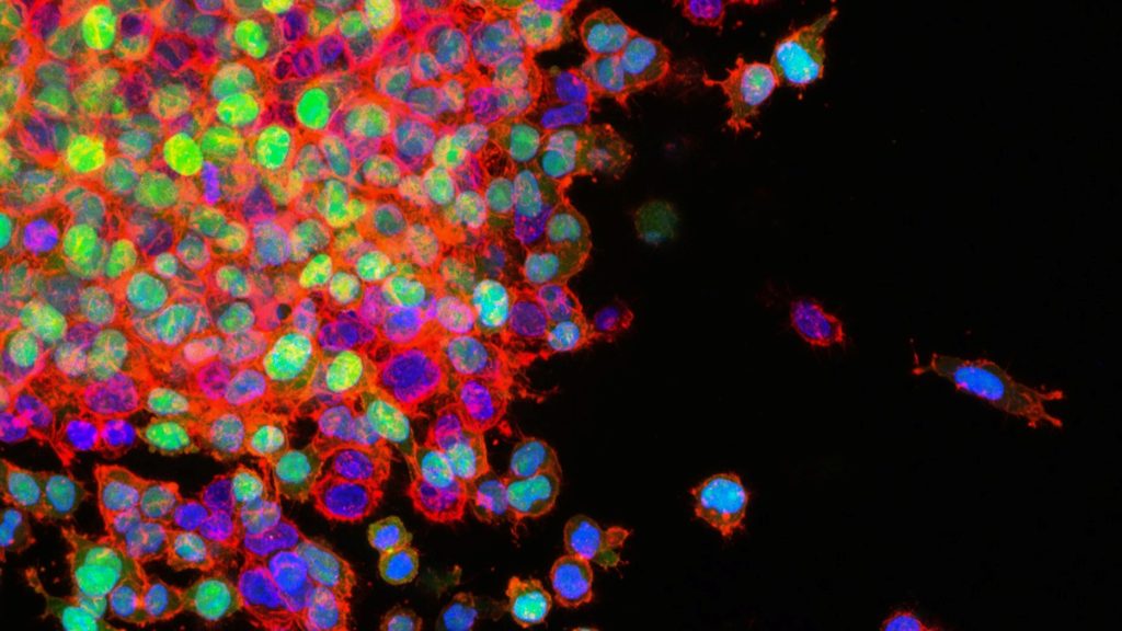
Colon cancer cells. Source: National Cancer Institute on Unsplash
A randomised study of northern European data shows that colonoscopy screening reduces the risk of colorectal cancer by 18%, much smaller than experts previously assumed. The results of the study appear in the New England Journal of Medicine.
Prior to the publication of this study, experts assumed that screening with colonoscopy had significantly better effect than screening with faecal tests. Faecal tests are used in colorectal cancer screening programs in many countries, and other countries have introduced screening with colonoscopy based on the fact that researchers via observational and modelling studies estimated that up to nine out of ten cases of colorectal cancer could be prevented with a colonoscopy screening. With faecal tests, similar models has estimated the effect to be two to three out of ten.
In the NordICC study, the researchers investigated the extent to which colonoscopy screening actually prevents colorectal cancer. Overall, 1.2% of those randomised to no screening were diagnosed with colon cancer during ten years, compared to 0.98% in those offered screening.
This translates to an 18% reduced risk of colorectal cancer among the participants who were offered colonoscopy screening. Furthermore, 455 colonoscopies were required to prevent one single case of colorectal cancer. Colonoscopy is fairly invasive and costly procedure, involving preparation, bowel prep with laxatives, and a 30-45 minute examination of the bowel with a camera inserted via the rectum. The figure of 455 procedures to prevent one case of cancer is certainly disappointing, Louise Emilsson concluded.
Colorectal cancer mortality was also found to be lower than expected in the NordICC study. Only three in a thousand died of the disease within ten years, regardless of whether they were offered screening or not, and thus, there was no significant difference between the groups in terms of mortality. The low mortality rate is however encouraging and likely caused by significantly improved treatment options over the past ten years.
Cambridge University scientists have discovered that cancer cells ‘hijack’ a process used by healthy cells to spread around the body, challenging how cancer metastasis is currently understood. Publishing in Nature Genetics, the team found that blocking the activity of a sodium channel protein in cells in mice with cancer triggers metastasis.
To their surprise, the investigators also discovered that this process extends beyond just cancer: when they removed NALCN from cancer-free mice, this caused their healthy cells to leave their original tissue and travel around the body where they joined other organs.
They found, for example, that healthy pancreas cells migrated to the kidney where they became healthy kidney cells. This suggests that metastasis isn’t an abnormal process limited to cancer as previously thought, but is in fact a normal process used by healthy cells that has been exploited by cancers to generate metastases.
Group Leader of the study, Professor Richard Gilbertson, said: “These findings are among the most important to have come out of my lab for three decades. Not only have we identified one of the elusive drivers of metastasis, but we have also turned a commonly held understanding of this on its head, showing how cancer hijacks processes in healthy cells for its own gains. If validated through further research, this could have far-reaching implications for how we prevent cancer from spreading and allow us to manipulate this process to repair damaged organs.”
Metastasis has been challenging to treat because of the difficulty in identifying key drivers of this process that could be targeted by drugs. Now that they have identified NALCN’s role in metastasis, the team are looking into various ways to restore its function, including using existing drugs on the market.
Lead researcher on the study Dr Eric Rahrmann, said: “We are incredibly excited to have identified a single protein that regulates not only how cancer spreads through the body, independent of tumour growth, but also normal tissue cell shedding and repair. We are developing a clearer picture on the processes that govern how cancer cells spread. We can now consider whether there are likely existing drugs which could be repurposed to prevent this mechanism from triggering cancer spreading in patients.”
NALCN stands for sodium (Na+) leak channel, non-selective. Sodium leak channels are expressed predominately in the central nervous system but are also found throughout the rest of the body, controlling the amount of sodium that goes in and out of the cell. Controlling this process also alters the balance of electricity across the cell membrane. It is still unclear why these channels seem to be implicated so directly in cancer metastasis.
Why breast cancer survivors experience troubling cognitive problems is a long-standing mystery, and for which inflammation is one possible culprit. A new long-term study of older breast cancer survivors published in the Journal of Clinical Oncology adds evidence to this link.
Higher levels of the inflammatory marker C-reactive protein (CRP) were related to older breast cancer survivors reporting cognitive problems in the new study.
“Blood tests for CRP are used routinely in the clinic to determine risk of heart disease. Our study suggests this common test for inflammation might also be an indicator of risk for cognitive problems reported by breast cancer survivors,” said study lead author Judith Carroll, an associate professor at UCLA.
The Thinking and Living with Cancer (TLC) Study is one of the first long-term efforts to examine the potential link between chronic inflammation and cognition in breast cancer survivors 60 and older, who make up a majority of the nearly 4 million breast cancer survivors in the United States. Previous research has focused largely on younger women and women immediately after therapy, making it difficult to draw conclusions about CRP’s role in long-term cognitive problems among older breast cancer survivors.
In TLC, teams of researchers from around the country talked to, and obtained blood samples from, hundreds of breast cancer survivors and women without cancer up to six times over the course of five years. The study was motivated by hearing from survivors and advocates that cognitive problems are one of their major worries.
“Cognitive issues affect women’s daily lives years after completing treatment, and their reports of their own ability to complete tasks and remember things was the strongest indicator of problems in this study,” said co-senior study author Dr Jeanne Mandelblatt, a professor of oncology at Georgetown University who is the lead of the TLC study.
“Being able to test for levels of inflammation at the same time that cognition was being rigorously evaluated gave the TLC team a potential window into the biology underlying cognitive concerns,” said Elizabeth C. Breen, a professor emerita of psychiatry and biobehavioral sciences at the Cousins Center for Psychoneuroimmunology at UCLA, who also served as co-senior study author.
The women’s cognition was evaluated through a commonly used questionnaire. The study found higher CRP levels among survivors were predictive of lower reported cognitive function among breast cancer survivors. There was no similar relationship between CRP levels and reported cognition in the women without cancer.
Cognitive performance, as measured by standardised neuropsychological tests, failed to show a link between CRP and cognition. The authors say this may indicate women are more sensitive to differences in their everyday cognitive function, self-reporting changes that other tests miss.
The authors said their study supports the need for research on whether interventions that can lower inflammation – including increased physical activity, better sleep, and anti-inflammatory medications – may prevent or reduce cognitive concerns in older breast cancer survivors.
Circadian rhythm disruptions have been linked to cancer but the connection has been poorly understood, even though shift workers and others with irregular schedules experience these disruptions regularly. A new Scripps Research article published in Science Advances shows that chronic circadian disruption significantly increased lung cancer growth in animal models.
By identifying the genes implicated, the researchers are illuminating the mysterious link between sleeping patterns and disease, which could help inform everything from developing more targeted cancer treatments to better monitoring high-risk groups.
“There has always been a lot of evidence that shift workers and others with disrupted sleep schedules have higher rates of cancer, and our mission for this study was to figure out why,” said senior author Katja Lamia, PhD, associate professor in the Department of Molecular Medicine.
The researchers used mice with KRAS genes. Half of the mice were housed in a “normal” light cycle, meaning 12 hours of light and 12 hours of darkness. The other half were housed in a light cycle meant to resemble that of shift workers’, where the light hours were moved earlier by eight hours every two or three days.
The findings agreed with predictions: mice that were exposed to the irregular, shifting light patterns had an increased tumour burden of 68%.
With RNA sequencing, they determined the different genes involved in the cancer growth – and were surprised to discover a subset in the heat shock factor 1 (HSF1) family of proteins.
“This is not the mechanism we were expecting to find here. HSF1 has been shown to increase rates of tumour formation in several different models of cancer, but it has never been linked to circadian disruption before,” Lamia says.
HSF1 genes are responsible for making sure proteins are still made correctly even when a cell is under extreme stress – in this case, when it experiences changes in temperature. The team suspects that HSF1 activity is increased in response to circadian disruption because changes in our sleep cycles disturb the daily rhythms of our bodies’ temperature.
“Normally, our body temperature changes by one or two degrees while we’re sleeping. If shift workers don’t experience that normal drop, it could interfere with how the HSF1 pathway normally operates – and ultimately lead to more dysregulation in the body,” A/Prof Lamia added. She believes cancer cells may exploit the HSF1 pathway to their own benefit and create mutant, misfolded proteins, but says this needs more research.
These findings could potentially point preventative strategies for at-risk groups. By non-invasive monitoring of body temperature, it may be possible to optimise shift workers’ schedules and even halt this type of dysregulation that can lead to cancer. The scientists are now evaluating if HSF1 signalling is required to increase tumour burden and isn’t solely just a correlation.
“Now that we know there’s a molecular link between HSF1, circadian disruption and tumour growth, it’s our job to determine how they’re all connected,” A/Prof Lamia said.
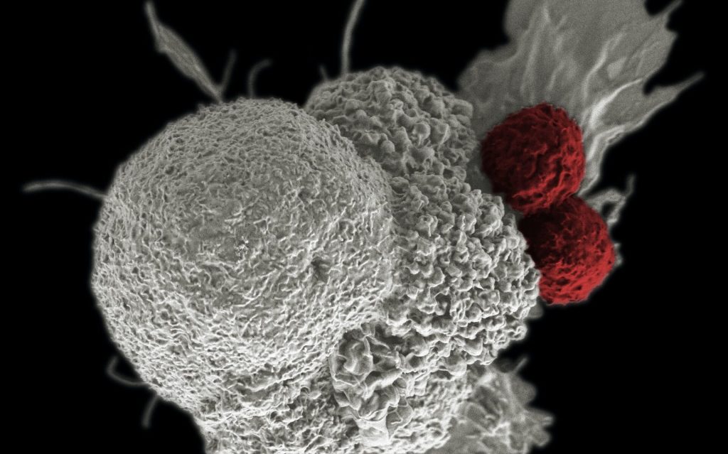
Shown here is a pseudo-colored scanning electron micrograph of an oral squamous cancer cell (white) being attacked by two cytotoxic T cells (red), part of a natural immune response. Photo by National Cancer Institute on Unsplash
Scientists have shown how tumour cells evade immunotherapy by forming temporary structures, where cells on the inside remain intact and can return to an individual state. These findings, published today in eLife, provide a novel theory as to how tumour cells avoid destruction by the immune system. They could also inform new treatments that combine immunotherapy with the timed inhibition of relevant signalling pathways in tumour cells.
“Cancer immunotherapy harnesses the body’s immune system to fight cancer. Despite its remarkable success, the majority of patients who receive immunotherapy will only see their tumours shrink in size temporarily before returning, and these relapsed tumours will likely be resistant to immunotherapy treatment,” said first author Amit Gutwillig, who was a PhD student at Tel Aviv University when the study was done.
To identify how tumours relapse after immunotherapy, Carmi and colleagues began by comparing the genetic sequences of whole genomes in primary and relapsed tumours in the same patient. Their analysis suggested that relapsed tumours do not change dramatically following immunotherapy.
Next, the team studied this process in breast cancer and melanoma, using mouse models in which immunotherapy-resistant tumours had relapsed. They administered the mice with cells from treated tumours and allowed these cells to reach a palpable size. The team found that the cells were equally susceptible to the same immunotherapy approach as the parent tumour, although they relapsed sooner.
To better characterise the tumour cells that survived in mice following immunotherapy, the researchers isolated and studied the live tumour cells. They found that most of the cells responded to the presence of T cells by organising into temporary formations. These were made up of clusters of several tumour cell nuclei, which are surrounded by a single, multilayered membrane and a meshwork of cortical actin filaments. The inner cell of the formation was dense and appeared to be compacted within another cell.
To show that this result was not due to the isolation of the melanoma cells, the team also analysed tumours with fluorescently labelled cell nuclei and membranes. They found that the cell-in-cell formation was more prevalent in immunotherapy-treated tumours, particularly in sites associated with tumour cell death. Further analysis indicated that roughly half of the tumour cells that survived immunotherapy were arranged in the cell-in-cell formation. Over time, these cells returned to a single-cell state, with similar structural features to those of the parental cell line.
The team next tested whether this phenomenon occurs in human cancers. To do this, they incubated tumour cell lines with pre-activated T cells from healthy donors. They discovered that the vast majority of breast, colon and melanoma tumour cells that survived T cell killing organised into the cell-in-cell structure. A three-day observation of T cells interacting with tumour cells showed that these structures were dynamic, with individual tumour cells constantly forming and disseminating from the structure.
Finally, they tested the clinical relevance of this discovery by analysing cancerous tissues from multiple organs of four stage 4 melanoma patients. These patients were undergoing surgical removal of primary and metastatic lymph nodes. The researchers found that in all four patients, the cell-in-cell formation was highly abundant in the T cell zone of the draining lymph nodes, but not in the primary tumours. Furthermore, in a patient with untreated recurrent melanoma, most of the cells in the primary tumour were single cells, whereas the recurrent tumours had an abundance of the cell-in-cell formations.
“This previously unknown mechanism of tumour resistance highlights a current limitation of immunotherapy,” said senior author Yaron Carmi at Tel Aviv University. “Over the past decade many clinical studies have used immunotherapy followed by chemotherapy. But our findings suggest that timed inhibition of relevant signalling pathways needs to occur alongside immunotherapy to prevent the tumour becoming resistant to subsequent treatments.”
Microscopy image of mouse sciatic nerves showing axons (red) wrapped by Schwann cells (green) with their nuclei depicted in blue.
Credit: A. Alvarez-Prats and T. Balla, Eunice Kennedy Shriver National Institute of Child Health and Human Development/NIH
In addition to forming the myelin sheath along peripheral nerves and supporting neighbouring neurons, Schwann cells have also been found to play an important immune modulating function, starting and shutting off inflammation. This function not only helps nerve repair, but may also turn nerve tumours benign. These new findings were reported in the journal Glia.
The research has revealed that Schwann cells produce signalling molecules that can activate other immune cells. In particular, however, they are able to stop inflammatory reactions in order to prevent excessive tissue damage and allow the nerve to regenerate.
“This is essential, because inflammation releases free radicals against which nerve fibers cannot protect themselves. Therefore, the inflammation must be cleared quickly, which is precisely what Schwann cells do,” explained study designer Dr. Sabine Taschner-Mandl, who designed the study and heads a research group at St. Anna CCRI.
Do Schwann cells protect against malignancy?
These findings also have implications for protection against malignancy After nerve injury, Schwann cells engage a ‘repair’ mode that is also found in benign infantile nerve tumours. There, it causes the tumour cells to mature and thus reach a stage where they lose their aggressive properties and no longer divide unchecked
“Based on the current results, we now suspect that the immune cell functions of Schwann cells also become effective in childhood nerve tumours. This is because in cancer, there is always a kind of inflammation bubbling away that never comes to a halt. In benign nerve tumours, ganglioneuromas, the accompanying chronic inflammation could be stopped by Schwann cells similar to nerve healing, because unlike malignancies, ganglioneuromas have many Schwann cells in their microenvironment. We also see that a lot of immune cells migrate into these tumours, for which the Schwann cells could also be responsible,” said Dr Taschner-Mandl.
Healthy Inflammation: First Activate, Then Shut Down
In particular, the current study shows that Schwann cells can influence T cells, which are key in cancer defence. Schwann cells – both those in nerve regeneration and those in benign tumours – carry MHC-I and MHC-II molecules on their surface that are important for T-cell regulation. Via these molecules, Schwann cells present recognition features of material they have previously taken up from their environment.
“We mimicked an inflammatory response in the laboratory and detected a whole range of additional stimulatory and inhibitory surface molecules that are also necessary for T cell activation,” explained Jakob Berner, MSc, co-first author of the study and interim PhD student at St. Anna CCRI. “Our experiments show that Schwann cells are able to take up large amounts of material via phagocytosis.”
As the first immune response to a nerve cut, Schwann cells secrete substances that attract T cells, macrophages and other immune cells. Now it turned out that not only a reaction between the classical immune cells takes place, but also between Schwann cells and T cells.
While Schwann cells initially fuel the inflammatory response by releasing interferon-gamma, they can later shut it down by up-regulating the T-cell inhibitory PD-L1 molecule.
“First activate, then shut down – that’s the normal process of an inflammatory response. If this were also the case in cancer, then it could curb cancer growth,” Dr Taschner-Mandl theorised. Researchers are now investigating whether and how these findings could be applied to cancer treatment.
Killer T cells about to destroy cancer cell (centre). Credit: NIH
While killer T cells have long been known to kill cancer cells with a cytotoxic weapon, new research has shown that they deliver the lethal strikes like expert warriors, grabbing and pulling the cancer cell’s membrane to more easily penetrate it like a dagger penetrating armour. The findings are detailed in a paper published in Developmental Cell.
Killer T cells are armed with lytic granules containing two key components for immune attack: a perforin proteins stabs a hole in the cell membrane, and the lethal granzymes which kill the cell.
T cells cosy up to targeted diseased cells in a close junction called the ‘cytotoxic immunological synapse’.
A research team at UNSW Sydney has found that mechanical forces generated by T cells influence how effectively perforin can punch through tumour cell membranes. The researchers describe the cell interactions and the integration of forces at both the front and rear of the cell.
The researchers detected physical forces within T cells that propel lytic granules toward the immunological synapse where their payloads are released. These forces also enable T cells to grab onto regions of the cancer cell membrane where the membranes of both immune and target cells are pulled and manipulated.
“It was very exciting to discover that, in addition to its mechanical tension and biochemical configuration, the shape of the target cell membrane plays an important role in T cell mediated cancer cell killing,” said Dr Daryan Kempe at UNSW Medicine & Health who co-led the research.
By stretching and bending the membranes of tumour cells in a certain direction, T cells made it easier for perforin to punch through, but only if the membranes were bent in the right direction.
Bias towards outwardly curved cell membranes
Using human melanoma cell lines, the researchers demonstrated that perforin preferentially perforated outwardly curved tumour cell membranes, rather than inwardly curved ones – which may help the T cells avoid being harmed by their own attack.
“As the granules arrive, their contents will be emptied at this region of the membrane that is very highly curved. That there was a bias between positively curved and negatively curved membranes was completely unexpected,” said senior Associate Professor Maté Biro at UNSW Medicine & Health.
Giving standard chemotherapy drugs in a specific sequence for certain types of metastatic breast cancer can cut costs while preserving quality of life, according to a study in theJournal of Clinical Oncology.
The study, led by researchers from UNC Lineberger Comprehensive Cancer Center and UNC Gillings School of Global Public Health, developed three different computer models to predict how a hypothetical set of 10 000 patients with specific types of metastatic breast cancer would respond to different sequences and types of chemotherapy. For this study, the patient’s cancer was either endocrine resistant or was triple-negative breast cancer.
Many chemotherapy choices are available to treat metastatic breast cancer. While oncologists may prefer certain drugs to use early in treatment, the best order in which to give the drugs is unclear. The researchers consulted oncologists and experts in the field to choose which chemotherapy drugs were preferred choices to include in the study.
Mimicking clinical practice, and based upon existing data, the researchers then assumed that if a person started treatment with one drug, they would change to a second-choice treatment after their cancer stopped responding to the first drug, or if the side effects weren’t tolerable. The purpose of the study was to test whether putting the drugs in one sequence compared to another could keep the patient on treatment for similar times while decreasing their side effect and/or cost burden.
“The cost of cancer drugs in the US has rapidly increased, even for generics. As a society, we urgently need more strategies to reduce cancer drug costs without compromising outcomes, and our analysis provides quantifiable evidence to help providers choose lower priced, but equally effective sequences of drugs,” said Stephanie B. Wheeler, PhD, MPH, professor of health policy & management at UNC Gillings and associate director of community outreach and engagement at UNC Lineberger and corresponding author of the article. “More spending on cancer care does not necessarily confer greater health benefits.”
The costs calculated in this study were inclusive of medical and nonmedical costs borne by patients, including lost productivity. In this simulation, after two years, nearly all women would have completed the first three sets of treatment, but the cancer would cause the death of about one-third of the women. Productivity days lost due to sickness were similar across chemotherapy sequences, so most of the cost difference was due to drug savings. In the simulation, patients were placed in three groups, depending on what treatments they had already received for earlier episodes of breast cancer.
Outcomes in the three groups were:
For people who had not previously received the common chemotherapy drug categories, including a taxane (e.g., paclitaxel) or an anthracycline (e.g., capecitabine), treatment with paclitaxel then capecitabine followed by doxorubicin corresponded to the highest expected gains in quality of life and lowest costs.
For people who had previously received a taxane and an anthracycline drug, treatment with carboplatin, followed by capecitabine, followed by eribulin, corresponded to the highest expected gains in quality of life and lowest costs.
For people who had previously received a taxane but not an anthracycline, treatment sequences beginning with capecitabine or doxorubicin, followed by eribulin, were most cost-effective.
“The drugs we studied are already recommended and reimbursed for the treatment of metastatic breast cancer, but the optimal sequencing of them has been unclear, which has led to considerable variation in physician preference and practice. Our study suggests that treatment sequencing approaches that minimise costs early may improve the value of care,” Wheeler said. “The implications of this study are fairly straightforward for medical oncologists and those developing value-based clinical pathways to implement in practice now.”
Associate professor Katherine E. Reeder-Hayes, one of the study’s authors, said the treatment choices for metastatic breast cancer are constantly changing, and new options for targeted therapy have emerged even since this study was conducted. “Many oncologists and patients find that there aren’t any more targeted therapies that fit the cancer’s molecular profiles, so they are left with the choice of a number of chemotherapy drugs that may feel pretty similar or have an unclear balance of pros and cons.
“In that scenario, I hope our study will help expand the framework that we use to make these decisions from one where we just think about the biologic action of the drug to one where we also consider the bigger picture of what the treatment experience is like for the patient, including their financial burden, investment of time, and side effects,” Reeder-Hayes added. “The most potent drug isn’t always the next best choice depending on what the patient values and wants to accomplish with their treatment.”
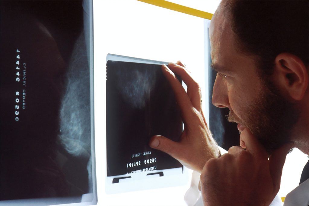
Researchers have developed a two-pronged approach to imaging breast density in mice, resulted in better detection of changes in breast tissue, including spotting early signs of cancer. If applied in humans, the technology may also help with prognosis of disease as density can be linked to specific patterns of mammary gland growth, including signs of cancer development. The findings appeared in the American Journal of Pathology.
“Having a means to accurately assess mammary gland density in mice, just as is done clinically for women using mammograms, is an important research advance,” said Priscilla A. Furth, MD, professor of oncology and medicine at Georgetown Lombardi Comprehensive Cancer Center, and corresponding author of the study. “This method has the benefit of being applicable across all ages of mice and mammary gland shapes, unlike some methods used in earlier studies.”
While working as an undergraduate in Furth’s lab, Brendan Rooney developed an innovative analytic computer program (C’20), which allowed for sorting of mammary gland tissue to one of two imaging assessments. At first, Rooney looked at younger mouse glands and found that a program that removed background ‘noise’ in those images helped boost detection of abnormalities in what are typically rounder, more lobular tissues. But as aging occurs and the chances of developing cancer increase, lobules diminish and ridges become more apparent, just as falling autumn leaves expose tree branches. The mammary ridges represent ducts that carry milk and other fluids. When the de-noising technique was applied to the images from the older mice, it was found to be less reliable in detecting ridges. Therefore Rooney and the team turned to a different imaging program, which has primarily been used to detect blood vessel changes in the eye’s retina.
“The idea for the analytic program came from routine visual observations of tissue samples and the challenges inherent in observing differences in breast tissue with just a microscope. We found that visual human observations are important but having another read on abnormalities from optimal imaging programs added validity and rigor to our assessments,” says Rooney, the lead author of the study. “Not only does our program result in a high degree of diagnostic accuracy, it is freely available and easy to use.”
Now that the broad strokes of the research have been laid down and proof-of-principle has been established, Rooney has started medical school with a possible eye toward specializing in oncology. Both Furth and Rooney believe that future studies will need to refine and streamline their research approach in mice, including better density measurements that could enable sorting of samples into higher and lower probabilities of cancer.