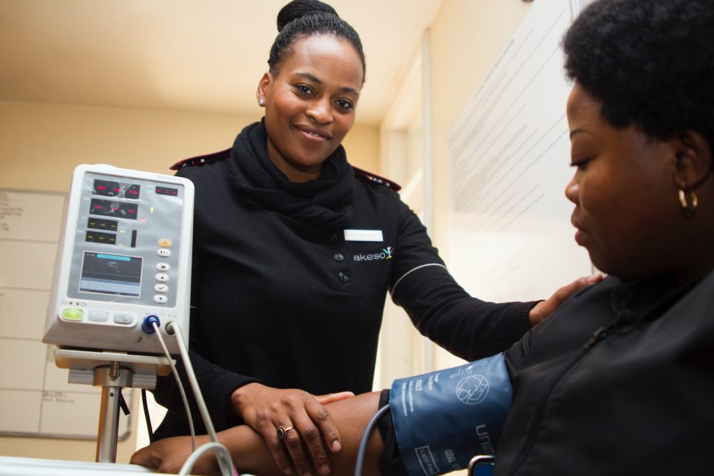
To significantly expand access to affordable, quality primary healthcare in underserved communities, the Cipla Foundation’s Sha’p Left initiative has partnered with the FirstRand Empowerment Foundation (FREF). The partnership aims to aggressively scale the cost-effective nurse-driven surgeries in local communities, across the Western Cape, KwaZulu-Natal and Gauteng.
HEALTHCARE CLOSER TO HOME
This collaboration will help to overcome systemic barriers to healthcare, particularly in terms of equitable access for low-income, uninsured individuals. For many people living in peri-urban and rural areas, access to quality primary healthcare services poses a significant challenge. Over-burdened State medical facilities are often congested, resulting in long waiting times for patients.
Sha’p Left is a patient-centred, cost-aware, nurse-driven primary healthcare service, in the heart of local communities. These nurse surgeries are located in easily accessible hubs such as busy taxi ranks to promote ease of access. The greatest benefit of Sha’p Left is that in addition to saving travel time, it helps to empower people both in terms of caring for their health, but also financially: the lack of queues mean that people don’t need to take a full day off work (resulting in a loss of income) to access basic healthcare.
Currently, Sha’p Left serves more than 5 000 patients monthly, with the patient profile comprising a 60% / 40% female / male split. The existing clinics are GMP compliant containerised solutions, as part of environmental sustainability initiatives and lowering overhead costs, solar solutions are being implemented at these clinics.
CHAMPIONING CHANGE
Strengthening community-based primary healthcare supports national health priorities by reducing the burden on State facilities, promotes preventative healthcare and creates an empowering, dignified experience for patients.
The investment by FREF will help Sha’p Left to deploy more nurse surgeries, and these solutions will ultimately help address inequality and reduce poverty as access to quality healthcare is basic human right. The partnership will scale Sha’p Left from 11 to 61 surgeries by the end of 2029.
SUSTAINABLE SOCIAL IMPACT
The business model involves enterprise development in conjunction with qualified, predominantly female clinical nurse practitioners (CNPs) and assists them to establish sustainable, owner-operated clinics in identified communities to provide affordable primary healthcare services.
This fee-for-service model, driven by the “entreprenurses”, provides a dignified and holistic patient experience. The surgeries have dispensing licenses and therefore a consultations includes the necessary medication required, up to Schedule 4 medicines.
The first three nurse surgeries being deployed in 2026, as part of this partnership, are in these areas:
· Senoane (Gauteng)
· KwaNyuswa (KZN)
· Verulam (KZN)
Blending social impact with sustainability creates a blueprint for scaling primary healthcare in South Africa. With FREF’s support, the Sha’p Left model will expand further into communities where access gaps remain widest, ensuring that more South Africans can easily receive the care they need. This investment ensures that good health is not merely a privilege for a select few people, but for all South Africans.
Ultra-sensitive food safety tests may drive food waste and unavailability with limited public health benefit, according to a Frontiers in Sciencestudy.
These food safety measures and ultra-sensitive tests may drive edible food being thrown away, excessive packaging, and extra costs for consumers.
The international team of researchers make it clear that food safety is an important concern, as foodborne pathogens account for approximately 420 000 deaths and 600 million cases of illness each year. However, the authors argue that food systems will be more sustainable, while continuing to protect public health, if “zero-detection” expectations are replaced with evidence-based targets for “sufficiently safe” food.
Their new article sets out how regulators might find trade-offs with other important factors, such as food supply security, sustainability, and nutritional health.
“Although the public expects food to be completely safe, there will always be some risk of foodborne illness. Zero risk doesn’t exist, and we shouldn’t be aiming for that either. Just as we don’t limit highway speeds to 10 miles per hour to minimize road deaths, we need to take a balanced approach that considers possible negative consequences of extreme food safety measures,” said lead author Prof Martin Wiedmann from Cornell University.
The study’s authors highlight several situations where excessive caution can cause harm.
Many rules and purchasing standards rely heavily on detecting a pathogen, sometimes treating any detection as unacceptable without fully considering dose, exposure, the food’s ability to support microbial growth, or who is most at risk.
For example, a food product might be considered contaminated if it tests positive for the bacterium Listeria monocytogenes, regardless of levels.
These alarms can result from ultra-sensitive tests detecting small amounts of microbes unlikely to cause disease in humans. In some cases, the concerns may come from bacteria that are not harmful themselves, but are an indirect indicator of contamination.
Throwing away such food reduces the available food supply and wastes resources. Similarly, recalling food products from consumers can damage consumer trust, pushing people away from otherwise healthy products.
Other protective measures, such as storage temperatures, packaging, and heat treatment, can waste energy, increase costs, and reduce nutritional content. While these are all important safety measures, they should only be applied if needed and associated trade-offs should be considered.
“A tremendous amount of food is wasted that would have been sufficiently safe to eat. Too often, trade-offs such as environmental or economic costs are only considered after a traditional microbial risk assessment. We cannot afford to carry on like this at a time when we desperately need to reduce our impact on the planet and assure not only food safety but food security,” said co-author Prof Sophia Johler at Ludwig Maximilian University of Munich, Germany.
Focus on risk rather than hazard
The current situation is driven by an emphasis on hazard-based assessments, according to the authors, where regulations focus on detecting pathogens, regardless of the threat to consumers. The researchers argue that the food system should move towards more flexible risk-based approaches, which assess the probabilities of harms and adjust the safety measures accordingly.
Regulations that overemphasize stringent corrective actions (such as recalls) when swab samples from a food-processing facility test positive for an indicator, for example, could lead to undue corrective actions in areas that are unlikely to contaminate the food. The authors explain that this could be an opportunity cost that diverts resources away from more effective interventions and control strategies in high-risk areas.
“There’s well-established evidence that focusing on end-product testing is generally ineffective to ensure safety. Overemphasis on end-product testing may distract from other food safety measures (e.g., applying validated and verified process controls), which can provide greater public health benefits,” said co-author Dr Sriya Sunil at Cornell University.
Better tools to assess priorities
Computational tools that incorporate vast amounts of information across the food production system could help with establishing acceptable risks.
One challenge is how to prioritize different hazards. For example, in the US, norovirus causes thousands of times more cases than Listeria monocytogenes, yet Listeria monocytogenes causes more deaths per year.
While there are trade advantages to having consistent international food safety standards, the balance between competing interests may vary between regions. This can become even more complex when factoring in the health and environmental implications of greenhouse gas emissions.
“Specialists across social sciences, economics, and life sciences must work together to establish values that align with consumers’ priorities. Together with advanced models that build on geographic information, AI, and genomics, we can assess, manage, and communicate risks far more accurately,” said Wiedmann.
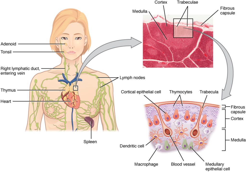
Mass General Brigham researchers used artificial intelligence to analyse routine medical scans, uncovering how the thymus impacts aging, cardiovascular risk, cancer incidence, and response to immunotherapy.
Two new studies from investigators at Mass General Brigham challenge a decades-old assumption that the thymus, an organ best known for its role in establishing immune function in childhood, becomes irrelevant in adulthood. Using artificial intelligence (AI) to analyse routine CT scans, researchers uncovered that adults with a healthy thymus had increased longevity and reduced risk for cardiovascular disease and cancer. In a separate study of patients with cancer, the researchers found that thymic health may influence response to immunotherapy – a treatment that depends on the strength of a patient’s immune system.
“The thymus has been overlooked for decades and may be a missing piece in explaining why people age differently, and why cancer treatments fail in some patients,” said Hugo Aerts, PhD, corresponding author on the papers and director of the Artificial Intelligence in Medicine (AIM) Program at Mass General Brigham. “Our findings suggest thymic health deserves much more attention and may open new avenues for understanding how to protect the immune system as we age.”
The thymus is a small organ in the chest that helps train T cells, priming the immune system to protect the body from infections and disease. For decades, doctors believed the organ was mostly inactive after puberty because it shrinks with age and produces fewer new T cells. As a result, its role in adult health has rarely been examined in large populations.
Previous research has linked T cell diversity to aging and immune decline, but most of those studies relied on small, blood-based analyses. In contrast, the new studies examined more than 25 000 adults in a national lung cancer screening trial and over 2500 participants in the Framingham Heart Study – a large, long-running population cohort of generally healthy adults.
The team analysed the size, shape, and composition of the thymus, generating a “thymic health” score. People with high thymic health scores had about a 50% lower risk of death, 63% lower risk of cardiovascular death, and 36% lower risk of developing lung cancer compared to those with low thymic health. These associations remained significant after adjusting for age and other health factors.
The researchers theorize that when thymic health and T cell diversity decline, the immune system may become less able to respond to new threats, like cancer or other diseases. Their analysis found that chronic inflammation, smoking, and high body weight were associated with poorer thymic health, suggesting that lifestyle and systemic inflammation may influence immune resilience across the lifespan.
In a second study, the researchers analysed CT scans and outcomes from more than 1200 immunotherapy-treated patients. Patients with stronger thymic health had about a 37% lower risk of cancer progression and a 44% lower risk of death, even after accounting for other patient, tumour, and treatment factors. These findings point to a previously underappreciated role the thymus may play in shaping how well patients respond to modern cancer immunotherapies.
The researchers caution that their findings will need to be confirmed in future studies, and the imaging method is not yet ready for routine clinical use. While lifestyle factors were linked to thymic health, the studies did not test whether modifying those factors can directly improve thymic function.
The team is currently leading additional research to investigate whether other care-associated factors may impact thymic health. In one study, they are examining whether unintended radiation exposure to the thymus in patients with lung cancer may affect outcomes.
“Improving our understanding and monitoring of thymic health could eventually help physicians better assess disease risk and guide treatment decisions,” said Aerts.
New TB tests have massive potential for South Africa’s struggle to get to grips with the age-old disease. Making the most of these new tests will require both ambition and smart implementation, argue Gaurang Tanna and Dr Yogan Pillay.
Every day, more than 140 people die from tuberculosis (TB) in South Africa, yet TB is both preventable and curable. Too many people are tested too late, allowing the disease to spread silently through communities and turning a curable illness into a fatal one.
Unlike most other diseases, anyone can contract TB – the bacteria are airborne and just the act of breathing makes us vulnerable to contracting TB. The risk of TB is higher for people with suppressed immunity, malnutrition, or living with cancer or HIV.
Reducing deaths from TB depends on earlier diagnosis, yet many people are diagnosed late, often after prolonged illness, and only once they reach hospitals with advanced disease. There are some opportunities for improvement. Firstly, we need to address persistent weaknesses in where and how TB tests are offered. Secondly, we need to address delays in care seeking, and missed opportunities for testing within health facilities. Finally, we need to close the operational barriers that impede testing. An added challenge that the TB disease presents is that it is often present without any symptoms.
In recent years, South Africa took important steps to strengthen its TB response and intensified efforts to find people with the TB disease through implementation of Targeted Universal TB Testing (TUTT). TUTT is a strategy that promotes systematic testing among high-TB risk groups, like people living with HIV, household contacts of individuals with TB, and people with previous TB, irrespective of symptoms.
South Africa now conducts approximately 3.6 million TB tests annually, representing a 50% increase compared to pre-COVID pandemic testing. However, we need to scale this up considerably if we are to reach the more than six million people living with HIV currently receiving HIV treatment in South Africa as well as all those with TB symptoms who are often missed at facilities.
Despite strong commitments, TB testing in South Africa continues to face several structural constraints.
First, the cost of molecular diagnostics limits the scale of testing. Current molecular TB tests cost approximately R230 per test.
Second, inefficient clinic workflows reduce testing coverage. In busy primary healthcare facilities, this leads to missed TB testing, contributing to prolonged diagnostic delays during which transmission continues and disease severity worsens.
Third, many patients, especially children and people living with HIV, can’t produce sputum, which current tests require, further reducing testing coverage.
Fourth, people with the highest burden of TB, particularly men, often do not attend government clinics. Men account for a disproportionate share of TB in South Africa but remain underrepresented in testing programmes, contributing to delayed diagnoses and ongoing transmission.
Evolving and strengthening testing capabilities in line with the ambitions of the next phase of TB control in South Africa requires leveraging emerging diagnostic tools and redesigning how TB testing is delivered.
New diagnostic tools create new opportunities
Just recently, the World Health Organization updated its recommendations on TB diagnostics, endorsing the use of near-point-of-care tests and use of tongue swabs for people who cannot produce sputum to expand access to TB diagnostics and improve diagnostic efficiency. These new tools provide an opportunity to rethink how testing is organised across the health system.
Tongue swabs offer a promising alternative sample type, enabling testing among patients who cannot produce sputum. It has also been demonstrated to be more acceptable for patients and providers and is easier to collect in clinics.
At the same time, near-point-of-care molecular platforms (such as Pluslife, a test that has been approved by the South Africa’s health products regulatory body) offer the potential to diagnose TB closer to the patient. It substantially reduces costs, to about one-third the cost of current molecular tests, while demonstrating comparable diagnostic performance for TB, making large-scale expansion of TB testing more accessible and affordable. By delivering results rapidly, within an hour, this technology could enable a test and treat approach. TB testing, diagnosis, and treatment initiation could all happen during a single primary healthcare visit. This would reduce the time to start treatment and limit the number of patients lost between diagnosis and treatment.
Clinic workflows need to be redesigned
Patients presenting with TB symptoms often move through multiple stages of the clinic process – registration, triage, waiting areas, and clinician consultations – before TB testing is considered. Improving TB testing requires services redesign for patient convenience and accessibility, and to be much more systematic. A few simple changes could be introduced.
Firstly, introduce a fast-track TB queue, allowing individuals to register digitally and drop off samples without completing a full clinic visit.
Secondly, embed TB symptom screening and sample collection at triage or vital-sign stations. Any patient reporting TB symptoms – cough, fever, night sweats, or weight loss – should have a sample collected while waiting to see a clinician.
Thirdly, for people living with HIV, introduce twin TB testing with annual viral load test (or CD4 for newly diagnosed patients) to systematically test all people living with HIV.
Lastly, we could equip facilities with a near-point-of-care testing platform, like Pluslife, to deliver results before the clinical consultation, allowing TB to be diagnosed rapidly and at lower cost to the health system. It would enable patients to start treatment on the same day.
These approaches could directly address the most persistent diagnostic and linkage gaps in South Africa’s TB programme.
Extending TB testing beyond clinics
New diagnostic platforms also enable TB testing to move beyond government clinics.
A substantial proportion of individuals with TB, particularly men, do not present to clinics and delay seeking care. Near-point-of-care molecular platforms could enable TB testing through alternative delivery channels, including community settings (such as taxi ranks), community pharmacies, workplace clinics, and households through community health worker programmes.
Expanding testing beyond clinics will help identify TB earlier among populations that remain underserved by current services.
From policy ambition to implementation
South Africa’s progress over the past four years demonstrates that intensified testing strategies such as TUTT can help increase TB diagnosis. Sustaining this momentum will require redesigning primary health care services to fully use these emerging diagnostic tools. Three priorities should guide this transition.
First, TB sample collection workflows in clinics should be redesigned to ensure that every symptomatic and at-risk person is tested for TB.
Second, new diagnostic tools should be deployed, including the use of tongue swabs for people who cannot produce sputum, as well as low cost near-point-of-care molecular tests to simplify testing and treatment initiation pathways.
Third, TB testing should be expanded through alternative delivery channels to reach people who do not routinely access government clinic services, especially men, who are less likely to seek care in these settings.
By aligning ambition and new technologies with service redesign, South Africa can significantly reduce diagnostic delays, decrease deaths due to TB and accelerate progress towards TB elimination.
*Tanna is a senior programme officer for TB, and Dr Pillay is the director of HIV and TB delivery at the Gates Foundation.
Disclosure: Spotlight receives funding from the Gates Foundation but is editorially independent – an independence that the editors guard jealously. Spotlight is a member of the South African Press Council.
Note: Spotlight aims to deepen public understanding of important health issues by publishing a variety of views on its opinion pages. The views expressed in this article are not necessarily shared by the Spotlight editors.
Trial provides new evidence to guide early treatment decisions for families and clinicians
An infant participating in the Baby CHAMP study raises both hands while seated in a stroller. The NIH-funded trial led by the Fralin Biomedical Research Institute at VTC examines early therapies designed to improve arm and hand function in young children with cerebral palsy affecting one side of the body. Credit: Jennifer Murray
Infants and toddlers with unilateral cerebral palsy, which affects the brain’s control of muscles on one side of the body, show lasting improvements in hand and arm function when they receive early, high-dose therapy, according to a new multisite clinical trial led by Virginia Tech researchers at the Fralin Biomedical Research Institute at VTC.
The Baby CHAMP (Children with Hemiparesis Arm-and-Hand Movement Project) study directly compared three therapist-delivered interventions: two forms of constraint-induced movement therapy, which limit the stronger arm to encourage use of the weaker one when combined with therapy, and bimanual therapy, which promotes coordinated use of both hands.
The researchers found that children ages 6 to 24 months showed similar gains whether therapy involved full-time casting, a splint worn during sessions, or bimanual training without constraining the stronger arm.
Published in Pediatrics Open Science, the study addresses a long-standing gap in clinical evidence.
“The brain in the first two years of life is remarkably plastic,” said Stephanie DeLuca, associate professor at the Fralin Biomedical Research Institute at VTC and co-principal investigator of the trial. “By delivering high-dose, play-based therapy early, we’re capitalizing on a window of opportunity when the nervous system is especially responsive to experience.”
While both constraint-induced movement therapy and bimanual therapy are widely recommended for children older than 2 years with unilateral cerebral palsy, limited data have been available to guide treatment decisions for infants and toddlers.
“This gives families and clinicians evidence-based options,” said Sharon Landesman Ramey, a Virginia Tech Distinguished Scholar, professor at the Fralin Biomedical Research Institute at VTC, and co-principal investigator of the Baby CHAMP trial. “The encouraging message is that early, intensive therapy works — and multiple approaches can help children build critical motor skills. Caregivers and families now have actionable evidence that can shape care during one of the most important periods of brain development.”
Unilateral cerebral palsy affects movement on one side of the body and can result in lifelong impairment of upper extremity function. Early intervention is considered critical because the brain is especially adaptable during the first two years of life.
DeLuca is director of the Fralin Biomedical Research Institute at VTC Neuromotor Research Clinic, which investigates novel treatments for children with a range of biomedical conditions and provides worldwide training for therapists to become certified in new evidence-based therapies.
All children received three hours of therapy per day, five days a week, for four consecutive weeks, totaling 60 hours of structured intervention. Parents also supported additional guided home practice.
Fifty-eight children were enrolled in the randomized controlled trial, funded by the Eunice Kennedy Shriver National Institute of Child Health and Human Development of the National Institutes of Health. Fifty-three completed treatment and end-of-therapy assessments, and 41 returned for evaluation six months later.
Across all three groups, children demonstrated significant improvements in the ability to use both hands, individually and together. Improvements were measured using standardized developmental assessments administered by evaluators who were unaware of each child’s treatment assignment.
Children also showed gains in fine motor skills in their less-affected arm. Improvements were most pronounced at the six-month follow-up, suggesting that benefits continued to build after formal therapy ended.
Researchers had hypothesized that bimanual therapy might lead to greater improvements in two-handed skills and that full-time casting might yield stronger gains in the affected arm. The data did not support those predictions. Instead, outcomes were broadly comparable across approaches.
The study also addressed concerns that constraining the stronger arm could impair its development. No evidence of harm was observed. In fact, children in the full-time cast group showed slightly greater gains in fine motor skills in their non-affected arm at six months compared with the bimanual group.
“This is important to the field because many people have worried that the use of a constraint might slow the developmental process of the less-affected arm,” DeLuca said. “Our findings confirm that this did not occur and this therapy may even help promote improvements in skills on the less-affected arm and hand.”
Some parents reported their child showed short-term frustration wearing a cast or splint, and minor skin irritation occurred in a small number of children using casts, but no were caused by the therapy itself.
The trial was conducted in collaboration with researchers at Virginia Tech, The Ohio State University, and Nationwide Children’s Hospital. Therapists were centrally trained to deliver structured, play-based interventions grounded in motor learning principles, including repetition, reinforcement, and progressively challenging activities.
Longer-term studies will be needed to better understand how early therapy influences development across many dimensions of a child’s life.
North Korean defectors who resettled in South Korea share genetics but markedly contrasting early-life exposures with South Korean residents. Research published in the Journal of Internal Medicine compared overall and site-specific cancer incidence rates between North Korean defectors and native South Koreans.
Breast cancer cells. Image by National Cancer Institute
Using the Korean National Health Insurance database, researchers matched 25 798 North Korean defectors and 1 276 601 South Korean residents. Defectors had higher risks of infection-related cancers (such as liver and cervical cancers) and lower risks of breast, colon, and prostate cancers (which are more prevalent in developed countries). Over time, though, their cancer profile changed, suggesting adaptation to South Korean society.
“The study provides a model for understanding how cancer epidemiology evolves in such transitions, offering lessons that may help guide prevention and health planning for other vulnerable groups in transition worldwide,” said corresponding author Sin Gon Kim, MD, PhD, of the Korea University College of Medicine.
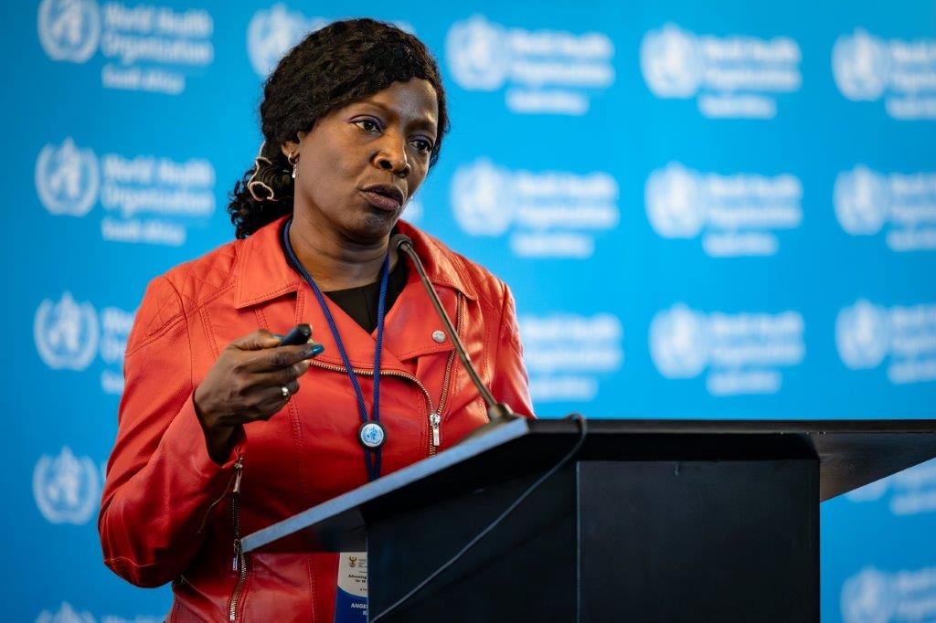
Associate Professor Angelique Kany Kany Luabeya speaks about TB vaccine trials and the introduction of TB vaccines in South Africa. (Photo: Supplied)
By Angelique Kany Kany Luabeya
The only tuberculosis vaccine we have is a century old and offers only limited efficacy in children. With leading South African researchers involved in the pivotal clinical trials of three new tuberculosis vaccine candidates, we are on the verge of a major breakthrough, writes Associate Professor Angelique Kany Kany Luabeya.
My uncle died of abdominal TB a few days ago, after facing repeated challenges in getting an accurate diagnosis. For him, the treatment started much too late. To many in his community, my uncle was a respected teacher, a breadwinner, a pillar of support and strength.
In 2026, why are people still dying from a preventable disease that continues to cause unnecessary deaths and hardship?
Why we urgently need a new TB vaccine should be obvious. For the millions who are sick, and for families living with the catastrophic loss of a loved one, the need is painfully clear.
Prior to the emergence of the SARS-CoV-2 virus, TB was the world’s deadliest infectious disease, killing more than 1.5 million people every year. While COVID-19 has since shown an epidemic downturn, TB’s toll remains devastatingly high.
Globally, an estimated 2 billion people are infected with the Mycobacterium tuberculosis that causes TB in humans. In this state, also known as latent TB infection, they do not have TB symptoms and are non-infectious, but the bacteria remain dormant in their bodies. Of these people, about 5 to 10% will go on to develop active TB when their immune system is no longer able to contain the bacteria. This means that they now have TB disease, sometimes without noticeable symptoms, and risks passing it to others. This could be a family member, a friend, or a stranger who happens to be nearby.
TB bacteria have coexisted with humans for millions of years. There is a cure, but treatment alone is not enough to stop transmission. TB mostly affects countries with limited resources because patients struggle to access care or are unable to complete treatment due to side effects or a lack of food to support the rigorous regimen of drugs they must take to cure them. In addition, the rise of multidrug-resistant tuberculosis is now fueling a global health crisis.
In South Africa, recent data from the World Health Organization’s (WHO) Global TB Report indicate progress, with a 57% reduction in new TB cases since 2015. However, TB mortality is still high and is concentrated mainly in poor and vulnerable communities. According to the WHO, TB still claims over 50 000 lives in South Africa every year. The burden is also unevenly distributed, with some geographic areas affected more than others.
A vaccine which prevents TB
Our hopes are now pinned on developing an efficacious vaccine which prevents people from developing TB disease. WHO modelling suggests that a vaccine which prevents most people with latent TB infection from progressing to active disease would have the most rapid impact on the epidemic in high‑burden countries.
The most urgent priorities for protection would be people living with HIV, healthcare workers at risk of workplace exposure, adolescents and young adults who are driving transmission, as well as those with comorbidities such as diabetes that increase their risk of TB diseases and negatively affect treatment outcomes.
The COVID-19 pandemic proved that when human survival is threatened, the scientific community can respond with breathtaking speed, developing multiple effective vaccines in under a year. Sadly, the urgency and resources allocated to finding an effective TB vaccine do not match the scale of its devastation.
For more than a century (since 1921), we have had only one licensed TB jab, which is the bacillus Calmette-Guérin (BCG) vaccine that is given at birth. Despite its limitations in preventing TB that infects the lungs – the main route of transmission – BCG remains a critical tool because it protects millions of babies from more serious forms of TB that can spread through the blood to the brain. But, clearly, the BCG vaccine is not enough.
Hope is on the horizon though, with several novel TB vaccines now in late-stage clinical trials. New vaccines or drugs are evaluated clinically in humans in steps, or phases, for safety, immunogenicity, and efficacy.
The most advanced is M72/AS01E (M72 for short), which is an adjuvanted subunit vaccine under development by the Gates Medical Research Institute and GlaxoSmithKline. In a phase 2 trial, this vaccine showed close to 50% efficacy in preventing TB disease in TB-infected people—the first time a vaccine has achieved this level of efficacy. A pivotal phase 3 trial of this vaccine has now completed enrolment of 20 000 volunteers, including 13 000 people in South Africa, with results expected in 2028. Developers typically apply for registration with regulatory authorities after successful phase 3 trials – so this study is the last big hurdle for this vaccine.
Another promising candidate is the MTBVAC vaccine, a live, whole, attenuated Mycobacterium tuberculosis vaccine developed by Biofabri, in partnership with the University of Zaragoza and sponsored by the International AIDS Vaccine Initiative. It is in a multi-country phase 2b trial in adults and adolescents and a phase 3 trial in newborns, including in South Africa.
BioNTech’s mRNA TB vaccine is also being evaluated in a phase 2a study in South Africa. Funded by BioNTech, this vaccine candidate harnesses mRNA technology, which has proved successful in the COVID-19 response.
Paving the way for acceptance and use
South African researchers are at the forefront of these TB vaccine efforts. Our strengths lie in our robust clinical trial capacity, world-class institutions, commitment to equitable solutions, and regulatory expertise, all of which help accelerate vaccine licensure. As a global policy leader, South Africa co-chairs the Finance and Access Working Group at the WHO TB Vaccine Accelerator Council, advocating for fair distribution and sustainable financing, and has recently co-hosted a vaccine preparedness workshop to position the country for the emergence of late-stage TB vaccines.
But the most important aspect to consider is the vaccine’s acceptability and uptake by a myriad of population groups at risk of TB. We learned from COVID-19 how misinformation can devastate vaccine uptake, leading to unnecessary morbidity and mortality. Confidence in new TB vaccines must be built to maximise impact. The context may be different—TB is an old, well-known enemy that affects people close to us. By involving South African communities in the early stages of vaccine trials, we can ensure their priorities are part of the development agenda.
While we continue to improve TB diagnosis and treatment, the hunt for an effective vaccine continues. After a century of fighting TB with only one vaccine and several antibiotics, we might be on the verge of a breakthrough that could finally shift the trajectory of this ancient and deadly disease.
*Associate Professor Angelique Kany Kany Luabeya is the clinical investigator on the M72 TB vaccine trials being conducted at the South African Tuberculosis Vaccine Initiative based at the University of Cape Town.
Disclosure: The Gates Medical Research Institute mentioned in this article is a non-profit organisation and subsidiary of the Gates Foundation. Spotlight receives funding from the Gates Foundation but is editorially independent – an independence that the editors guard jealously. Spotlight is a member of the South African Press Council.
Note: Spotlight aims to deepen public understanding of important health issues by publishing a variety of views on its opinion pages. The views expressed in this article are not necessarily shared by the Spotlight editors.
Credit: Darryl Leja National Human Genome Research Institute National Institutes Of Health
A major UK clinical trial co-led by researchers at UCL will test whether lower doses of common hormone therapies can treat advanced prostate cancer while reducing the severe side effects many patients experience.
The ENHANCE trial, jointly funded by Cancer Research UK and Prostate Cancer UK, will recruit 1,500 men with advanced prostate cancer from hospitals across the UK to compare standard and half-dose treatment using four commonly prescribed hormone drugs. UCL is sponsoring the trial and Professor Allan Hackshaw (Director of CRUK & UCL Cancer Trials Centre, UCL Cancer Institute) is joint-lead investigator, alongside Professors Ananya Choudhury and Peter Hoskin at the University of Manchester.
Men diagnosed with advanced prostate cancer, where the disease has spread beyond the prostate, are typically treated with powerful hormone therapies that slow tumour growth. While these drugs can extend life, they can also cause debilitating side effects such as extreme fatigue, hot flushes and high blood pressure, which can make it difficult for some patients to remain on treatment.
Researchers hope the £3.2 million ENHANCE trial will show that lower doses of these drugs can be just as effective while improving patients’ quality of life. The trial has not yet been launched and is awaiting the relevant regulatory and ethics approvals.
Professor Allan Hackshaw said: “Although focus is often on cancer trials to improve survival, we also need to find more tolerable ways of treating cancer without compromising survival. Side effects of cancer therapies matter a lot to patients, especially when they are frequent.
“We believe that several modern cancer drugs can be given at a much lower dose than what they were licensed for. Not only would this improve patient’s quality of life, but healthcare costs would be lower allowing more access especially in countries that cannot afford these drugs at their current high dose. The ENHANCE study can therefore influence clinical practice worldwide.
“Furthermore, Black men are more likely to get prostate cancer, and they too suffer from the same side effects of hormone treatment. They are often under-represented in clinical trials, so the ENHANCE study is a good opportunity to show that they get the same benefits from lowering the treatment dose as other ethnic groups.”
ENHANCE will compare full and half-dose treatment across four hormone therapies widely used for advanced prostate cancer: abiraterone, enzalutamide, darolutamide and apalutamide. If successful, the findings could influence prostate cancer treatment guidelines in the UK and internationally as early as 2030, improving care for thousands of men and reducing costs for the NHS.
Dr Ian Walker, Executive Director of Policy at Cancer Research UK, said: “Thanks to research, there’s been huge progress in prostate cancer treatments. Today, more than 8 in 10 men diagnosed with the disease in the UK will survive for 10 years or more. There’s more that can be done to save even more lives though, and in addition to finding more effective treatments, we need to find kinder ones too. The ENHANCE trial is looking to do just that and could mean that men affected by prostate cancer live not just longer lives but have a better quality of life.”
Dr Matthew Hobbs, Director of Research at Prostate Cancer UK, said: “No man should be forced to compromise between survival and their day-to-day wellbeing. This is a crucial issue for men with prostate cancer. That’s why Prostate Cancer UK is thrilled to be working alongside Cancer Research UK, pooling our resources and expertise to deliver the impact men need by funding this bold new trial that puts men’s wellbeing at its centre.”
Prostate cancer is now the most common cancer in UK men. Around 55 900* men are diagnosed each year. While survival rates have tripled since the 1970s, many men still face difficult side effects from treatment.
Retired solicitor Jonathan Edwards, 80, from Cheshire, experienced severe side effects after starting the hormone-blocking drug enzalutamide following his prostate cancer diagnosis in 2024, but when his nurse reduced the dose, his cancer remained under control and his quality of life improved dramatically.
Jonathan said: “It was such a shock when I was diagnosed. I had several health issues and after many tests was eventually told that I was suffering from prostate cancer and that it had spread beyond the prostate wall to my bones. I was referred to The Christie Hospital for treatment and was prescribed hormone blockers. The side effects made me extremely tired; I was sleeping through the day on and off and I had frequent hot flushes and generally felt weak.
“When the nurse suggested lowering the dose I was not sure what to expect. The difference soon became apparent and I felt normal again. I know that I will stay on the medication for as long as it is effective but, in the meantime, I am able to live a pretty normal life. I now exercise more and do not usually need an afternoon sleep. Happily, my PSA level started to go down until, after a few months, it was undetectable and has so far remained undetectable.
“My life has been transformed by the medication, my energy levels are higher, and I can socialise as normal. Traveling was a problem but now I can plan trips as long as I work around the 12-week cycle of injections and consultations. I am delighted that this trial has the potential to help other men going through the same thing in the future by enabling them to be treated for prostate cancer with their quality of life still largely intact.”
At least 10 per cent of trial participants will be Black men. Historically under-represented in clinical trials, Black men are often treated based on data that may be less applicable to them. Although data shows Black men are more likely to develop prostate cancer, more evidence is needed to understand their risk of aggressive disease and the role of overdiagnosis.
Alongside testing lower doses, the trial will collect tissue, blood and urine samples to identify biomarkers that could help determine which men are most suitable for reduced-dose treatment, shaping more personalised care in the future.
*Based on the average annual number of new cases of prostate cancer (ICD10 C61) diagnosed in the UK in the years 2018-2019, 2021.
View of the spinal cord. Credit: Scientific Animations CC4.0
Mechanoreceptors are present in the spinal cord from birth, are sensitive to mechanical stimuli, and play an important role in triggering the pathological events that follow trauma. What happens if they are blocked? The extent of the damage decreases. This is the finding of a new study published inThe Journal of Physiology and conducted by a team at Scuola Internazionale Superiore di Studi Avanzati (SISSA), led by Professor Giuliano Taccola, with Atiyeh Mohammadshirazi as first author.
Everything happens within the very first milliseconds after the trauma, the scientists explained. It is during this brief time window that these spinal mechanoreceptors become active, triggering an impairment of the electrical signals that underlies normal neural communication. This initial event sets off a cascade of neurotoxic factors known as secondary damage, which amplifies and spreads the original traumatic lesion over the following hours and days.
Understanding the role of these receptors, according to the authors, is important not only for clarifying what happens during spinal shock. As demonstrated in the experiments, when their activity is blocked, the functional damage is also reduced. For this reason, spinal mechanoreceptors may represent a potential target for strategies aimed at reducing the disabling consequences of spinal cord injury.
Physical trauma disrupts electrical signaling
“It is well known that physical trauma to the spinal cord disrupts the flow of electrical signals that underlie the functioning of our nerve fibers. This phenomenon is known as DIP (Depolarizing Injury Potential). It begins almost immediately after trauma and continues propagating the primary damage over the following weeks, progressively worsening the lesion,” explain Atiyeh Mohammadshirazi and Giuliano Taccola. “However, the origins of this phenomenon are not yet fully understood.”
Mechanoreceptors and their role in the spread of damage
Receptors are cellular structures that respond to specific signals. Among them are mechanoreceptors, specialized proteins located on the cell membrane of sensitive cells that act as sensors for mechanical forces such as compression. Mechanoreceptors are found throughout the body, including around the spinal cord and within its central canal. In this environment, according to the SISSA research, they appear to play an important role in the propagation of injury.
The two authors explain, “In the progression of damage, depolarization precedes other well-known events such as the release of neurotoxic agents, and the inflammatory response that ultimately leads to cell death, the transient spinal hypoxia, and the rapid cell neuronal loss in the area of the primary lesion.” In this context, mechanoreceptors seem to contribute to initiating the depolarization process.
Mohammadshirazi and Taccola confirm: “When we blocked their activity in our experiments, we observed that the functional damage was significantly contained and limited.”
A possible avenue for reducing trauma-induced damage
“Our work,” conclude Giuliano Taccola and Carmen Falcone, who contributed to the histological analysis of the study, “explored what happens at the cellular level immediately after spinal trauma. As we explained, these injuries do not only involve the initial mechanical damage; they also trigger a cascade of complex neurotoxic events that amplify and worsen cellular damage and disrupt communication between neurons.”
They conclude: “With our laboratory model experiments, we demonstrated that blocking mechanosensitive receptors can effectively reduce the immediate pathological effects of spinal trauma. Our research is basic research, of course, and practical applications are still far away. Nevertheless, it may open a promising path to explore in the future to reduce spinal shock and the damage that follows trauma.”
By Kerissa Varma, Microsoft Chief Security Advisor, Africa
Africa’s healthcare sector is facing a silent emergency. Many healthcare operators, facilities and doctors across Africa already grapple with the challenges of under-resourced environments, an uneven distribution of resources and massive demand for services. Now, healthcare administrators must turn their attention to a relatively new and extremely urgent concern. While doctors fight to save lives, cybercriminals are infiltrating hospitals, laboratories, and clinics, turning life-saving environments into digital battlegrounds.
A growing epidemic
World Health Organization director-general Tedros Adhanom Ghebreyesus noted that the digital transformation of healthcare, combined with the high value of health data, has made the sector a prime target for cybercriminals, commenting that “At best, these attacks cause disruption and financial loss. At worst, they undermine trust in the health systems on which people depend, and even cause patient harm and death.”
Recent attacks have exposed the fragility of Africa’s medical infrastructure. In May 2025, Mediclinic Southern Africa was hit by a cyber extortion attack, compromising sensitive HR data. Later in 2025, Lancet Laboratories faced a regulatory penalty for failing to notify patients about data breaches under South Africa’s POPIA law, while a ransomware strike on the National Health Laboratory Service disrupted blood test processing nationwide, delaying critical care for millions.
M-Tiba, a Kenyan digital health platform managed by CarePay and backed by Safaricom, suffered a significant cyberattack and data breach in late 2025, while earlier this year Pharmacie.ma, a Moroccan pharmaceutical platform, was reportedly the target of an alleged data leak incident that allegedly involved the unauthorised export of a customer database. And recent research indicates that Nigeria’s private healthcare sector is now one of the most targeted on the African continent, with attacks increasing at an alarming rate.
Many incidents also go unreported, as hospitals and healthcare facilities rarely disclose them publicly, yet these incidents are not isolated, with ransomware dominating the threat landscape. Africa’s healthcare sector is heavily targeted by cybercriminals, with healthcare organisations facing an average of 3575 weekly attacks in 2025, a 38% surge from the previous year, with encryption of patient data, temporary loss of access to hospital systems and the risk of data appearing on the dark web cited as potential impacts.
Why healthcare is a prime target
The healthcare industry in Africa, particularly in the public sector, is working with legacy systems, fragmented infrastructure, and underfunded IT teams, all of which combine to make the sector an easy target for unscrupulous bad actors.
Many medical institutions are adopting open-source AI tools for diagnostics and patient management. While cost-effective, these platforms often lack enterprise-grade security, leaving sensitive data exposed. Combined with fragmented storage of paper and electronic patient records – often unencrypted and scattered across multiple systems – the risk of breaches multiplies.
Hospitals and healthcare facilities cannot afford downtime. Every minute offline risks lives, making them more likely to pay ransoms in an attempt to regain control of their systems. Cyber insurers indicate that in 2 of 5 cases of a ransom being paid, data and operations still cannot be recovered. Additionally, in instances where some or all of the seized data is recovered after paying a ransom, the attacker goes on to request further payments.
Medical records are also a premium target for cybercriminals. In the USA, researchers found that patient records, insurance details, and research data fetch premium prices on the dark web – up to 10 times higher than financial data, according to cybersecurity analysts. A single stolen medical record can sell for $260–$310, compared to $30–$50 for a credit card, because unlike credit cards, medical records never expire and medical information cannot be easily changed, making it useful for years. Medical records frequently include personal identifiers, insurance details, and sometimes biometric data, enabling identity theft and fraud, while criminals use medical data for fake insurance claims, prescription fraud, and targeted scams. Microsoft believes cybersecurity needs to be embedded into every technology implementation. This should be a key priority, especially with sensitive medical data and operations.
How healthcare can use modern technology safely
As Africa’s healthcare systems digitise and embrace AI, protecting the digital lifeline must become as critical as protecting the physical one. Key steps can secure healthcare organisations and facilities like laboratories and diagnostic services’ systems.
Include cybersecurity in your resilience planning
Medical professionals and healthcare facilities often prioritise the resilience of physical capabilities. Power backups, multiple devices should equipment fail, and a standby roster in the event of a practitioner being unavailable are all practices that save lives. Equally cybersecurity and safeguarding online systems needs to be built into the overall resilience planning of medical facilities and services.
Investing in cybersecurity technology that can quickly identify and contain attacker activity before it leads to system downtime or data theft can save lives. Having a response plan that is practiced and maintained in the event of a cyber breach and ensuring strong data backups could mean the difference between a total failure of health services or a minor incident. Ensuring incident response plans are aligned with local compliance laws such as South Africa’s POPIA, and Kenya and Nigeria’s Data Protection Acts is critical for healthcare providers to meet both their resilience and compliance objectives.
Prepare for AI-driven attacks that are going to increase attacker speed and success
Threat actors are increasingly exploiting the interconnectedness of modern software ecosystems and operational structures to conduct malicious activity, so regular auditing of third-party integrations, especially those involving AI or cloud services, is critical.
Adversaries are using AI to scale and tailor operations, with AI-driven phishing being 4.5x more effective than traditional phishing. However, in equal measure, AI is transforming cyber defence – it automates response and containment, detects threats faster and more accurately, and identifies detection gaps and adapts to attacker behaviour. Healthcare organisations should invest in AI-driven threat detection for faster response and anomaly detection and must also take steps to secure AI models and data pipelines by implementing robust access controls, vulnerability scanning, and regular patching for open-source tools.
Remote and wider access to patient records requires strong identity practices
As both patients and medical professionals start accessing patient records digitally, strong means of identification, verification and authentication are critical. The Microsoft Digital Defense Report 2025 notes that the abuse of valid accounts is a frequent occurrence, with malicious actors gaining access to user credentials (usernames and passwords) and using them to infiltrate systems without triggering traditional security alerts. Therefore, organisations must deploy phishing-resistant multifactor authentication (MFA) and conditional access to strengthen user defences.
Invest in people and skills
People are at the heart of robust cybersecurity measures, so it is vital to train staff against common tactics such as phishing, which is the most common entry point for attackers, and apply role-based access controls for both clinical and research data to prevent privilege misuse.
Cybersecurity is no longer an IT issue – it’s a patient safety issue. Healthcare services and providers must treat digital resilience with the same urgency as infection control. By investing in comprehensive cybersecurity strategies and leveraging AI-powered defences, Africa’s healthcare sector can position itself as a crucial front line against emerging threats and help build stronger, more resilient digital ecosystems.


