Year: 2025

Spotting Blood Clots Before They Strike

Thrombophilia. Credit: Scientific Animations CC4.0.

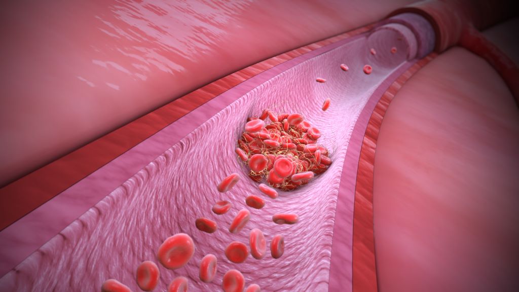
Researchers from the University of Tokyo have found a way to observe clotting activity in blood as it happens – without needing invasive procedures. Using a new type of microscope and artificial intelligence (AI), their study shows how platelet clumping can be tracked in patients with coronary artery disease (CAD), opening the door to safer, more personalised treatment.

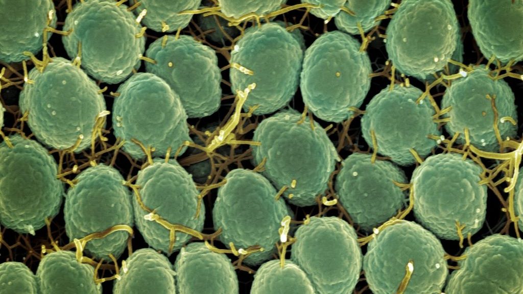
“Platelets play a crucial role in heart disease, especially in CAD, because they are directly involved in forming blood clots,” explained Dr Kazutoshi Hirose, an assistant professor at the University of Tokyo Hospital and lead author of the study in Nature Communications. “To prevent dangerous clots, patients with CAD are often treated with antiplatelet drugs. However, it’s still challenging to accurately evaluate how well these drugs are working in each individual, which makes monitoring platelet activity an important goal for both doctors and researchers.”

That challenge pushed Hirose and his collaborators to develop a new system for monitoring platelets in motion, using a high-speed optical device and artificial intelligence.

“We used an advanced device called a frequency-division multiplexed (FDM) microscope, which works like a super high-speed camera that takes sharp pictures of blood cells in flow,” said co-author Yuqi Zhou, an assistant professor of chemistry at the University of Tokyo . “Just like traffic cameras capture every car on the road, our microscope captures thousands of images of blood cells in motion every second. We then use artificial intelligence to analyse those images. The AI can tell whether it’s looking at a single platelet (like one car), a clump of platelets (like a traffic jam), or even a white blood cell tagging along (like a police car caught in the jam).”

The research team applied this technique to blood samples from over 200 patients. Their images revealed that patients with acute coronary syndrome had more platelet aggregates than those with chronic symptoms – supporting the idea that this technology can track clotting risk in real time.

“Part of my scientific curiosity comes from the recent advances in high-speed imaging and artificial intelligence, which have opened up new ways to observe and analyse blood cells in motion,” said Keisuke Goda, a professor of chemistry at the University of Tokyo who led the research team. “AI can ‘see’ patterns beyond what the human eye can detect.”

One of the most important findings was that a simple blood drawn from the arm – rather than from the heart’s arteries – provided nearly the same information.

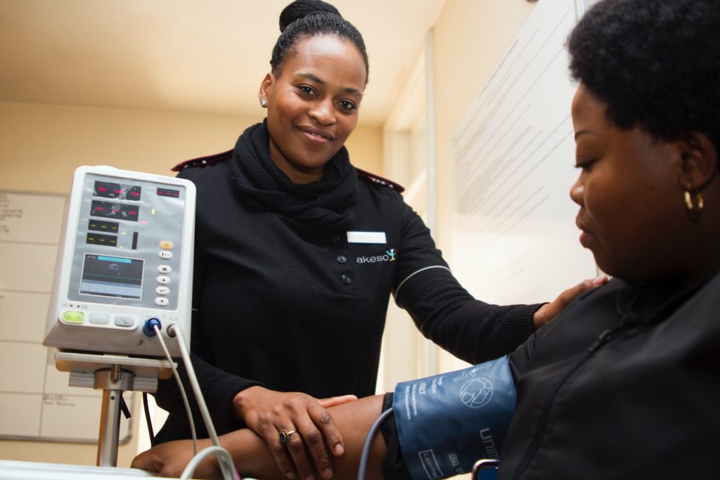
“Typically, if doctors want to understand what’s happening in the arteries, especially the coronary arteries, they need to do invasive procedures, like inserting a catheter through the wrist or groin to collect blood,” said Hirose. “What we found is that just taking a regular blood sample from a vein in the arm can still provide meaningful information about platelet activity in the arteries. That’s exciting because it makes the process much easier, safer and more convenient.”

The long-term hope is that this technology will help doctors better personalise heart disease treatment.

“Just like some people need more or less of a painkiller depending on their body, we found that people respond differently to antiplatelet drugs. In fact, some patients are affected by recurrent thrombosis and others are suffering from recurrences of bleeding events even on the same antiplatelet medications,” said Hirose. “Our technology can help doctors see how each individual’s platelets are behaving in real time. That means treatments could be adjusted to better match each person’s needs.”

“Our study shows that even something as small as a blood cell can tell a big story about your health,” Zhou added.

Source: University of Tokyo

Language Shapes how Sensory Experiences are Stored in the Brain

A study in stroke patients shows the brain’s vision-language connection shapes object knowledge

A schematic view of the main findings, adapted from a brain figure in the study. Image credit: Adapted from Liu Bet al., 2025, PLOS Biology, CC-BY 4.0

Our ability to store information about familiar objects depends on the connection between visual and language processing regions in the brain, according to a study published May 20th in the open-access journal PLOS Biology by Bo Liu from Beijing Normal University, China, and colleagues.

Seeing an object and knowing visual information about it, like its usual colour, activate the same parts of the brain. Seeing a yellow banana, for example, and knowing that the object represented by the word “banana” is usually yellow, both excite the ventral occipitotemporal cortex (VOTC). However, there’s evidence that parts of the brain involved in language, like the dorsal anterior temporal lobe (ATL), are also involved in this process – dementia patients with ATL damage, for example, struggle with object colour knowledge, despite having relatively normal visual processing areas. To understand whether communication between the brain’s language and sensory association systems is necessary for representing information about objects, the authors tested whether stroke-induced damage to the neural pathways connecting these two systems impacted patients’ ability to match objects to their typical colour. They compared colour-identification behaviour in 33 stroke patients to 35 demographically-matched controls, using fMRI to record brain activity and diffusion imaging to map the white matter connections between language regions and the VOTC.

The researchers found that stronger connections between language and visual processing regions correlated with stronger object color representations in the VOTC, and supported better performance on object color knowledge tasks. These effects couldn’t be explained by variations in patients’ stroke lesions, related cognitive processes (like simply recognizing a patch of color), or problems with earlier stages of visual processing. The authors suggest that these results highlight the sophisticated connection between vision and language in the human brain.

The authors add, “Our findings reveal that the brain’s ability to store and retrieve object perceptual knowledge – like the colour of a banana – relies on critical connections between visual and language systems. Damage to these connections disrupts both brain activity and behaviour, showing that language isn’t just for communication – it fundamentally shapes how sensory experiences are neurally structured into knowledge.”

Provided by PLOS

Undervalued but Unshaken: Fundi Steps up to Celebrate South Africa’s Nurses Amid Global Aid Cuts

Photo by Hush Naidoo on Unsplash

As the world marks Nurses Day this month, this annual anniversary has shone a sharp spotlight on the realities facing this group of South African heroes and heroines. With many battling burnout, bracing for further staff shortages and trying to find ways to absorb the impact of global funding cuts, Fundi is inviting the country to get behind our nurses and celebrate their daily untold contribution to our local communities.

Recent announcements by the United States to slash foreign aid to global health programmes (including PEPFAR, which funds major HIV initiatives in South Africa) sent shockwaves through our local healthcare sector.

“For nurses already stretched to breaking point, this was yet another blow,” notes Mary Maponya, Fundi Executive Head: Lending. “This isn’t just about money disappearing from a spreadsheet. It’s about support being taken from clinics, treatment delays growing longer and nurses being asked to pick up even more of the slack. We need to acknowledge that nurses are the pulse of public healthcare in South Africa – and that pulse is under pressure.”

Maponya says this is why it was so important for Fundi to find ways to add its voice of thanks and appreciation for nurses this month – including sponsoring and attending Denosa events in Limpopo, Mpumalanga and KwaZulu-Natal. “Department of Health employees make up 16% of our total loan book, with a high proportion of these being nurses,” she explains. “They are a growing sector; continuously investing in their own self-development as a means of deepening their vocations and contribution.”

Fundi’s presence at these events made it possible to engage meaningfully with nurses on the ground; building relationships and understanding how best to continue to provide support around career advancement in particular.

“We are also be running a social media campaign until 23 May where frontline nurses can win free lunch for the week – with Uber Eats delivering it straight to their workplace to lighten the load. Small acts of kindness and appreciation make all the difference,” says Maponya. “And that’s exactly what we’re hoping this campaign will do!”

According to the South African Nursing Council, South Africa has just over 280 000 registered nurses[1], with many nearing retirement age. Meanwhile, nurse emigration is accelerating as we struggle to keep our local nurses employed and supported[2]. “It is estimated that our country will need over 100 000 new nurses by 2030 to maintain even basic healthcare coverage[3],” Maponya explains. “With the bulk of patient care still falling on nurses – from vaccinations and chronic disease management to trauma response – the pressure on these individuals is relentless.”

Amid these systemic failings, one thing is clear however: South Africa’s nurses continue showing up with grace and courage. “This was perhaps the most important take-out from the DENOSA engagement. Our nurses show-up each day – safeguarding the life and health of their patients. These remarkable individuals remain our first line of defence during pandemics, pregnancies, mental health emergencies and more. If we want a healthier South Africa, we must protect the protectors. We need to continue investing in our nurses not just during Nurses Month, but every single day,” she concludes.

[1] https://www.sanc.co.za/

[2] https://www.nursingservices.co.za/news/addressing-south-africas-nursing-shortage

[3] https://www.news24.com/southafrica/news/nursing-in-sa-is-changing-but-is-it-enough-to-avert-an-anticipated-crisis-20240928

After Cardiac Event, Excessive Sedentary Time Led to Increased Risk of Another Event

People who sit or remain sedentary for more than 14 hours a day, on average, may have a higher risk of a cardiovascular event or death in the year after treatment at a hospital for symptoms of a heart attack such as chest pain, according to new research published today in the American Heart Association’s peer-reviewed scientific journal Circulation: Cardiovascular Quality and Outcomes.

Previous research from the study authors found that people who had experienced a heart attack were spending up to 12 to 13 hours each day being sedentary, defined as any awake activity that involved little-to-no physical movement. In this study, the researchers used a wrist accelerometer to track the amount of time each participant spent moving or being sedentary for a median of 30 days after discharge from a hospital’s emergency department. 

Wrist accelerometers measure the acceleration of motion in three directions: forwards and backwards, side-to-side, and up and down. These measurements allowed the researchers to infer the intensity of a participant’s physical activity, and they provide more accurate measurements of the participants’ time spent moving, rather than asking participants to remember. Some examples of moderate intensity physical activities are brisk walking, water aerobics, dancing, playing doubles tennis or gardening, and examples of vigorous-intensity activities are running, lap swimming, heavy yardwork such as continuous digging or hoeing, playing singles tennis or jumping rope.

”Current treatment guidelines after a cardiac event focus mainly on encouraging patients to exercise regularly,” said study lead author Keith Diaz, Ph.D., the Florence Irving Associate Professor of Behavioral Medicine at Columbia University Medical Center in New York City, a certified exercise physiologist and a volunteer member of the American Heart Association’s Physical Activity Science Committee. “In our study, we explored whether sedentary time itself may contribute to cardiovascular risk.”

Researchers followed more than 600 adults, ages 21 to 96, treated for a heart attack or chest pain in the emergency department at a single hospital system in New York City. Participants wore a wrist accelerometer for a median of 30 consecutive days after hospital discharge to measure the amount of time they spent sitting or being inactive each day. Additional cardiac events and deaths were evaluated one year after hospital discharge via phone surveys with patients, electronic health records and the Social Security Death Index. The study was focused on understanding the risk of sedentary behavior and identifying modifiable risk factors that may improve long-term outcomes in this high-risk group.

The analysis found:

  • Compared to participants in the group with the highest physical activity level, those in the group with the lowest activity level had a 2.58 times higher risk of having another heart problem or dying within the next year.
  • Replacing 30 minutes of sedentary time with 30 minutes of moderate to vigorous physical activity, daily, reduced the risk of adverse cardiovascular events or death by 61%; replacing the sedentary time with light-intensity physical activity reduced risk by 50%; and replacing the sedentary time with 30 minutes of sleep lowered risk by 14%.
  • According to accelerometer data, participants in the most physically active group had average daily physical activity measures of 143.8 minutes of light physical activity; 25 minutes of moderate-to-vigorous physical activity; 11.7 hours spent sedentary; and 8.4 hours of sleep.
  • Participants in the least physically active group had daily averages of 82.2 minutes each day of light physical activity; 2.7 minutes of moderate-to-vigorous physical activity; 15.6 hours spent sedentary; and 6.6 hours of sleep.
  • Participants in the group between most and least physically active had daily averages of 109.2 minutes of light intensity physical activity; 11.4 minutes of moderate-to-vigorous intensity physical activity; 13.5 hours spent sedentary and 7.8 hours of sleep.

“We were surprised that replacing sedentary time with sleep also lowered risk. Sleep is a restorative behavior that helps the body and mind recover, which is especially important after a serious health event like a heart attack,” Diaz said. “Our study indicates that one doesn’t have to start running marathons after a cardiovascular event to see benefits. Sitting less and moving or sleeping a little more can make a real difference. More physical activity and more sleep are healthier than sitting, so we hope these findings support health professionals to move toward a more holistic, flexible and individualized approach for physical activity in patients after a heart attack or chest pain.”

Physical activity and sleep are both key components of the American Heart Association’s Life’s Essential 8, a list of health behaviours and factors that support optimal cardiovascular health. Poor sleep is a known risk factor for cardiovascular disease, which claims more lives each year in the U.S. than all forms of cancer and chronic lower respiratory disease combined, according to the American Heart Association’s 2025 Statistical Update. In addition to sleep duration, a recent scientific statement from the Association highlighted the importance of sleep continuity, sleep timing, sleep satisfaction, sleep regularity, sleep-related daytime functioning and sleep architecture in cardiometabolic health.

The study had several limitations, including that the definition of sedentary behaviour was based only on the intensity level of physical movement, meaning that the study may have overestimated the time participants spent in sedentary behaviour. Additionally, there was no information about participants’ income and characteristics of the neighbourhoods where they live, which limits the study’s ability to account for social and environmental factors, including participants’ risk of one-year cardiac events and deaths. Also, hospital discharge information about whether patients were sent home, referred to rehabilitation or referred to other care centres such as skilled nursing facilities were not collected. This limited the study’s ability to fully assess whether the patients’ settings had an impact on their recovery.

Source: American Heart Association

Amid Surge in Cases, UK’s NHS to Offer Gonorrhoea Vaccine


Neisseria gonorrhoeae Bacteria Scanning electron micrograph of Neisseria gonorrhoeae bacteria, which causes gonorrhoea. Captured by the Research Technologies Branch (RTB) at the NIAID Rocky Mountain Laboratories (RML) in Hamilton, Montana. Credit: NIAID. Photo by National Institute of Allergy and Infectious Diseases on Unsplash

In the midst of a record high in gonorrhoea cases, the NHS is to offer a gonorrhoea vaccine to gay and bisexual men with a history of multiple partners or a sexually transmitted infection (STI), the BBC reports. The gonorrhoea vaccination, which is actually a repurposed meningococcal vaccine, is estimated only to be 30–40% effective. Research shows, however, that this will be sufficient to reduce cases and their attendant costs to the NHS.

Gonorrhoea is caused by the bacterium Neisseria gonorrhoeae and is typically transmitted by having intercourse without a condom. It can cause pain, unusual discharge, genital inflammation and infertility. Evidence has shown that the MeNZB and four-component serogroup B meningococcal (4CMenB) vaccines, designed against Neisseria meningitidis, can also offer protection against gonorrhoea.

In 2023, there were more than 85 000 cases – the highest since records began in 1918. A study published in The Lancet estimates that gonorrhoea vaccination would prevent 100 000 cases, saving the NHS £7.9 million over the next decade.

While gonorrhoea is treatable with antibiotics, resistance is growing and there is concern that it may eventually become untreatable. According to The Guardian, some cases are now “extensively drug resistant” (XDR) – not responsive to ceftriaxone or the second line of treatment. There were 17 cases of ceftriaxone-resistant gonorrhoea between January 2024 and March 2025, the UK Health Security Agency (UKHSA) reported.

Over the same period, nine XDR cases were reported, while between 2022 and 2023, there were only five.

The people most affected by gonorrhoea in the UK are the 16 to 25 age group, gay and bisexual men, and those of black and Caribbean ancestry. The study’s scenario for vaccinating at-risk populations included those who had more than five sexual partners per year or who had a positive gonorrhoea test.

The vaccine, costing about £8 per dose, is cost-effective when administered to this at-risk group of men, rather than adolescents. Despite this, clinicians will be able to offer the vaccine to anyone who, in their judgment, would benefit from it. Other vaccines such as for mpox – another STI with high transmission between gay and bisexual men – and hepatitis will also be offered.

Controlling 8 Risk Factors may Eliminate Early Death Risk from Hypertension

Pexels Photo by Thirdman

A new study led by researchers at Tulane University suggests that people with hypertension can significantly reduce, and possibly eliminate, their increased risk of premature death by controlling several key health risk factors at once.

The study, appearing in Precision Clinical Medicine, tracked more than 70 000 people with hypertension and over 224 000 without it, using data from the UK Biobank. Researchers followed participants for nearly 14 years to understand how managing these risk factors affected early mortality, defined as dying before age 80.

The eight health risk factors evaluated in the study include: blood pressure, body mass index, waist circumference, LDL “bad” cholesterol, blood sugar, kidney function, smoking status and physical activity. Notably, researchers found that hypertensive patients who had addressed at least four of these risk factors had no greater risk of an early death than those without hypertension. 

“Our study shows that controlling blood pressure is not the only way to treat hypertensive patients, because high blood pressure can affect these other factors,” said corresponding author Dr Lu Qi, professor of epidemiology at Tulane University. “By addressing the individual risk factors, we can help prevent early death for those with hypertension.” 

Hypertension, defined as a blood pressure of 130mmHg or higher, is the leading preventable risk factor for premature death worldwide. 

The study found that addressing each additional risk factor was associated with a 13% lower risk of early death, 12% lower risk of early death due to cancer and 21% lower risk of death due to cardiovascular disease, the leading cause of premature death globally.

“Optimal risk control” – having 7 or more of the risk factors addressed – was linked to 40% less risk of early death, 39% less risk of early death due to cancer and 53% less risk of early death due to cardiovascular disease. 

“To our knowledge, this is the first study to explore the association between controlling joint risk factors and premature mortality in patients with hypertension,” Qi said. “Importantly, we found that any hypertension-related excess risk of an early death could be entirely eliminated by addressing these risk factors.” 

Only 7% of hypertensive participants in the study had seven or more risk factors under control, highlighting a major opportunity for prevention. Researchers say the findings underscore the importance of personalised, multifaceted care – not just prescribing medication for blood pressure, but addressing a broader range of health behaviours and conditions.

Source: Tulane University

Baby with Rare, Incurable Disease is First to Receive Personalised Gene Therapy

NIH-supported gene-editing platform lays groundwork to rapidly develop treatments for other rare genetic diseases.

Photo by Sangharsh Lohakare on Unsplash

A research team supported by the National Institutes of Health (NIH) has developed and safely delivered a personalised gene editing therapy to treat an infant with a life-threatening, incurable genetic disease. The infant, who was diagnosed with the rare condition carbamoyl phosphate synthetase 1 (CPS1) deficiency shortly after birth, has responded positively to the treatment.

The process, from diagnosis to treatment, took only six months and marks the first time the technology has been successfully deployed to treat a human patient. The technology used in this study was developed using a platform that could be tweaked to treat a wide range of genetic disorders and opens the possibility of creating personalised treatments in other parts of the body.

A team of researchers at the Children’s Hospital of Philadelphia (CHOP) and the Perelman School of Medicine at the University of Pennsylvania (Penn) developed the customised therapy using the gene-editing platform CRISPR. They corrected a specific gene mutation in the baby’s liver cells that led to the disorder. CRISPR is an advanced gene editing technology that enables precise changes to DNA inside living cells. This is the first known case of a personalised CRISPR-based medicine administered to a single patient and was carefully designed to target non-reproductive cells so changes would only affect the patient.

“As a platform, gene editing – built on reusable components and rapid customisation – promises a new era of precision medicine for hundreds of rare diseases, bringing life-changing therapies to patients when timing matters most: Early, fast, and tailored to the individual,” said Joni L. Rutter, Ph.D., director of NIH’s National Center for Advancing Translational Sciences (NCATS).

CPS1 deficiency is characterized by an inability to fully break down byproducts from protein metabolism in the liver, causing ammonia to build up to toxic levels in the body. It can cause severe damage to the brain and liver. Treatment includes a low protein diet until the child is old enough for a liver transplant. However, in this waiting period there is a risk of rapid organ failure due to stressors such as infection, trauma, or dehydration. High levels of ammonia can cause coma, brain swelling, and may be fatal or cause permanent brain damage.

The child initially received a very low dose of the therapy at six months of age, then a higher dose later. The research team saw signs that the therapy was effective almost from the start. The six-month old began taking in more protein in the diet, and the care team could reduce the medicine needed to keep ammonia levels low in the body. Another telling sign of the child’s improvement to date came after the child caught a cold, and later, had to deal with a gastrointestinal illness. Normally, such infections for a child in this condition could be extremely dangerous, especially with the possibility of ammonia reaching dangerous levels in the brain.

“We knew the method used to deliver the gene-editing machinery to the baby’s liver cells allowed us to give the treatment repeatedly. That meant we could start with a low dose that we were sure was safe,” said CHOP pediatrician Rebecca Ahrens-Nicklas, MD, PhD.

“We were very concerned when the baby got sick, but the baby just shrugged the illness off,” said Penn geneticist and first author Kiran Musunuru, MD, PhD. For now, much work remains, but the researchers are cautiously optimistic about the baby’s progress.

The scientists announced their work at the American Society of Gene & Cell Therapy Meeting on May 15th and described the study in The New England Journal of Medicine.

Source: NIH/Office of the Director

Key Player in Childhood Food Allergies Identified: Thetis Cells

Photo by cottonbro studio

A decade ago, a clinical trial in the UK famously showed that children who were exposed to peanuts in the early months of life had reduced risk of developing a peanut allergy compared with children who avoided peanuts.

Now, researchers at Memorial Sloan Kettering Cancer Center (MSK) have a likely answer as to why that’s the case: Thetis cells.

This recently discovered class of immune cells, which were first described by MSK researchers in 2022, plays an essential and previously unknown role in suppressing inflammatory responses to food, according to findings published in Science, one of the world’s premier scientific journals.

Moreover, the study, which was conducted in mouse models, points to a critical window in the early months of life for training the immune system not to overreact to food allergens, termed “oral tolerance.”

The study also opens the door to new therapeutic possibilities, the researchers say.

“This is a great example of how clinical studies can reveal clues to fundamental mechanisms in biology,” says physician-scientist Chrysothemis Brown, MBBS, PhD, the study’s senior author. “These new understandings can pave the way for new treatment strategies for food allergies, which are desperately needed.”

The research was led by co-first authors from the Brown Lab: paediatric hematologist-oncologist Vanja Cabric, MD, and research assistant Yollanda Franco Parisotto, PhD.

Thetis Cells Train the Immune System To Tolerate Helpful Outsiders

Thetis cells are a type of antigen-presenting cell, whose job is to present foreign substances (antigens) to other immune cells. Antigen-presenting cells must educate the immune system. These cells provide signals that tell the immune system to attack foreign bacteria and viruses – or instruct it to tolerate harmless proteins in the foods we eat.

Previous research led by Dr Brown and immunologist Alexander Rudensky, PhD, Chair of the Immunology Program at MSK’s Sloan Kettering Institute, identified a window in early life where a “developmental wave” of Thetis cells within the gut creates an opportunity for developing immune tolerance.

“We previously showed that Thetis cells train the immune system not to attack the helpful bacteria in the digestive system. So we wondered whether these cells might also be important for preventing inflammatory responses to food, and whether the increased abundance of the cells during early life would result in increased protection against food allergy,” says Dr. Brown, whose lab is in MSK’s Human Oncology and Pathogenesis Program (HOPP).

The new study found that Thetis cells not only help to broker peace accords with “good” bacteria, but also with proteins in foods that can act as allergens, such the Ara h proteins found in peanuts (though they weren’t specifically tested in the study) or the ovalbumin found in eggs.

Thetis cells got their name because they share traits with two different types of antigen-presenting cells: medullary thymic epithelial cells and dendritic cells, just as Thetis in Greek mythology had shape-shifting attributes.

A Key Role for Gut-Draining Lymph Nodes

The research team used a variety of genetically engineered mouse models to investigate oral tolerance. They attached a fluorescent dye to ovalbumin in order to visualise which cells in the gut interacted with it.

And this showed that a subset of Thetis cells, the same ones that regulated tolerance to healthy gut bacteria, took up the protein. This allowed Thetis cells to program another type of immune cell called regulatory T cells to suppress the immune response to the egg protein, essentially telling the body it was safe.

“This process is often studied in adult models, but by examining what happens when mice first encounter food proteins at the time of weaning, we could see which specific cells were critical to generating tolerance to food during early life,” Dr. Cabric says.

Although Thetis cells could also induce tolerance throughout life, there was a significant difference in the immune response when the egg protein was introduced later.

Source: Memorial Sloan Kettering Cancer Center

The Effect of Physical Fitness on Mortality is Overestimated

Photo by Ketut Subiyanto on Unsplash

Many observational studies have shown that people who exercise more and have good cardiorespiratory fitness early in life are at lower risk of premature death from causes such as cancer and cardiovascular disease. But a new study published in the European Journal of Preventive Cardiology suggests that the association between physical fitness and a reduced risk of mortality may be misleading.

“We found that people with high fitness levels in late adolescence had a lower risk of dying prematurely, for example from cardiovascular disease, compared to those with low fitness levels. But when we looked at their risk of dying in random accidents, we found an almost similarly strong association. This suggests that people with high and low fitness levels may differ in other important ways, which is something that previous studies have not fully taken into account,” says Marcel Ballin, associated researcher in epidemiology and lead author of the study.

Conscription data from over 1 million men

In the study, the researchers leveraged data from 1.1 million Swedish men who were conscripted for military service between the years 1972 and 1995. The men, who were on average 18 years old at the time of conscription, were divided into five groups based on their fitness level at the time. They were then followed until their 60s or until they died. With access to the National Cause of Death Register, the researchers were able to see their cause of death. They subsequently used different methods to study the association between fitness level in late adolescence and premature death.

The researchers started with a traditional analysis of mortality from cardiovascular disease, cancer and from all causes, as in previous observational studies. They adjusted their statistical models for factors such as BMI, age at conscription, year of conscription, and parents’ income and education level. The results showed that the group with the highest fitness level had a 58% lower risk of dying from cardiovascular disease, a 31% lower risk of dying from cancer, and a 53% lower risk of dying from all causes, compared with the group with the lowest fitness level.

Very similar risk of dying in random accidents

Next, the researchers examined how fitness was associated with the risk of dying in random accidents such as car accidents, drownings and homicides. They chose random accidents because they assumed that there ought to be no association between the men’s fitness in late adolescence and the risk of dying in random accidents. This method is called negative control outcome analysis and involves testing the validity of your results for a primary outcome by comparing them with an outcome where no association ought to be found. If, however, an association is found, it may indicate that the groups studied are not actually comparable, and that the study suffers from what is typically referred to as confounding. The researchers found that men with the highest fitness levels had a 53% lower risk of dying in random accidents. Yet, it is unlikely that the men’s fitness would have such a big effect on their risk of dying in random accidents.

These results were also confirmed when the researchers used the sibling comparison design. Using this method, the researchers compared the risk of premature death between siblings with different fitness levels to control for all the factors that the siblings share such as behaviours, environmental factors, and some genetic factors.

“It surprised us that the association with accidental mortality reflected the other associations, even after we controlled for all the factors that siblings share. This underlines how strong the assumptions are that you make in observational studies, since it appears to be very difficult to create comparable groups. The consequences may be that you overestimate the magnitudes of the effects you find,” says Marcel Ballin.

Picture confirmed in other studies

The study is one of the largest of its kind in which researchers used negative control outcomes to investigate whether the associations between fitness and mortality are in fact valid. The results in this study are also supported by other research.

“That the effects of good cardiorespiratory fitness may be overstated might sound controversial to some, but the fact is that if you look at the results from studies others than traditional observational studies, a more nuanced picture does emerge. A number of twin studies for example have found similar results. Some genetic studies also suggest that there are genes that affect both the propensity to be physically active or have a good fitness level, and the risk of developing diseases such as cardiovascular disease.”

Important to base interventions on correct estimates

Marcel Ballin also argues that there are many different reasons for promoting physical activity. However, large-scale interventions or policy changes intended to apply to the entire population must be based on reliable estimates – otherwise there is a risk of expecting effects that have in fact been overestimated.

“Our results should not be interpreted as if physical activity and exercise are ineffective or that you should not try to promote it. But to create a more nuanced understanding of how big the effects of fitness actually are on different outcomes, we need to use several different methods. If we just ask the question in the same way, we will always get the same answer. It’s only when we get the same answer to a question that we have asked in slightly different ways that we can be sure that the findings are accurate,” says Marcel Ballin.

Source: Uppsala University

Loss of Lung Capacity Starts Between the Ages of 20 and 25

Photo by Ketut Subiyanto on Pexels

A study led by the Barcelona Institute for Global Health (ISGlobal), a centre supported by ”la Caixa” Foundation, in collaboration with the Clínic-IDIBAPS, has shown for the first time, how lung capacity evolves from childhood to old age. The findings, published in The Lancet Respiratory Medicine, provide a new basic framework for assessing lung health.

Until now, it was thought that lung function increased until it peaked at around 20–25 years of age, after which it stabilised. It was also thought that in later adulthood, lung function begins to decline as the lungs age. However, this model was based on studies that did not cover the whole life course.

In contrast, the current study used an “accelerated cohort design”, meaning data from several cohort studies were combined to cover the desired age range. “We included more than 30 000 individuals aged 4 to 82 years from eight population-based cohort studies in Europe and Australia,” explains Judith Garcia-Aymerich, first author of the study and co-director of the ISGlobal programme on Environment and Health over the Life Course. Lung function and lung capacity parameters were assessed using forced spirometry, a test in which the patient exhales all the air as quickly as possible after taking a deep breath. Data on active smoking and asthma diagnosis were also collected.

Two growth phases and an early decline

The study showed that lung function develops in two distinct phases: a first phase of rapid growth during childhood and a second phase of slower growth until peak lung function is reached. Lung function was assessed using two parameters: forced expiratory volume in one second (FEV1), which measures the amount of air expelled in the first second of a forced breath after a deep inhalation; and forced vital capacity (FVC), which is the maximum amount of air a person can breathe out without a time limit after a deep inhalation. 

In women, FEV1 peaks around the age of 20, while in men it peaks around the age of 23. Surprisingly, the study found no evidence of a stable phase following this peak. “Previous models suggested a plateau phase until the age of 40, but our data show that lung function starts to decline much earlier than previously thought, immediately after the peak,” explains Garcia-Aymerich.

Source: Barcelona Institute for Global Health (ISGlobal)