Year: 2025

Research Supports Continued Use of Nasogastric Tube After Oesophageal Cancer Surgery

To the researchers’ surprise, it was not without risk to omit the tube after this surgery. Illustration: Jakob Hedberg

In the largest Nordic study to date concerning oesophageal cancer surgery, the researchers found clear evidence that decompression with a nasogastric tube is associated with less serious complications. Their results challenge a trend of declining use of the nasogastric tube after major surgical procedures. The study was led from Uppsala University and has now been published in Lancet Regional Health Europe.

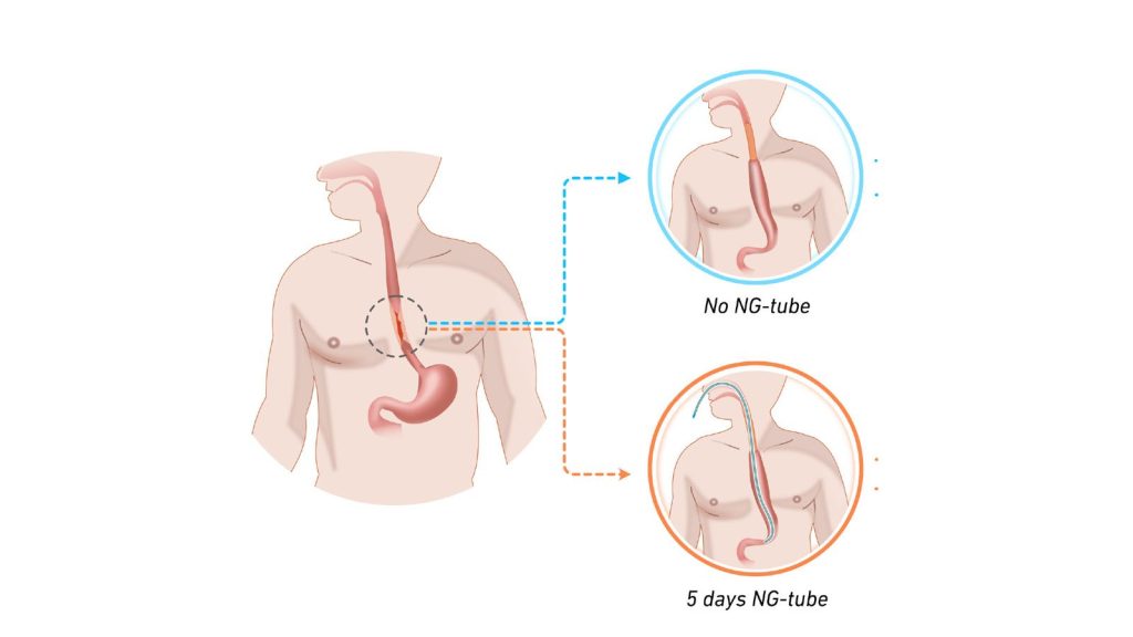
A number of small studies had previously suggested that it is safe to abandon the tradition of leaving in a decompressing – but for many patients unpleasant – nasogastric tube after surgery to remove oesophageal cancer (gullet cancer). The tube is plastic and runs from the nose down to the stomach, and its use in this particular context is to relieve and reduce pressure in this newly operated area. When the question was discussed in a Nordic research collaboration, it was concluded that these smaller studies lacked sufficient statistical power to justify a change in care. Subsequently, a randomised trial was carried out at 12 university hospitals across Sweden, Norway, Denmark and Finland, where patients were randomised to have or not have a decompressing nasogastric tube in their oesophagus following this type of surgery.

Patients without the tube experienced leakage

To the researchers’ surprise, it was not without risk to omit the tube after this surgery, as more patients without the tube experienced leakage in the anastomosis created during the operation. Leakage must be treated immediately, often with interventions under general anaesthesia, resulting in suffering for the patient and a longer length of hospital stay.

Although no differences in survival rates or other complications were found, this new knowledge may help to reduce suffering for patients in the future.

“Oesophageal cancer is an uncommon form of cancer, with only about 200 operations of this type being performed per year in Sweden. National and international cooperation is therefore absolutely necessary in order to conduct sufficiently large trials to answer the research questions we have. The fact that in just over two years, almost 450 patients have been recruited for the trial surpassed our expectations and represents a great success for this network,” says Jakob Hedberg, surgical oncologist, associate professor at Uppsala University and consultant surgeon at Uppsala University Hospital who is also principal investigator for the study.

“Strong interest has been shown at international conferences where our preliminary results have been presented, and the principle of building surgical care on solid evidence has allowed us to provide the best care to our patients. Another important effect of this successful collaboration is that we can build more clinical trials within the Nordic network which has now been consolidated. In fact, the next clinical trial is already under development,” says Jakob Hedberg.

Source: Uppsala University

Good Prognosis for Men with Prostate Cancer Treated According to Guidelines

Credit: Darryl Leja National Human Genome Research Institute National Institutes Of Health

Most men who are treated for prostate cancer according to modern guidelines have good survival rates and the majority of these men will die of causes other than prostate cancer. This is revealed in a new study from Uppsala University published in the Journal of the National Comprehensive Cancer Network.

“We were surprised by how much life expectancy affected the prognosis. This shows the importance of a thorough assessment of the general health of a man with newly diagnosed prostate cancer. The patient’s life expectancy has a substantial impact on the choice of appropriate treatment strategy,” says Marcus Westerberg, researcher at the Department of Surgical Sciences at Uppsala University, who led the study.

In prostate cancer, the disease progression often takes decades and the risk of dying from prostate cancer therefore depends on both the characteristics of the cancer and life expectancy based on the man’s age and other diseases at the time of diagnosis. Recommendations in guidelines and care programmes are therefore also based on both cancer characteristics and life expectancy. This means that the recommended initial treatment can range from active monitoring for low-risk cancer to combinations of local and systemic treatment for high-risk cancer.

High average age at disease onset

As the average age at diagnosis of prostate cancer is often high and the cancer often progresses very slowly, it is particularly important to know the long-term risk of death from prostate cancer in order to choose the best treatment for patients. Previously, not much has been known about this.

“We wanted to fill that knowledge gap, so we looked at outcomes up to 30 years after the men were diagnosed. In all cases, we had information about the characteristics of the cancer, treatment and the patient’s life expectancy based on age and comorbidity,” says Westerberg.

The researchers used data from the Prostate Cancer Database Sweden (PCBase), which contains information from the National Prostate Cancer Register (NPCR) and other health data registers. They focused on men who had received the recommended treatment for prostate cancer that had not spread in the body. Using statistical modelling, the researchers estimated the lifetime risk of dying from prostate cancer and other causes.

11 per cent risk of dying of cancer

For men with low-risk cancer and short life expectancy (less than 10 years), the risk of dying from prostate cancer was 11% and the risk of dying from other causes was 89% within 30 years of diagnosis.

For men with high-risk cancer (eg stage T3, PSA 30ng/mL and Gleason score 8) and long life expectancy (over 15 years), the risk of dying from prostate cancer was 34% and the risk of dying from other causes was 55% within 30 years of diagnosis.

“We hope that our results will be used to provide a realistic picture of the prognosis for men with prostate cancer. Our study shows that most men who receive the recommended treatment have a good prognosis,” Westerberg concludes.

Life expectancy was based on age and comorbidity. Examples of low-risk cancers are stage T1, PSA 5ng/mL and Gleason score 6. Examples of high-risk cancers are stage T3, PSA 30ng/mL and Gleason score 8.

Source: Uppsala University

Hibernation ‘Superpowers’ May Be Hidden in Human DNA

Photo by Sangharsh Lohakare on Unsplash

Animals that hibernate are incredibly resilient. They can spend months without food or water, muscles refusing to atrophy, body temperature dropping to near freezing as their metabolism and brain activity slow to a crawl. When they emerge from hibernation, they recover from dangerous health changes similar to those seen in type 2 diabetes, Alzheimer’s disease, and stroke.

New genetic research suggests that hibernating animals’ superpowers could lie hidden in human DNA – with clues on how to unlock them, perhaps one day leading to treatments that could reverse neurodegeneration and diabetes.

Two studies describing the results are published in Science.

The genetics of metabolism and obesity

A gene cluster called the “fat mass and obesity (FTO) locus” plays an important role in hibernators’ abilities, the researchers found. Intriguingly, humans have these genes too. “What’s striking about this region is that it is the strongest genetic risk factor for human obesity,” says Chris Gregg, PhD, professor in neurobiology and human genetics at University of Utah Health and senior author on the studies. But hibernators seem able to use genes in the FTO locus in new ways to their advantage.

The team identified hibernator-specific DNA regions that are near the FTO locus and that regulate the activity of neighbouring genes, tuning them up or down. The researchers speculate that adjusting the activity of neighbouring genes, including those in or near the FTO locus, allows hibernators to pack on the pounds before settling in for the winter, then slowly use their fat reserves for energy throughout hibernation.
 
Indeed, the hibernator-specific regulatory regions outside of the FTO locus seem crucial for tweaking metabolism. When the researchers mutated those hibernator-specific regions in mice, they saw changes in the mice’s weight and metabolism. Some mutations sped up or slowed down weight gain under specific dietary conditions; others affected the ability to recover body temperature after a hibernation-like state or tuned overall metabolic rate up or down. 

Intriguingly, the hibernator-specific DNA regions the researchers identified weren’t genes themselves. Instead, the regions were DNA sequences that contact nearby genes and turn their expression up or down, like an orchestra conductor fine-tuning the volume of many musicians. This means that mutating a single hibernator-specific region has wide-ranging effects extending far beyond the FTO locus, explains Susan Steinwand, research scientist in neurobiology at U of U Health and first author on one of the studies.  “When you knock out one of these elements – this one tiny, seemingly insignificant DNA region – the activity of hundreds of genes changes,” she says. “It’s pretty amazing.”
 
Understanding hibernators’ metabolic flexibility could lead to better treatments for human metabolic disorders like type 2 diabetes, the researchers say. “If we could regulate our genes a bit more like hibernators, maybe we could overcome type 2 diabetes the same way that a hibernator returns from hibernation back to a normal metabolic state,” says Elliott Ferris, MS, bioinformatician at U of U Health and first author on the other study.

Uncovering the regulation of hibernation

Finding the genetic regions that may enable hibernation is a problem akin to excavating needles from a massive DNA haystack. To narrow down the regions involved, the researchers used multiple independent whole-genome technologies to ask which regions might be relevant for hibernation. Then, they started looking for overlap between the results from each technique.

First, they looked for sequences of DNA that most mammals share but that had recently changed in hibernators. “If a region doesn’t change much from species to species for over 100 million years but then changes rapidly and dramatically in two hibernating mammals, then we think it points us to something that is important for hibernation, specifically,” Ferris says.

To understand the biological processes that underlie hibernation, the researchers tested for and identified genes that turn up or down during fasting in mice, which triggers metabolic changes similar to hibernation. Next, they found the genes that act as central coordinators, or “hubs,” of these fasting-induced changes to gene activity.

Many of the DNA regions that had recently changed in hibernators also appeared to interact with these central coordinating hub genes. Because of this, the researchers expect that the evolution of hibernation requires specific changes to the controls of the hub genes. These controls comprise a shortlist of DNA elements that are avenues for future investigation.

Awakening human potential

Most of the hibernator-associated changes in the genome appeared to “break” the function of specific pieces of DNA, rather than confer a new function. This hints that hibernators may have lost constraints that would otherwise prevent extreme flexibility in the ability to control metabolism. In other words, it’s possible that the human “thermostat” is locked to a narrow range of continuous energy consumption. For hibernators, that lock may be gone.

Hibernators can reverse neurodegeneration, avoid muscle atrophy, stay healthy despite massive weight fluctuations, and show improved aging and longevity. The researchers think their findings show that humans may already have the needed genetic code to have similar hibernator-like superpowers—if we can bypass some of our metabolic switches. 

“Humans already have the genetic framework,” Steinwand says. “We just need to identify the control switches for these hibernator traits.” By learning how, researchers could help confer similar resilience to humans.

Source: University of Utah Health

Eliminating the Risk of Anaphylaxis from Children’s Peanut Allergy Desensitisation

Photo by Corleto on Unsplash

Oral immunotherapy helps many children with peanut allergy – but for some, it can also trigger severe allergic reactions. In the journal Allergy, a team led by Young-Ae Lee explains what might be behind these differences and how treatment could become more personalised.

Peanut allergy is one of the most common – and most dangerous – food allergies. Tiny amounts of the protein-rich legumes can be enough to cause allergic reactions like itching and swelling, or even life-threatening anaphylaxis. For a long time, the only solution was to avoid peanuts as vigilantly as possible. Since many foods may contain traces of peanuts, that’s still a major challenge, especially for parents of affected children. Emergency medication must always be close at hand.

Recently, oral desensitisation has become available for children with peanut allergies. “Some children respond well to this treatment, but others don’t benefit at all,” says Professor Young-Ae Lee, Group Leader of the Molecular Genetics of Allergic Diseases lab at the Max Delbrück Center. “In some cases, the therapy – based on gradually increasing doses of peanut allergens – can even trigger anaphylactic reactions.”

A team led by Lee and Professor Kirsten Beyer, Head of the Pediatric Allergy Clinical Research Center at Charité – Universitätsmedizin Berlin, has now investigated why children respond so differently to the therapy and how to make it safer and more effective. Their study, published in “Allergy,” was led by first author Dr Aleix Arnau-Soler, a scientist in Lee’s lab. “We looked for molecular changes in the immune systems of children undergoing oral immunotherapy ¬– and we found them,” explains Arnau-Soler.

Gut immune cells play a key role

For their study, the researchers analyzed blood samples from 38 children, with an average age of seven, who were undergoing oral desensitization for peanut allergy at Charité. The team measured levels of immunoglobulins, which are allergy-related antibodies, and cytokines, which are inflammatory messengers, before and after therapy. 

Our results open the door to personalised approaches to treating peanut allergy – which affects three per cent of all children in industrialised countries – more effectively and safely in the future.

Young-Ae LeeHead of the “Molecular Genetics of Chronic Inflammation and Allergic Disease” lab

They also assessed how much peanut protein each child could tolerate before and after treatment – essentially, how successful the desensitization was. To delve deeper, they used modern omics technologies to identify which genes in the children’s immune cells were activated when they were exposed to peanut proteins in the lab.

“Children who responded well to the therapy already had a less reactive immune system before treatment began. Their blood showed lower levels of immunoglobulins and cytokines,” explains Arnau-Soler. These findings could help identify in advance which children are most likely to benefit from desensitization – and those who are at higher risk of side effects.

The team also found consistent differences in gene expression and DNA methylation patterns between children who responded well and those who didn’t. Methylation plays a key role in regulating gene activity. “These differences were particularly pronounced in certain immune cells that are rarely found in the blood, but more common in the gut, where they perform important functions,” says Arnau-Soler. These included both specialized T cells, part of the adaptive immune system, and cells involved in the body’s innate defenses.

New biomarkers pave the way for personalized therapy

“Our results open the door to personalized approaches to treating peanut allergy – which affects three percent of all children in industrialized countries – more effectively and safely in the future,” says Lee. “We now have potential biomarkers to find out how well a child will respond to the therapy and what risks are associated with it in each individual case, even before the therapy begins.” It may soon be possible to tailor the length of treatment and the amount of peanut allergen given to each child’s unique immune profile.

The team is currently working to validate their findings in a follow-up study. They also plan to further investigate the gut-associated immune cells found in blood. “At the same time, we’re developing a predictive model so that in the future we can use a simple blood test to better tailor oral desensitization to the individual child,” adds Arnau-Soler. That could make peanut allergy far less frightening for families.

Source: Max Delbrück Center for Molecular Medicine

​​​​Artificial Sweetener May Be a Greater Diabetes Risk than Sugar

Photo by Breakingpic on Pexels

An Australian study has found that drinking just one can of artificially sweetened soft drink a day may increase the risk of developing type 2 diabetes by 38 per cent.​​​

​​​​Surprisingly, that risk for artificially sweetened soft drink is even higher than for those who consume sugar-sweetened beverages, such as regular soft drinks, where the risk was found to be 23 per cent higher.​​​

​​​​The research, conducted by a team from Monash University together with RMIT University and the Cancer Council Victoria, followed more than 36 000 Australian adults over nearly 14 years.​​​

​​​​The study – led by Distinguished Professor Barbora de Courten from Monash University and RMIT University, Associate Professor Allison Hodge, from the Cancer Council Victoria, and Monash PhD student Robel Hussen Kabthymer, and published in ​​Diabetes & Metabolism​​ – adds to growing global concern about the health effects of both sugary and artificially sweetened drinks.​​​

​​​​“Drinking one or more of these beverages each day – whether sweetened with sugar or artificial substitutes – was linked to a significantly higher chance of developing type 2 diabetes,” said Mr Hussen Kabthymer.​​​

​​​​Professor de Courten, senior author on the study, said the findings challenge the common assumption that artificially sweetened beverages are a safer choice.​​​

​​​​“Artificial sweeteners are often recommended to people at risk of diabetes as a healthier alternative, but our results suggest they may pose their own health risks,” she said.​​​

​​​​While the link between sugary drinks and diabetes could largely be explained by obesity, the connection between artificially sweetened drinks and type 2 diabetes remained strong even after adjusting for body weight, suggesting a potentially direct effect on metabolism.​​​

​​​​Professor de Courten said the findings have important implications for public health policy.​​​

​​​​“We support measures like sugary drink taxes, but our study shows we also need to pay attention to artificially sweetened options. These are often marketed as better for you; yet may carry their own risks. Future policies should take a broader approach to reducing intake of all non-nutritive beverages.”​​​

​​​​The study analysed data from the long-running Melbourne Collaborative Cohort Study, also known as Health 2020, involving participants aged 40–69 years, and adjusted for diet, exercise, education, and health history.​​​

Source: Monash University

Surrogates Have an Increased Risk of a Mental Illness Diagnosis

Source: Pixabay CC0

People who are gestational carriers (or “surrogates”) appear more likely to be diagnosed with a new mental illness during and after pregnancy, according to new research from ICES, McGill University, and the Research Institute of the McGill University Health Centre. 

As the number of births by surrogacy increases, this is the first large study using Ontario-based health data to explore the mental health challenges faced by some surrogates. 

Despite guidelines requiring mental health screening, the researchers found that nearly 1 in 5 gestational carriers had a prior mental illness diagnosis before pregnancy—including some with serious conditions that may have made them ineligible to carry a pregnancy for someone else. 

“Our findings underscore the importance of adequate screening and counselling of potential gestational carriers before pregnancy about the possibility of a new-onset mental illness, or exacerbation of a prior mental-illness during or after pregnancy,” says lead author Dr. Maria Velez, an adjunct scientist at ICES, associate professor at McGill University, and scientist at the Research Institute of the McGill University Health Centre. 

The study, published in JAMA Network Open, included 767 406 births at more than 20 week’s gestation in Ontario, Canada between 2012 and 2021 among women without known mental illness before pregnancy. Comparison groups included 97.6% (748,732) who were conceived without assistance, 2.3% (17,916) by IVF and 0.1% (758) using gestational carriers. 

Gestational carriers were more likely to have previously given birth, resided in a lower-income area, and had higher rates of obesity and chronic hypertension. 

Adequate mental health support needed 

The incidence rate per 100 person-years of new-onset mental illness was 5.2 among non-gestational carriers with unassisted conception, 5.0 among non-gestational carriers who conceived by IVF, and 6.9 among gestational carriers. 

The findings were even more pronounced for mental illness that was newly diagnosed through an emergency department encounter or a hospitalisation, compared to an outpatient mental illness diagnosis. 

“Unfortunately, fewer than half of those who visit the emergency room for mental health concerns after childbirth receive timely follow-up care which leaves many, especially gestational carriers, at risk during a vulnerable period,” says Velez. 

The authors say that they hope this study will inform future guidelines that ensure adequate mental health support for gestational carriers during and after pregnancy. 

Source: ICES

New Technique Could Increase the Number of Hearts Available for Transplant

The method, rapid recovery with extended ultra-oxygenated preservation, involves flushing the donor heart with a cold oxygenated preservation solution after death.

Photo by Natanael Melchor on Unsplash

Vanderbilt University Medical Center researchers have developed a groundbreaking new method for the recovery of hearts from deceased organ donors after circulatory death (DCD).

The method, rapid recovery with extended ultra-oxygenated preservation (REUP), involves flushing the donor heart with a cold oxygenated preservation solution after death. This avoids the disadvantages of two existing preservation methods, both of which reanimate the heart, one that has ethical questions and another that is expensive.

The former method known as normothermic regional perfusion (NRP) involves reanimating the heart in the deceased donor’s body, which raises ethical concerns for some and is not allowed in all states or countries. The latter uses ex situ perfusion systems that are costly and laborious, and provide an imperfect and less physiologic reanimation of the heart.

The new method has similar outcomes to existing methods but is simpler and much less expensive, said first author Aaron Williams, MD, in an article just published in the New England Journal of Medicine. He said the technique has great potential to expand the number of donor hearts available by making organ preservation technology more widely available worldwide and expanding the use of DCD hearts.

“It’s something that has never been done in the field of heart transplantation with success,” he said. “I think this is really going to be a game changer. This is going to be a technique that’s going to essentially have worldwide applicability.”

The VUMC team was successful in deploying the method in donor hearts used in three transplants, starting in November 2024. The technique consists of the use of a flush circuit to oxygenate two litres of cold preservation solution that includes packed red cells, del Nido cardioplegia and other additives. To date, VUMC has used the method for 20 transplants, Williams said, with excellent outcomes – similar to, if not better than, the existing techniques.

“This arose out of the problems with the other two methods; the ethical issues with the one and the cost with the other,” Williams said. “We have all been thinking about these issues for some time now. We, as a team, came up with this cardiac preservation solution and technique that helps to resuscitate and protect these DCD hearts well so they can be used for transplantation.”

Williams said the technique has been successful in preserving hearts for more than four hours and to as many as eight.

The use of DCD hearts has changed the transplant field significantly. Over the last five years, Vanderbilt’s heart transplant program has been a leader in utilising hearts from DCD donors, hearts that were previously discarded because they were determined to be too injured and too high risk for subsequent problems. Special preservation techniques that Vanderbilt uses have made it possible to recover DCD hearts and support them for up to 10 hours prior to transplantation. This allows Vanderbilt thoracic organ recovery teams to travel farther in search of organs and add hundreds of organs to the donor pool.

Prior to 2020, Vanderbilt only transplanted organs from DBD (donation after brain death) donors. Like DCD donors, DBD donors have sustained devastating, non-recoverable neurologic injury. Unlike DBD donors, however, DCD donors don’t yet meet formal brain death criteria – as such, the methods that are used for withdrawal of donor life support and surgical retrieval of DCD versus DBD organs differs.

Williams said the new technique described in the paper has only been used on donor hearts, and further study is needed to see if it can be applied to other donor organs, such as livers, kidneys, pancreas and lungs. The technique could also be applied to paediatric transplants. All told, it could increase the pool of available donors and save lives through transplantation.

Source: Vanderbilt University Medical Center

How Aging Quiets Lupus and Brings Relief to Some Older Patients

A woman with Systemic Lupus Erythematosus. Source: Wikimedia CC0

Researchers have found that certain antiviral genes become less active over time in lupus, revealing why some patients see their symptoms fade as they age.

It causes the immune system’s first-line viral defences, known as interferons, to attack the body. Nearly every organ is at risk, leading to conditions like kidney and heart disease.

But unlike many other autoimmune or chronic illnesses, lupus can improve as patients reach their 60s and 70s. University of California San Francisco researchers have now found a potential explanation.

“I see my younger lupus patients in their 20s, 30s, and 40s every few months, monitoring them closely for signs of severe disease, but many of my older patients just once a year to touch base,” said Sarah Patterson, MD, assistant professor of medicine in the division of rheumatology at UCSF. “If patients make it through those risky decades, they sometimes see a dramatic improvement.”

Now, Patterson and colleagues have published a study in Science Translational Medicine that reveals how this works.

By analysing blood samples from patients across the age spectrum, the team discovered that aging turns down the activity of certain immune genes in people with lupus, leading to fewer interferons and other inflammatory proteins in the body.

The study found that in healthy adults, inflammation-related genes and proteins rose slowly over the years, a process that has been dubbed “inflammaging.” In patients with lupus, however, the expression of these genes and proteins were abnormally high in mid-life but decreased as the decades went by.

“Inflammaging seemed to be reversed in the lupus patients,” said Chaz Langelier, MD, PhD, associate professor of medicine at UCSF and senior author of the paper. “But it wasn’t fully reversed. The lupus patients still had a greater level of inflammatory signaling compared to healthy adults in older age.”

That reversal reflected what Patterson has seen in her patients — a return to something approaching healthy older age.

Next, the team intends to test whether drugs that block interferons are more or less effective in lupus patients at different ages. They also hope to extend the approach to understand other inflammation-related conditions, such as rheumatoid arthritis, COPD, and atherosclerosis.

Source: University of California San Francisco

Breakthrough in Sepsis Research: Vitamin B1 Stops Production of Lactate

Photo by National Cancer Institute on Unsplash

Scientists in Ghent, Belgium have made a major breakthrough in sepsis research. In a study on mice, the researchers demonstrate that vitamin B1 (thiamine pyrophosphate, TPP) restores mitochondrial energy metabolism, drastically reduces lactate production, and increases survival rates in sepsis. The study results were published in Cell Reports.

Sepsis, the body’s runaway reaction to an infection, affects vital organs such as the heart, lungs, liver, and kidneys, while patients experience an excessive buildup of lactic acid in the blood.

Each year, sepsis affects 49.5 million people worldwide and claims 11 million lives. To date, there is still no targeted treatment for this condition. New research from the VIB-UGent Center for Inflammation Research may now represent a breakthrough. In a study led by Professor Claude Libert, a Ghent-based research team has discovered a simple yet powerful therapeutic approach: a combination of vitamin B1 and glucose.

Vitamin deficiency causes an energetic blackout 

In 2021, the same research group had already shown that lactic acid accumulates in the blood of sepsis patients because the body can no longer efficiently clear it. Lactic acid is a metabolite that builds up in our muscles after intense physical exercise. Under normal circumstances, lactic acid is processed by the liver, but in sepsis patients, this process comes to a halt. When too much lactic acid remains in the bloodstream, the patient’s blood pressure plummets rapidly, often with fatal consequences.

With a new study, the research group has now uncovered why lactic acid is produced in such large quantities in the first place and how this can be counteracted. The answer turns out to be remarkably simple and clinically relevant: an acute shortage of vitamin B1 in the mitochondria – the cell’s energy factories – forces another molecule, pyruvate, to be converted into lactic acid.

“For the first time, we’ve been able to show that the problem in sepsis is not so much a lack of oxygen, but a fundamental biochemical defect caused by vitamin B1 deficiency,” explains Louise Nuyttens, lead author of the study. “This shuts down the entire energy network in the body and creates a vicious cycle of lactic acid production and organ damage.”

An effective treatment for sepsis 

As the next step, the researchers investigated whether they could restore energy metabolism by administering vitamin B1. In mouse models, they observed that such treatment drastically reduced lactic acid production and improved survival rates. But the real breakthrough came when they combined vitamin B1 with glucose.

“Although it seems logical to give severely ill patients extra glucose, this often leads to more lactic acid production, which is undesirable in sepsis patients. Thanks to vitamin B1, however, we were able to reprogram glucose metabolism. Glucose was safely converted into pyruvate and then into energy, rather than into toxic lactic acid,” explains Louise Nuyttens.

“The results are truly spectacular,” says Prof. Claude Libert. “In our severe sepsis animal models, nearly all mice survived with the combination of vitamin B1 and glucose. This is one of the most powerful metabolic interventions we’ve ever seen, acting on very simple mechanisms that make it quickly translatable to intensive care.”

Bad blood 

Beyond its scientific impact, the societal relevance is also significant. These new insights may offer a path toward a globally applicable therapy for a condition as deadly as heart attacks or strokes, but far less recognised.

The research group now plans further preclinical studies in larger animal models to test whether this therapy also works in patients already in an advanced stage of sepsis.

Source: VIB (the Flanders Institute for Biotechnology) 

Impact over Volume: South Africa’s Path to Value-based Healthcare

Photo by Hush Naidoo on Unsplash

As healthcare reform gains momentum in South Africa, value-based care is becoming a hot topic – but meaningful progress has yet to take hold. The biggest hurdle? How care is purchased. Despite clear signs of stagnation, most funders remain committed to the same failed approach and have yet to drive the change that is needed.

“It’s encouraging to see a move away from fee-for-service thinking and a growing focus on value-based care. But to turn that interest into action will require real system reform – starting with strategic approaches to purchasing care that support system reengineering,” says Lungile Kasapato, CEO of PPO Serve, a healthcare management company that has been implementing value-based care in South Africa for more than a decade.

At its core, value-based care flips the script on how private healthcare is purchased in South Africa. Instead of rewarding volume, it prioritises prevention, puts patients at the centre, and ties payment to measurable outcomes. This stands in stark contrast to the dominant fee-for-service model, where doctors and hospitals are incentivised to provide more services rather than focus on delivering effective care.

A leading example of value-based care in practice is The Value Care Team, operationally supported by PPO Serve. This GP-led multidisciplinary programme broadens access while keeping costs in check. Teams are paid a risk-adjusted global fee to provide holistic patient care, along with substantial incentives tied to improved outcomes. The result is a model that aligns payment with patient outcomes – not the volume of services delivered.

To put it simply, Kasapato explains; “With value-based care, you don’t pay for every kilometre run, you pay to cross the finish line. And that finish line means improved health outcomes, prevention, and system efficiency.” For patients, primary healthcare under The Value Care Team looks and feels completely different. With no scheme benefit limits to navigate, patients are supported by a dedicated care coordinator who guides them through decisions made by their nominated GP and allied professionals. Each clinical team member has a complete picture of the patient’s health, working collaboratively rather than competitively to share accountability for delivering better outcomes.

This new approach to delivering primary care in the private sector isn’t just an isolated test run – it’s being developed and refined in real time. “We’re not just talking about value-based care, we’re implementing it,” says Kasapato. “At PPO Serve, we partner with practices to navigate day-to-day challenges, while working with medical schemes to design payment models that enable strategic purchasing. The Value Care Team is proof that value-based contracting isn’t just possible – it’s already happening in South Africa’s healthcare system.”

For this approach to take root and scale, medical schemes and state funders must take the lead by creating the market incentives that encourage providers and hospitals to adopt new ways of working. The Competition Commission’s 2019 Health Market Inquiry warned that without bold reform, South Africa’s private healthcare sector could face collapse – a warning that remains just as relevant today. But there is still an opportunity to change course. By embracing a strategic purchasing role, funders can help drive the system-wide transformation that’s urgently needed.

The Value Care Team is already leading by example, with a presence across Gauteng and KwaZulu-Natal, as well as in Bloemfontein and Gqeberha. Recognised by the World Health Organisation and featured in international peer-reviewed research as a breakthrough case study in emerging markets, the programme is actively driving real change – improving care coordination, cutting waste, and reducing unnecessary hospital admissions. Even so, Kasapato points out, the journey is far from over; “There is still a lot to learn from and with others as we move from talking about value to actually implementing it.”