Day: November 17, 2025

Metabolic Hormone Found to Boost Resilience Against Flu Symptoms

Photo by Andrea Piacquadio on Pexels

A hormone known for regulating energy balance also helps the body cope with influenza by triggering protective responses in the brain, a study led by UT Southwestern Medical Center researchers shows. The findings, published in the Proceedings of the National Academy of Sciences (PNAS)suggest that targeting this pathway could offer a new pharmacological approach for treating the flu.

“Our work demonstrates that FGF21, a stress-induced hormone that regulates whole-body metabolism, acts on the brain to protect against the hypothermia and weight loss caused by influenza infection,” said senior author Steven Kliewer, PhD, Professor of Molecular Biology and Pharmacology at UT Southwestern.

The study found that levels of fibroblast growth factor 21 (FGF21) rose in both humans and mice during flu infection. In mice, the hormone activated a brain region that regulates the noradrenergic nervous system, prompting heat production from tissues that help regulate body temperature in mice.

This thermogenic response helped stabilise body temperature and improved the response to flu infection. Mice lacking FGF21 or its receptor in these neurons recovered more slowly, while treatment with pharmacologic FGF21 improved recovery. The hormone did not change viral levels, indicating that it protects the body by mitigating the physiological stress of infection rather than directly targeting the virus. Collectively, these results suggest FGF21 could help the body respond more effectively to a range of infections, not just influenza. 

“For serious cases of influenza infection, the care is mostly supportive,” Dr Kliewer said. “Our findings suggest a new pharmacological approach for treating the flu. Further studies are required to determine if these findings are applicable to other infections.”

The research builds on decades of work from the Mangelsdorf/Kliewer Lab at UTSW, which previously identified FGF21 as a hormone produced by the liver in response to metabolic stresses such as fasting and alcohol exposure. The new study extends that work to infection, showing that FGF21 uses the same liver-to-brain signalling pathway to help the body maintain metabolic balance during illness. 

“These findings demonstrate that the immune system is not the only critical part of the response to infection,” said corresponding author Kartik Rajagopalan, M.D., Ph.D., Assistant Professor of Internal Medicine in the Division of Pulmonary and Critical Care Medicine and in Children’s Medical Center Research Institute at UT Southwestern. “There are signals that are sent to the brain that reprogram metabolism for an optimal response.”

Source: UT Southwestern Medical Center

Controlling Inflammation from Sunburn May Prevent Skin Cancer

Photo by Rfstudio on Pexels

In a new study published in Nature Communications, researchers at the University of Chicago have discovered how prolonged exposure to ultraviolet (UV) radiation can trigger inflammation in skin cells through degradation of a key protein called YTHDF2. This protein acts as a gatekeeper in preventing normal skin cells from becoming cancerous. The finding reveals that YTHDF2 plays a crucial role in regulating RNA metabolism to keep cells in a healthy state and opens the door to developing potential new approaches to skin cancer prevention and treatment.

Uncontrolled inflammation triggers skin cancer

Each year, nearly 5.4 million people in the United States are diagnosed with skin cancer, with more than 90% of cases attributed to excessive UV exposure. UV rays can damage DNA and cause oxidative stress and inflammation in skin cells — leading to redness, pain and blistering, commonly known as sunburn.

“We’re interested in understanding how inflammation caused by UV exposure contributes to the development of skin cancer,” said Yu-Ying He, PhD, Professor of Medicine in the Section of Dermatology at the University of Chicago.

RNA or ribonucleic acid is an essential molecule that helps convert genetic information into proteins. A special class known as non-coding RNAs regulates gene expression without producing proteins. These molecules typically function in either the nucleus, where a cell’s DNA is stored or the cytoplasm, where most cellular activity occurs.

Low levels of YTHDF2 turn normal skin cells cancerous

He’s laboratory studies how environmental stressors, such as UV radiation or arsenic in drinking water, affect molecular pathways and damage cellular systems, leading to cancer. Through screening various enzymes, the researchers found that UV exposure causes a marked decrease in levels of YTHDF2, a “reader” protein that specifically binds to RNA sequences marked with a chemical tag known as N6-methyladenosine (m6A).

“When we removed YTHDF2 from skin cells, we saw that UV-triggered inflammation was much worse,” He said. “This suggests that the YTHDF2 protein plays a key role in suppressing inflammatory responses.”

Although inflammation is essential for fighting off infections, it also plays a major role in causing life-threatening diseases, including cancer. However, the molecular mechanisms that regulate this response, especially after UV damage, are not well understood.

YTHDF2 in regulation of non-coding RNA interactions

Using multi-omics analysis and additional cellular assays, the research team found that YTHDF2 binds to a specific non-coding RNA known as U6, which is modified by m6A and classified as a small nuclear RNA (snRNA). Under UV stress, cancer cells showed increased levels of U6 snRNA, and these modified RNAs were found to interact with toll-like receptor 3 (TLR3), an immune sensor known to activate inflammatory pathways linked to cancer.

Surprisingly, these interactions occurred within endosomes, where cellular compartments are typically involved in recycling materials, not where U6 snRNA is usually located.

“We spent a lot of time figuring out how these non-coding RNAs get to the endosome, since that’s not where they usually reside,” He explained. “For the first time, we showed that a protein called SDT2 transports U6 into the endosome, and YTHDF2 travels with it.”

Once both YTHDF2 and m6A-modified U6 RNA arrive at the endosome, YTHDF2 blocks the RNA from activating TLR3. However, when YTHDF2 is absent – such as after UV damage, the RNA freely binds to TLR3, triggering harmful inflammation.

“Our study uncovers a new layer of biological regulation, a surveillance system through YTHDF2 that helps protect the body from excessive inflammation and inflammatory damage,” He said.

The findings could open the door to new strategies for preventing or treating UV-induced skin cancer by targeting the RNA-protein interactions that regulate inflammation.

Source: University of Chicago Medicine

The study, “YTHDF2 regulates self non-coding RNA metabolism to control inflammation and tumorigenesis,” was supported by grants from the National Institutes of Health, the University of Chicago Medicine Comprehensive Cancer Center, the ChicAgo Center for Health and EnvironmenT (CACHET), and the University of Chicago Friends of Dermatology Endowment Fund.

Old Blood Pressure Drug, New Tumour-fighting Tricks

A Penn-led team has revealed a how hydralazine, one of the world’s oldest blood pressure drugs and a mainstay treatment for preeclampsia, works at the molecular level. In doing so, they made a surprising discovery – it can also halt the growth of aggressive brain tumours.

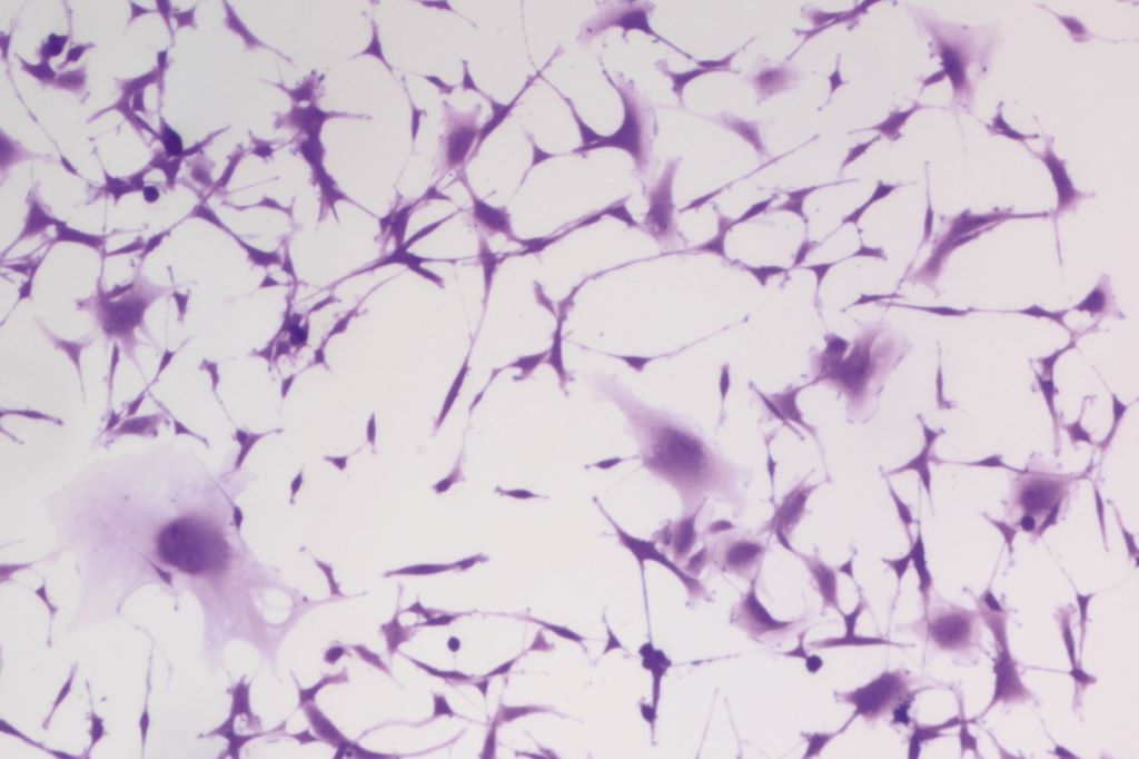
Reseachers from the Megan Matthews lab at Penn treated human glioblastoma brain tumour cells with hydralazine, one of the oldest-known blood pressure drugs and a first-line treatment for preeclampsia, for three days. At day three (imaged), more cells become enlarged and flattened – a hallmark of senescence. (Image: Courtesy of Kyosuke Shishikura)

Over the last 70 years, hydralazine has been an indispensable tool against life-threatening high blood pressure, especially during pregnancy. But despite its essential role, a fundamental mystery has persisted: No one knows its mechanism of action, which allows for improved efficacy, safety, and what it can treat.

“Hydralazine is one of the earliest vasodilators ever developed, and it’s still a first-line treatment for preeclampsia – a hypertensive disorder that accounts for 5-15% of maternal deaths worldwide,” says Kyosuke Shishikura, a physician-scientist at the University of Pennsylvania. “It came from a ‘pre-target’ era of drug discovery, when researchers relied on what they saw in patients first and only later tried to explain the biology behind it.”

Now Shishikura, his postdoctoral adviser at Penn Megan Matthews, and collaborators have solved this long-standing puzzle.

In a paper published in Science Advances, the team uncovered the method of action of hydralazine, and in doing so, revealed an unexpected biological link between hypertensive disorders and brain cancer. The findings highlight how long-established treatments can reveal new therapeutic potential and could help in the design of safer, more effective drugs for both maternal health and brain cancer.

“Preeclampsia has affected generations of women in my own family and continues to disproportionately impact Black mothers in the United States,” Matthews says. “Understanding how hydralazine works at the molecular level offers a path toward safer, more selective treatments for pregnancy-related hypertension—potentially improving outcomes for patients who are at greatest risk.”

Hydralazine blocks an oxygen-sensing enzyme

The team found that hydralazine blocks an oxygen-sensing enzyme called 2-aminoethanethiol dioxygenase (ADO) – a molecular switch for blood vessels contraction.

“ADO is like an alarm bell that rings the moment oxygen starts to fall,” Matthews says. “Most systems in the body take time; they have to copy DNA, make RNA, and build new proteins. ADO skips all that. It flips a biochemical switch in seconds.”

Hydralazine acts by binding to and blocking ADO – effectively “muting” that oxygen alarm. Once the enzyme was silenced, the signaling proteins it normally degrades – called regulators of G-protein signaling (RGS) – remained stable.

The buildup of RGS proteins, says Shishikura, tells the blood vessels to stop constricting, effectively overriding the “squeeze” signal. This reduces intracellular calcium levels, which he calls the “master regulator of vascular tension.” As calcium levels fall, the smooth muscles in blood vessel walls relax, causing vasodilation and a drop in blood pressure.

From preeclampsia to brain cancer: A common target

Prior to this study, cancer researchers and clinicians had begun to suspect that ADO was important in glioblastoma, where tumours often have to survive in pockets of very low oxygen, Shishikura explains. Elevated levels of ADO and its metabolic products had been linked with more aggressive disease, suggesting that shutting this enzyme down could be a powerful strategy, but no one had a good inhibitor to test that idea.

To see if hydralazine was a contender, Shishikura worked closely with structural biochemists at the University of Texas, who used X-ray crystallography to visualise hydralazine bound to ADO’s metal centre, and with neuroscientists at the University of Florida, who tested the drug’s effects in brain cancer cells.

They found that the ADO pathway that regulates vascular contraction also helps tumour cells survive in low-oxygen environments. Unlike chemotherapy, which aims to kill all cells outright, hydralazine disrupted that oxygen-sensing loop, triggering cellular senescence.

Unlocking the potential for other lifesaving treatments

Their findings highlight how long-established treatments can reveal new therapeutic potential and could help in the design of safer, more effective drugs for both maternal health and brain cancer.

They say the next step is to push the chemistry further building new ADO inhibitors that are more tissue specific and better at crossing, or exploiting weak points in, the blood-brain barrier so they hit tumour tissue hard while sparing the rest of the body.

Matthews is also working to continue engineering the next generation of medical solutions by revealing the mechanics of clinically tested, long-known treatments.

“It’s rare that an old cardiovascular drug ends up teaching us something new about the brain,” Matthews says, “but that’s exactly what we’re hoping to find more of – unusual links that could spell new solutions.”

Source: University of Pennsylvania

Babies Learning to See After Being Born Blind

Photo by Jeffrey Riley on Unsplash

A study conducted by University of Louvain (UCLouvain), published in Nature Communications, shows that part of the brain of babies born blind is permanently altered, while another part remains surprisingly intact. Babies’ brains are much more adaptable than previously thought: even if they cannot see at the very beginning of life, they can later learn to recognise the world around them.

Some babies are born with early blindness due to dense bilateral congenital cataracts, requiring surgery to restore their sight. This period of several months without vision can leave a lasting mark on how the brain processes visual details, but surprisingly little on the recognition of faces, objects, or words.

Using brain imaging, the researchers compared adults who had undergone surgery for congenital cataracts as babies with people born with normal vision. The results are striking: in people born with cataracts, the area of the brain that analyses small visual details (contours, contrasts, etc.) retains a lasting alteration from this early blindness. On the other hand, the more advanced regions of the visual brain, responsible for recognising faces, objects, and words, function almost normally. These “biological” results have been validated by computer models involving artificial neural networks. This distinction between altered and preserved areas of the brain paves the way for new treatments. In the future, clinicians may be able to offer visual therapies that are better tailored to each patient.

“Babies’ brains are much more adaptable than we thought,” explains Olivier Collignon, Professor at University of Louvain (UCLouvain). “Even if vision is lacking at the very beginning of life, the brain can adapt and learn to recognise the world around it even on the basis of degraded information.”

These findings also challenge the idea of a single “critical period” for visual development. Some areas of the brain are more vulnerable to early vision loss, while others retain a surprising capacity for recovery. “The brain is both fragile and resilient,” adds Olivier Collignon. “Early experiences matter, but they don’t determine everything.”

Source: Université catholique de Louvain

Right to Life Hangs in the Balance for Two SA Girls This World Children’s Day

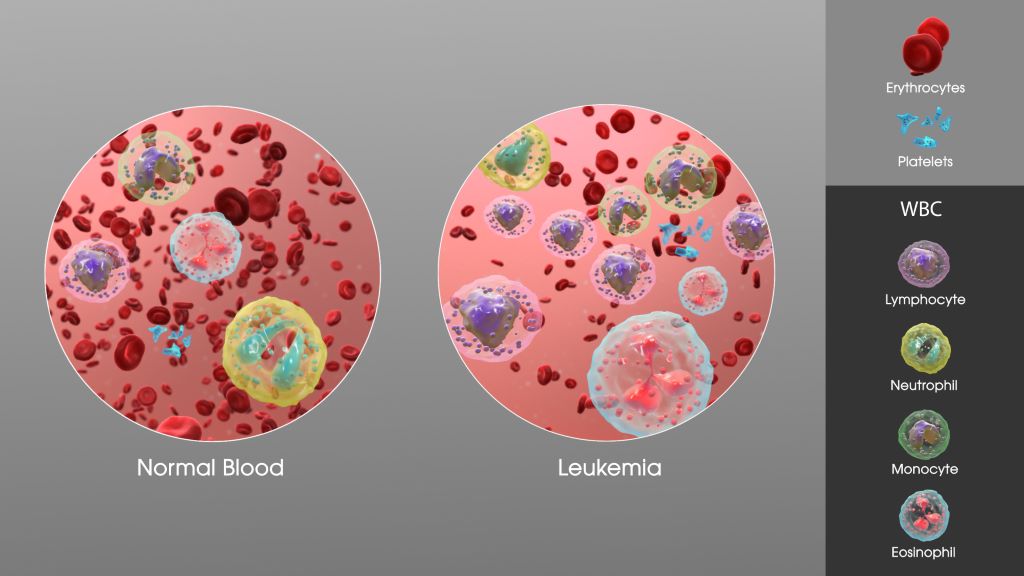
SAG Leukaemia. Credit: Scientific Animations CC0

At four years old, a child’s world should be full of crayons, playgrounds, and fairy tales. For Dianty and Nqobimpi, it has become a world of chemotherapy, blood transfusions, and sterile hospital rooms. As the world observes World Children’s Day on 20 November and its theme of “My day, my rights,” their fight for the basic right to life casts a light on the most critical need of all: a second chance, which can only come from a matching stem cell donor.

A Donor for Dianty

Hailing from Randfontein, Dianty is a bright and joyful four-year-old whose smile can light up any room. Yet, behind her cheerful spirit lies a harrowing health battle. It began last year with inexplicable fevers and stomach pains, initially dismissed as a sinus issue and later as growing pains. When Dianty suddenly lost the ability to walk, her mother, Claudine, knew something was terribly wrong.

After she was admitted to the hospital, blood tests revealed the devastating truth: Dianty had leukaemia. Chemotherapy began immediately. “The ups and downs were relentless,” Claudine explains. Despite initial progress, Dianty relapsed just after her fourth birthday this year, forcing her family back to square one.

The search for a stem cell donor has been fraught with disappointment. With no suitable match found within her family, Dianty’s hope now rests on the public registry. “You never think it will happen to you until it does,” says Claudine. “I encourage every single person to go and do the cheek swab… You can save the life of a little child. You can give them a second chance.”

Nqobimpi Needs a Match

Nqobimpi from Pretoria is facing a similarly terrifying reality. Her journey began with persistent nosebleeds and terrifying episodes of vomiting blood, which left her weak and pale. Her mother rushed her to a clinic, which led to a referral and eventually a diagnosis of T-Cell Acute Lymphoblastic Lymphoma (T-Cell ALL).

Expressing the shock that rocked their family, her mother shares, “I wasn’t aware children could get cancer.” Nqobimpi was pulled out of crèche, and her life became a cycle of month-long hospital stays for treatment. The strain has rippled through their family, affecting her siblings’ ability to focus at school as they worry constantly about their baby sister. All Nqobimpi wants is to return home and play with her dolls, a simple childhood pleasure now dependent on finding a selfless stranger willing to register as a donor.

The Power to Save a Life

“For children like Dianty and Nqobimpi, a stem cell transplant from a matching donor is their only hope for a cure,” highlights Palesa Mokomele, Head of Community Engagement and Communications at DKMS Africa. Every new person who registers brings fresh hope to them and the many other patients waiting for a lifeline.”

“On this World Children’s Day, as we reflect on the rights of every child, consider giving the ultimate gift: the right to life,” she concludes.

Register today at https://www.dkms-africa.org/save-lives.