Talk to your patients about safe, effective use of medicines
Photo by Cottonbro on Pexels
Approximately one in ten patients experience an adverse drug reaction during their care1. This can lead to serious harm or even death. Sanofi is committed to reducing these numbers by working with healthcare practitioners to create a culture of patient safety.
“Patient safety is a top priority for Sanofi,” says Yusuf Dawood, Multi-Country Safety Head for Sanofi Southern Africa. “We believe that patients should be essential partners in their healthcare journeys, and we are committed to working with healthcare professionals alongside their patients to ensure optimal therapeutic outcomes. We call on all healthcare practitioners to join us in raising awareness of patient safety. By working together, we can advocate for improved communication and reduce patient harm.”
Here are some key tips for healthcare practitioners on how to improve patient safety:
Ask patients about their concerns and listen to their feedback. They can provide valuable insights into their own health and well-being and by engaging them, healthcare practitioners can ensure that potential issues are detected as soon as possible and handled appropriately.
Provide patients with clear and concise information about their care. Patients need to understand what their diagnosis is, what treatment options are available, and what the benefits and risks of each option are. They also need to know what to expect during and after their treatment, and how to manage any side effects or complications. By giving patients accurate and easy-to-understand information, healthcare practitioners can empower them to make informed choices about their care.
Communicate with patients and other members of the healthcare team. Use simple and unambiguous language, avoid jargon and acronyms, and confirm that the patient has understood the information they have been given. Use tools such as checklists, handovers, and feedback loops to ensure that the information they share is complete and accurate.
Follow safety protocols and procedures. Healthcare practitioners need to adhere to guidelines, policies, protocols, best practices and standards of care established by professional bodies and regulatory authorities, which have been designed to prevent or minimise harm to patients.
Report issues immediately. Report any patient safety issues to the appropriate authorities in the interest of public safety. Report any medication-related patient safety issues to the relevant pharmaceutical companies. This enables companies to continuously monitor the benefit-risk profile of their products and ensure the safe use of medicines.
“Patient safety should be a top priority for healthcare professionals and pharmaceutical companies because the goal of both sectors is to improve and protect the well-being of individuals,” says Dawood. “When safety is compromised, it not only jeopardises the health and trust of patients but also undermines the credibility and integrity of the entire healthcare system. By working with pharmaceutical companies like Sanofi, healthcare professionals can provide real-world feedback on drug efficacy and side effects. This collaborative approach ensures that treatments are both safe and effective.
Join Sanofi in championing patient care. Let’s collaborate, communicate, and make every patient’s journey safer.
Reference 1. Ribeiro, M. et al. (2018) ‘Increase of 10% in the rate of adverse drug reactions for each drug administered in hospitalized patients’, Clinics, 73, pp. 1–6. doi:10.6061/clinics/2018/e185.
Relief could be on the way for people with painful hand osteoarthritis after a new study found an affordable existing drug can help. Until now there has been no effective treatment.
Published in The Lancet, the paper investigated methotrexate, a low-cost, effective treatment for inflammatory joint conditions such as rheumatoid arthritis and psoriatic arthritis. It has been widely used in Australia and globally since the early 1980s.
Researchers led by Monash University and Alfred Health found that methotrexate reduced symptoms in those with hand osteoarthritis (OA). A 20mg weekly oral dose over six months had a moderate effect in reducing pain and stiffness in patients with symptomatic hand OA.
Hand OA is a disabling condition that causes pain and affects function, impeding daily activities such as dressing and eating. It can significantly reduce quality of life. About one in two women and one in four men will experience symptoms from hand OA by the time they turn 85.
About half will have inflamed joints, which cause pain and are associated with significant joint damage. Despite the high prevalence and disease burden, there are no effective medications.
Senior author Professor Flavia Cicuttini said that the study identified the role of inflammation in hand OA and the potential benefit of targeting patients who experience painful hand OA.
“In our study, as with most studies of osteoarthritis, both the placebo group and methotrexate groups’ pain improved in the first month or so,” Professor Cicuttini said.
“However, pain levels stayed the same in the placebo group but continued to decrease in the methotrexate group at three and six months, when they were still decreasing. The pain improvement in the methotrexate group was twice as much as in the placebo group.
“Based on these results, use of methotrexate can be considered in the management of hand osteoarthritis with an inflammatory pattern. This provides clinicians with a treatment option for this group, which tends to get more joint damage.”
Professor Cicuttini said in patients with hand OA and inflammation, the effects of methotrexate were present at about three months and by six months it was very clear if it worked.
“At that time patients and their doctors can decide whether to continue or stop it,” she said. “This is very similar to what we currently do with other forms of inflammatory arthritis.”
The randomised, double-blind, placebo-controlled trial of 97 people with hand OA and MRI-detected inflammation assessed whether 20mg of methotrexate weekly reduced pain and improved function compared to placebo in patients with symptomatic hand OA and synovitis (inflammation) over six months.
Professor Cicuttini said the results could provide relief for people with hand OA inflammation, which was particularly common in women as they experienced menopause.
“Further trials are needed to establish whether the effect of methotrexate extends beyond six months, for how long we need to treat patients, and whether methotrexate reduces joint damage in patients with hand osteoarthritis and associated inflammation,” she said.
Professor Cicuttini now plans to conduct an extension trial to address these questions, in particular whether women who develop hand OA around menopause and often have severe pain and joint damage may benefit.
Reducing overall calorie intake may rejuvenate muscles and activate biological pathways important for good health, according new study, published in the journal Aging Cell. Calorie restriction, which cuts intake of calories but not essential nutrients, has long been known to delay the progression of age-related diseases in animal models. This finding, by researchers at the National Institutes of Health and their colleagues, suggests the same biological mechanisms may also apply to humans.
Researchers analysed data from participants in the Comprehensive Assessment of Long-Term Effects of Reducing Intake of Energy (CALERIE), a study supported by the National Institute on Aging (NIA) that examined whether moderate calorie restriction conveys the same health benefits seen in animal studies. They found that during a two-year span, the goal for participants was to reduce their daily caloric intake by 25%, but the highest the group was able to reach was a 12% reduction. Even so, this slight reduction in calories was enough to activate most of the biological pathways that are important in healthy aging.
“A 12% reduction in calorie intake is very modest,” said corresponding author and NIA Scientific Director Luigi Ferrucci, MD, PhD. “This kind of small reduction in calorie intake is doable and may make a big difference in your health.”
The research team next sought to understand the molecular underpinnings of the benefits seen in limited, previous research of calorie restriction in humans. One study showed that individuals on calorie restriction lost muscle mass and an average of 20 pounds of weight over the first year and maintained their weight for the second year. However, despite losing muscle mass, calorie restriction participants did not lose muscle strength, indicating calorie restriction improved the amount of force generated by each unit of muscle mass, called muscle specific force.
For the current study, scientists used thigh muscle biopsies from CALERIE participants that were collected when individuals joined the study and at one-year and two-year follow ups.
To figure out which human genes were impacted during calorie restriction, the scientists isolated messenger RNA (mRNA), a molecule that contains the code for proteins, from muscle samples. The team determined the protein sequence of each mRNA and used the information to identify which genes originated specific mRNAs. Further analysis helped the scientists establish which genes during calorie restriction were upregulated, meaning the cells made more mRNA; and which were downregulated, meaning the cells produced less mRNA. The researchers confirmed calorie restriction affected the same gene pathways in humans as in mice and non-human primates. For example, a lower caloric intake upregulated genes responsible for energy generation and metabolism, and downregulated inflammatory genes leading to lower inflammation.
“Since inflammation and aging are strongly coupled, calorie restriction represents a powerful approach to preventing the pro-inflammatory state that is developed by many older people,” said Ferrucci.
A groundbreaking, easy-to-use 3D printable finger prosthesis created by a recent University of Houston graduate could offer amputees a low-cost solution to restore finger functionality. David Edquilang first designed Lunet, which doesn’t need metal fasteners, adhesives or special tools to assemble, as an undergraduate student at the Gerald D. Hines College of Architecture and Design. While standard prostheses can cost thousands of dollars, Edquilang aims to make his design open access on the internet, instead of selling it.
Edquilang explains: “Lunet began when I decided to design and 3D print prototype finger mechanisms for a prosthetic hand for fun in my free time. 2 weeks and 18 prototypes later, I created a mechanism and finger structure that closely replicated the range of motion of real fingers.”
Edquilang’s mentor at UH was Associate Professor Jeff Feng, co-director of UH’s Industrial Design program. Through a partnership with Harris Health System, Feng learned of a patient who had her fingers amputated due to frostbite. Inspired by working on an upper limb prosthesis Edquilang previously developed with student Niell Gorman, working closely with Professor Feng, Edquilang created prosthetic fingers that returned mobility to the patient, allowing her to pick up objects again.
Edquilang continues: “My professor and I were then referred to a finger amputee who lost 3 of her fingers. I applied the mechanism I created to design a finger prosthesis for her. Nearly 40 design iterations and multiple rounds of patient testing were performed to ultimately create a functional prosthesis that fit her.
His “breakthrough” came from a literal break in his design.
“After we finished working with this amputee patient, I continued to tinker with my finger designs. I intentionally broke one of my finger prototypes to see where its structural weakpoint is. It broke at the distal knuckle. This led to me having a breakthrough in the design. I added a linkage that replaces the previously rigid distal knuckle, and I stumbled upon inventing a novel finger mechanism that was more flexible and nearly unbreakable. I then set on refining the design to be more functional, easily 3D printable, and more visually appealing. Inspiration from cyberpunk art and fighter jets influenced the design. 28 design iterations and a myriad of prototypes later resulted in Lunet.”
“It feels great knowing you have the capability to positively impact people’s lives and give them help they otherwise wouldn’t be able to get,” said Edquilang.
“Not every good idea needs to be turned into a business. Sometimes, the best ideas just need to be put out there ,” said Edquilang, who graduated with a Bachelor of Science in Industrial Design last year. “Medical insurance will often not cover the cost of a finger prosthesis, since it is not considered vital enough compared to an arm or leg. Making Lunet available online for free will allow it to help the greatest number of people.”
Lunet wins awards
The prosthetic design garnered Edquilang a 2023 Red Dot: Luminary award, the highest level of recognition accorded at the Red Dot Award: Design Concept. He and Feng took home the coveted accolade at Red Dot’s ceremony last month in Singapore.
“Good results come from dedication. Extraordinary results come from experimentation. Incredible results come from a combination of both,” he said upon winning the award. He has also received a number of other accolades, including iFDesign, and national runner up for the James Dyson Award.
“David’s recent success in winning the most prestigious design awards across the world is the best manifestation of the unparalleled education and training students experience in our Industrial Design program,” Feng said. “Built upon a belief that every student is a creative individual, the program pedagogy focuses on methods of cultivating innovative minds, which is enforced with rigorous professional training.”
Lunet’s geometry inspired its name
Lunet is made up of two common types of 3D printed plastics: polylactic acid and thermoplastic polyurethane. Each finger is made up of four parts held together by plastic pins. Edquilang describes arcs and circular orbits as the foundation for the motion of the finger mechanism. The geometric basis of the design evoked the idea that the prosthesis orbits around the user’s joints like a moon, or lunet, hence the name.
Another element of Lunet’s uniqueness is that it is nearly impossible to break; other finger prosthetics can be complicated and require many parts.
“The problem with higher mechanical complexity is that these designs are less durable,” Edquilang said. “The more parts you have, the more points of failure. You need to make prosthetic fingers robust and as strong as possible, so it doesn’t break under normal use, yet you want the design to be simple. This was one of the greatest challenges in making Lunet.”
He encourages other design students not to be afraid to experiment and fail because that is often how one can learn to improve the most.
“Where the world has an abundance of problems, designers have an abundance of talent, and we should not be selfish with it,” Edquilang said.
Researchers at the University of Oxford have produced an engineered tissue representing a simplified cerebral cortex by 3D printing human stem cells. The results, published in the journal Nature Communications, showed that, when implanted into mouse brain slices, the structures became integrated with the host tissue.
The breakthrough technique could lead to tailored repairs for brain injuries. The researchers demonstrated for the first time that neural cells can be 3D-printed to mimic the architecture of the cerebral cortex.
Brain injuries, including those caused by trauma, stroke and surgery for brain tumours, typically result in significant damage to the cerebral cortex. For example, each year, around 70 million people globally suffer from traumatic brain injury (TBI), with 5 million of these cases being severe or fatal. Currently, there are no effective treatments for severe brain injuries, leading to serious impacts on quality of life.
Tissue regenerative therapies, especially those in which patients are given implants derived from their own stem cells, could be a promising route to treat brain injuries in the future. Up to now, however, there has been no method to ensure that implanted stem cells mimic the architecture of the brain.
In this new study, the University of Oxford researchers fabricated a two-layered brain tissue by 3D printing human neural stem cells. When implanted into mouse brain slices, the cells showed convincing structural and functional integration with the host tissue.
Lead author Dr Yongcheng Jin (Department of Chemistry, University of Oxford) said: ‘This advance marks a significant step towards the fabrication of materials with the full structure and function of natural brain tissues. The work will provide a unique opportunity to explore the workings of the human cortex and, in the long term, it will offer hope to individuals who sustain brain injuries.’
The cortical structure was made from human induced pluripotent stem cells (hiPSCs), which have the potential to produce the cell types found in most human tissues. A key advantage of using hiPSCs for tissue repair is that they can be easily derived from cells harvested from patients themselves, and therefore would not trigger an immune response.
The hiPSCs were differentiated into neural progenitor cells for two different layers of the cerebral cortex, by using specific combinations of growth factors and chemicals. The cells were then suspended in solution to generate two ‘bioinks’, which were then printed to produce a two-layered structure. In culture, the printed tissues maintained their layered cellular architecture for weeks, as indicated by the expression of layer-specific biomarkers.
When the printed tissues were implanted into mouse brain slices, they showed strong integration, as demonstrated by the projection of neural processes and the migration of neurons across the implant-host boundary. The implanted cells also showed signalling activity, which correlated with that of the host cells. This indicates that the human and mouse cells were communicating with each other, demonstrating functional as well as structural integration.
The researchers now intend to further refine the droplet printing technique to create complex multi-layered cerebral cortex tissues that more realistically mimic the human brain’s architecture. Besides their potential for repairing brain injuries, these engineered tissues might be used in drug evaluation, studies of brain development, and to improve our understanding of the basis of cognition.
The new advance builds on the team’s decade-long track record in inventing and patenting 3D printing technologies for synthetic tissues and cultured cells.
Senior author Dr Linna Zhou (Department of Chemistry, University of Oxford) said: “Our droplet printing technique provides a means to engineer living 3D tissues with desired architectures, which brings us closer to the creation of personalised implantation treatments for brain injury.”
Senior author Associate Professor Francis Szele (Department of Physiology, Anatomy and Genetics, University of Oxford) added: “The use of living brain slices creates a powerful platform for interrogating the utility of 3D printing in brain repair. It is a natural bridge between studying 3D printed cortical column development in vitro and their integration into brains in animal models of injury.”
Senior author Professor Zoltán Molnár (Department of Physiology, Anatomy and Genetics, University of Oxford) said: “Human brain development is a delicate and elaborate process with a complex choreography. It would be naïve to think that we can recreate the entire cellular progression in the laboratory. Nonetheless, our 3D printing project demonstrates substantial progress in controlling the fates and arrangements of human iPSCs to form the basic functional units of the cerebral cortex.”
Dr Bukiwe Spondo recently received the Rural Doctor of the Year award at the Rural Health Conference held in Chintsa in the Eastern Cape. PHOTO: Supplied
By Biénne Huisman for Spotlight
Describing the rutted gravel road between Butterworth and Tafalofefe District Hospital in the Eastern Cape, Dr Bukiwe Spondo uses the word “terrible” at least eighteen times. Dipping through the Amatole District, the 55-kilometre journey can take several hours. With heavy rain, tractors may be required to dislodge ambulances and often even staff have difficulty getting to work because of the mud.
Since 2007, Spondo and her colleagues have offered a multitude of services at Tafalofefe in the lush but impoverished Centane village. First off, she moved the hospital’s ARV clinic from an out-building to inside the premises – reducing stigma – “because if patients went into that building on the outside, automatically everyone knew,” she says.
In 2012, having observed how patients stopped taking treatment due to travel costs, she started driving up to 40 kilometres a day twice weekly to nine clinics in the area, where up to fifty patients would be queuing to see her. To make life easier for patients, she started pre-packing medication to take to them at the clinics. Later she opened a CHAMP (Clinical HIV /AIDS Management Programme) site at Tafalofefe to see complicated cases referred from the clinics, and a multi-drug-resistant TB (MDR-TB) review clinic in conjunction with Butterworth Provincial Hospital.
“As a rural doctor, you become a social worker, a pharmacist, a priest – you do everything,” she says, laughing.
Rural doctor of the year
Spondo’s efforts have not gone unnoticed. Last month at the Rural Doctor’s Association of South Africa (RuDASA’s) annual Rural Health Conference, she received the Rural Doctor of the Year award. RuDASA chairperson Dr Lungile Hobe conferred the award at the event hosted near Chintsa. Spondo is quick to point out that she also won an Amatole District leadership award last year.
Speaking to Spotlight over Zoom, she says, “So the roads here at Centane are terrible. It becomes a challenge to get ambulances through and the chopper cannot fly either when it’s raining. I mean, the other day a truck was stuck, crossing the road so the ambulance couldn’t pass. We had to take a private car from the hospital to go meet the ambulance halfway.”
She adds that the community hoped that roads would be improved after a devastating accident five kilometres from Tafalofefe in 2020 when an overloaded 65-seater bus plunged into a gorge, causing 25 deaths and 62 injuries. But, she says, the improvements never come.
At Tafalofefe, the two nearest referral hospitals are Cecilia Makiwane and Frere Provincial in East London, situated an additional 110 kilometres or 90-minute drive from Butterworth along the N2 highway. Housed in a pale building, Tafalofefe has 160 beds served by 41 professional nurses and seven doctors – including three community service doctors who joined last year. The additions have increased capacity, for example, emergency caesareans are now available around the clock.
Taking healthcare to the people
The hospital has three 4×4 bakkies [pick-ups] for visiting or transporting patients. It is in one of these that Spondo travels to see patients in remote corners between the Kobonqaba and Kei Rivers on Tuesdays and Thursdays.
“Clinics are part of decentralised primary healthcare goals,” she says. “But the problem was that if there were complicated cases – like if a patient is taking ARVs and then develop side effects, the sisters are not equipped to handle that. For example, if there is a kidney problem, they [cannot] do anything about that.
“And in time, I realised that for these people traveling to the hospital costs too much money. Let’s say, for example, the clinic at Qolora – for a person to travel from Qolora to Tafalofefe is R100. A return ticket is R200. And you know, most people here are unemployed. They can’t afford this. By the time they have saved up enough money to travel to the hospital, it’s too late. Like it would be the end stage of their kidney problem. You could not send this patient for dialysis, nothing could be done to help them. This is why I started my outreach trips.”
In motivating for Spondo to receive the RuDASA award, Tafalofefe’s CEO Masizakhe Madlebe pointed out how her work days start at 7am, only finishing once all patients had been seen, whether at the hospital or at one of the local clinics. In addition, he notes how, over the years, Spondo has mentored youth in the area, including children whose parents had succumbed to AIDS, and school girls on topics like life goals and contraceptives. He adds that Spondo even reached into her own pocket to pay school fees for children without parents.
Spondo relays how she noticed girls as young as twelve years old in their maternity ward, giving birth. “Myself and some nurses we went to two schools in the area to educate them, to discuss goals and contraceptives,” she says. “We started with grade 12 pupils. No teachers were present. It was just us and them. And I was surprised at how free they were talking. I said to them education is more important. I said to them – You see me? I am a doctor. One day you can be a doctor too, but you need to be educated. I told them they could come to Tafalofefe any time if they needed to talk, that I could help them apply for tertiary degrees, to college or to university.”
Spondo has kept a close eye on children orphaned by AIDS in the area. “I tell them to bring me their June, September, and December school reports, so I can see how they’re doing, so I can motivate them,” she says.
“These kids, I’ve seen them grow up. Some of them I saw angry – with everyone, with their own deceased parents. And I explained to them, don’t be angry. It’s not your mother’s fault. It’s not your father’s fault. It was the government’s fault for not giving your parents access to ARVs. But now, take your own ARVs and you will be fine. Some of them have passed high school with distinction, some even now have access to universities.”
Bringing her skills back home
Alongside two brothers whom she describes as “wonderful”, Spondo grew up in the village of Nqamakwe, on the opposite side of Butterworth. Her parents have passed away, but she still considers Nqamakwe her home. Here her family’s farming interests include cattle, goats, and sheep.
She attended Blythswood Secondary School in Nqamakwe – excelling at biology and physics, even though maths was hard work. “Becoming a doctor was just something I always wanted,” she says, relaying how in her formative years she had been a sickly child who often required medical care. This changed, she says, as she cannot remember ever being sick as an adult.
Spondo graduated from medical school at the University of KwaZulu-Natal in 2002, completing her internship at Cecilia Makiwane and her community service at Tafalofefe and Frere in 2004.
Speaking with rapid enthusiasm, she says how happy she is to bring her healthcare skills back home to serve the community that shaped her own humanity.
“I mean, I know these people inside out. I was born in front of them, raised in front of them,” she says. “These are our relatives, our aunts, our grannies. It’s giving back to them, to the community that raised you, that has done everything for you. Who supported you through all these years.”
She adds that Tafalofefe’s clinical manager, Sambona Ntamo, grew up near Butterworth too.
“Who would look after these people if we didn’t?” she asks.
Where does she find the resilience that drives her passion to care for sick people, often queuing at the end of long rutted roads?
“Lots of exercise,” she says, smiling.
At Tafalofefe there is a staff gym with a treadmill, a bicycle, weight lifts, and pilates balls.
“I tell the guys after work it’s gym time, it’s gym time, it’s gym time!” she says. “We’ve got a key and everyone knows that even if they want to go to the gym after midnight, they may get the key and go.”
Photographs capture an air of camaraderie at Tafalofefe. Staff sharing a meal of tripe and creamed spinach on heritage day, a farewell gathering for a retiring nurse with balloons and huge gifts in silver wrapping, [and] women knitting countless bright beanies for babies delivered in the maternity ward. A picture inside the hospital’s paediatric room shows youngsters on plastic motorbikes and mothers holding toddlers wrapped in blankets.
Spondo and her own eight-year-old son, Lutho desperately – which means the greatest one – live in a doctor’s house on the hospital’s premises. They travel to their family home in Nqamakwe over weekends.
For Spondo, being a doctor does not feel like a job. “When you do something you love, it doesn’t feel like a job,” she says. “Being a doctor is something I look forward to every morning. When patients return to me, saying they feel better with a smile on their faces, saying thank you for the treatment – that just makes my day.”
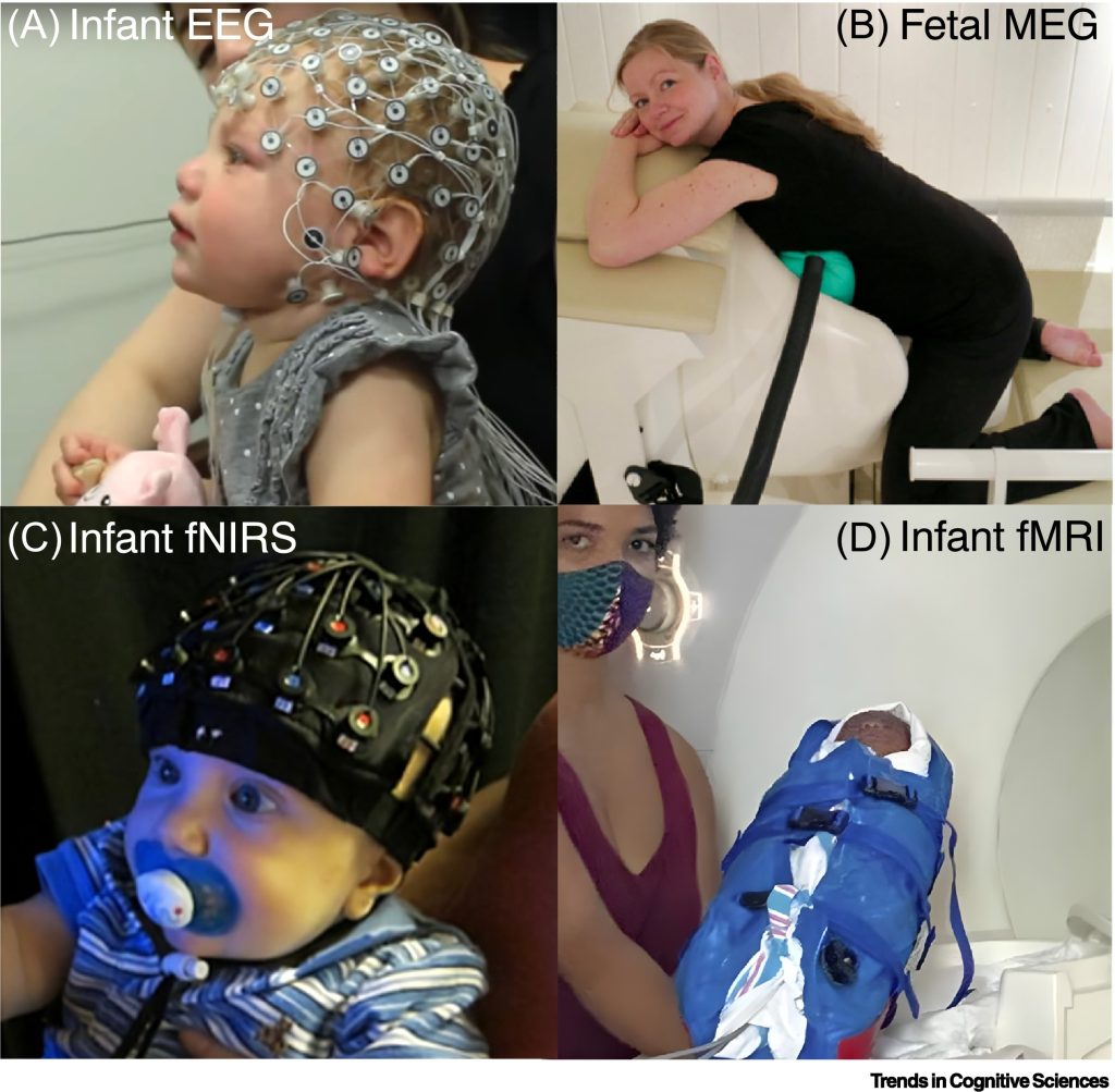
Figure I Neural measurement tools for studying the emergence of consciousness. Examples of techniques for recording brain activity and/or neuroimaging in infants and foetuses. (A) Infant electroencephalography (EEG) with a geodesic electrode net. (B) Foetal magnetoencephalography (MEG) recorded from a pregnant woman. (C) Infant functional near infrared spectroscopy (fNIRS) recording with multichannel optode cap. (D) An infant is prepared for functional magnetic resonance imaging (fMRI). Source: Bayne et al., 2023
There is evidence that some form of conscious experience is present by birth, and perhaps even in late pregnancy, an international team of researchers has found. The findings, published today in Trends in Cognitive Science, have important clinical, ethical and potentially legal implications, according to the authors.
Converging evidence from studies of functional network connectivity, attention, multimodal integration, and cortical responses to global oddballs suggests that consciousness is likely to be in place in early infancy and may even occur before birth. Over the decades, theorists have argued that consciousness emerges from anywhere from 30 to 35 weeks of pregnancy (based on EEG of the foetus’s brain) to 12 to 15 months of age (based on higher-order representational theory).
In the study, the researchers argue that by birth the infant’s developing brain is capable of conscious experiences that can leave a lasting imprint on their developing sense of self and understanding of their environment.
The team comprised neuroscientists and philosophers from Monash University, in Australia, University of Tübingen, in Germany, University of Minnesota, in the USA, and Trinity College Dublin.
Although each of us was once a baby, infant consciousness remains mysterious, because infants cannot tell us what they think or feel, explains one of the two lead authors of the paper Dr Tim Bayne, Professor of Philosophy at Monash University.
“Nearly everyone who has held a newborn infant has wondered what, if anything, it is like to be a baby. But of course we cannot remember our infancy, and consciousness researchers have disagreed on whether consciousness arises ‘early’ (at birth or shortly after) or ‘late’ – by one year of age, or even much later.”
To provide a new perspective on when consciousness first emerges, the team built upon recent advances in consciousness science. In adults, some markers from brain imaging have been found to reliably differentiate consciousness from its absence, and are increasingly applied in science and medicine. This is the first time that a review of these markers in infants has been used to assess their consciousness.
Co-author of the study, Lorina Naci, Associate Professor in the School of Psychology, who leads Trinity’s ‘Consciousness and Cognition Group, explained: “Our findings suggest that newborns can integrate sensory and developing cognitive responses into coherent conscious experiences to understand the actions of others and plan their own responses.”
The paper also sheds light into ‘what it is like’ to be a baby. We know that seeing is much more immature in babies than hearing, for example. Furthermore, this work suggests that, at any point in time, infants are aware of fewer items than adults, and can take longer to grasp what’s in front of them, but it is easier for them to process more diverse information, such as sounds from other languages.
Researchers at Monash University have identified a new way of mapping ‘phosphenes’ – the visual perception of the bright flashes we see when no light is entering the eye – to improve the outcome of surgery for patients receiving a cortical visual prosthesis.
Cortical visual prostheses are devices implanted onto the brain with the aim of restoring sight by directly stimulating the area responsible for vision, the visual cortex, bypassing damage to the retina of the eye or the optic nerve. Phosphenes, apparent flashes and patterns of lights, were described by the ancient Greeks and can be elicited by pressure, injury, disease, certain medications or direct electrical stimulation.
A typical prosthesis consists of an array of fine electrodes, each of which is designed to trigger a phosphene. Given the limited number of electrodes, understanding how electrodes can best be placed to generate useful perceived images becomes critical.
Published in the Journal of Neural Engineering, the study presents a more realistic simulation for cortical prosthetic vision.
As part of this researchers from the Department of Electrical and Computer Systems Engineering at Monash University, led by Associate Professor Yan Tat Wong, are honing in on the ideal distribution of phosphenes.
“Phosphenes are likely to be distributed unevenly in an individual’s visual field, and differences in the surface of the brain also affect how surgeons place implants, which together result in a phosphene map unique to each patient,” Associate Professor Wong said.
The study used a retinotopy dataset based on magnetic resonance imaging (MRI) scans, consulting with a neurosurgeon about realistic electrode implantation sites in different individuals, and applying a clustering algorithm to determine the most suitable regions to present stimuli.
Sighted participants recruited for the study were asked to test and verify the phosphene maps based on visual acuity and object recognition.
“We’re proposing a new process that incorporates our simulation paradigm into surgical planning to help optimise the implantation of a cortical prosthesis,” Associate Professor Wong said.
The process would begin with an MRI scan to plot the recipient’s brain surface in the area of the visual cortex. Potential implant locations would then be identified, and the simulation developed in the Monash research would be used to plot phosphene maps.
“We can use the metrics we computed to find practical implant locations that are more likely to give us a usable phosphene map, and we can verify those options through psychophysics tests on sighted participants using a virtual reality headset,” Associate Professor Wong said.
“We believe this is the first approach that realistically simulates the visual experience of cortical prosthetic vision.”
In a study published in Current Biology, people with early Alzheimer’s disease were found to have difficulty turning when walking. The new study used virtual reality and a computational model to further explore the intricacies of navigational errors previously observed in Alzheimer’s disease.
Researchers, led by Professor Neil Burgess and colleagues in the Space and Memory group at the UCL Institute of Cognitive Neuroscience, grouped participants into three categories: healthy younger participants (31 total), healthy elderly participants (36 total) and patients with mild cognitive impairment (43 total). They then asked them to complete a task while wearing virtual reality goggles, which allowed them to make real movements.
In the trial, participants walked an outbound route guided by numbered cones, consisting of two straight legs connected by a turn. They then had to return to their starting position unguided.
The task was performed under three different environmental conditions aimed at stressing the participant’s navigational skills: an unchanged virtual environment, the ground details being replaced by a plain texture, and the temporary removal of all landmarks from the virtual reality world.
The researchers found that people with early Alzheimer’s consistently overestimated the turns on the route and showed increased variability in their sense of direction. However, these specific impairments were not observed in the healthy older participants or people with mild cognitive impairment, who did not show underlying signs of Alzheimer’s.
This suggests that these navigational errors are specific to Alzheimer’s disease – rather than an extension of healthy ageing or general cognitive decline – and could help with diagnosis.
Joint first author, Dr Andrea Castegnaro (UCL Institute of Cognitive Neuroscience), said: “Our findings offer a new avenue for the early diagnosis of Alzheimer’s disease by focusing on specific navigational errors. However, we know that more work is needed to confirm these early findings.
Dr Castegnaro added, “Cognitive assessments are still needed to understand when the first cognitive impairments develop, and when it comes to existing spatial memory tests used in clinics, those often rely on verbal competence. Our tests aim to offer a more practical tool that doesn’t rely on language or cultural background.”
People who carry three gene variants that have bene inherited from Neanderthals are more sensitive to some types of pain, according to a new study co-led by UCL researchers. The findings, published in Communications Biology, are the latest findings to show how past interbreeding with Neanderthals has influenced the genetics of modern humans.
The researchers found that people carrying three so-called Neanderthal variants in the gene SCN9A, which is implicated in sensory neurons, are more sensitive to pain from skin pricking after prior exposure to mustard oil.
Previous research has identified three variations in the SCN9A gene – known as M932L, V991L, and D1908G – in sequenced Neanderthal genomes and reports of greater pain sensitivity among humans carrying all three variants. However, prior to this study the specific sensory responses affected by these variants was unclear.
An international team measured the pain thresholds of 1963 people from Colombia in response to a range of stimuli.
The SCN9A gene encodes a sodium channel that is expressed at high levels in sensory neurons that detect signals from damaged tissue. The researchers found that the D1908G variant of the gene was present in around 20% of chromosomes within this population and around 30% of chromosomes carrying this variant also carried the M932L and V991L variants.
The authors found that the three variants were associated with a lower pain threshold in response to skin pricking after prior exposure to mustard oil, but not in response to heat or pressure. Additionally, carrying all three variants was associated with greater pain sensitivity than carrying only one.
When they analysed the genomic region including SCN9A using genetic data from 5971 people from Brazil, Chile, Colombia, Mexico and Peru, the authors found that the three Neanderthal variants were more common in populations with higher proportions of Native American ancestry, such as the Peruvian population, in which the average proportion of Native American ancestry was 66%.
The authors propose that the Neanderthal variants may sensitise sensory neurons by altering the threshold at which a nerve impulse is generated. They speculate that the variants may be more common in populations with higher proportions of Native American ancestry as a result of random chance and population bottlenecks that occurred during the initial occupation of the Americas. Although acute pain can moderate behaviour and prevent further injury, the scientists that say additional research is needed to determine whether carrying these variants and having greater pain sensitivity may have been advantageous during human evolution.
Diagram comparing the nose shape of a Neanderthal with that of a modern human by Dr Macarena Fuentes-Guajardo.
Previous research by co-corresponding author Dr Kaustubh Adhikari (UCL Genetics, Evolution & Environment and The Open University) has shown that humans also inherited some genetic material from Neanderthals affecting the shape of our noses.
Dr Adhikari commented: “In the last 15 years, since the Neanderthal genome was first sequenced, we have been learning more and more about what we have inherited from them as a result of interbreeding tens of thousands of years ago.
“Pain sensitivity is an important survival trait that enables us to avoid painful things that could cause us serious harm. Our findings suggest that Neanderthals may have been more sensitive to certain types of pain, but further research is needed for us to understand why that is the case, and whether these specific genetic variants were evolutionarily advantageous.”
First author Dr Pierre Faux (Aix-Marseille University and University of Toulouse) said: “We have shown how variation in our genetic code can alter how we perceive pain, including genes that modern humans acquired from the Neanderthals. But genes are just one of many factors, including environment, past experience, and psychological factors, which influence pain.”