Year: 2023

Genetic Analysis Reveals Secrets of Vlad Dracula the Impaler

Mediaeval tyrant and inspiration for vampires, protein analysis reveals health secrets about Vlad the Impaler

New research analysing ancient protein residues left in letters written by the sadistic 15th century tyrant – and vampire inspiration – Vlad Dracula the Impaler suggests that he suffered from a number of health conditions. One of these conditions seemingly confirms one of the more outlandish tales about him – that he cried tears of blood.

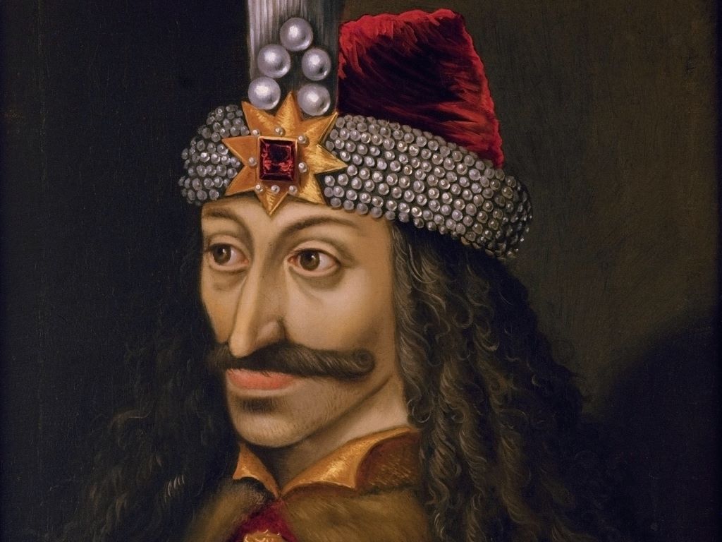
Vlad the Impaler got his nickname because he impaled thousands of people on stakes: enemies (mainly the Ottoman Empire), criminals and anyone suspected of conspiring against his rule. He was eventually defeated in 1460, but the newly invented printing press spread the tale of his gruesome deeds all over Europe. Tales surrounding him may have inspired the iconic character of Bram Stoker’s Count Dracula in 1897. Nevertheless, more modern vampire stories such as Netflix’s ‘Castlevania’ make use of Vlad as inspiration.

This terrifying reputation made him an interesting topic for a bit of genetic archaeology in a paper published in Analytical Chemistry. Using sophisticated proteomic techniques, scientists analysed three letters written in 1457 and 1475 by the voivode of Wallachia, Vlad III, also known as Vlad the Impaler, or Vlad Dracula. This allowed them to tease out information about the man who wrote the letters as well as general information about the environmental conditions of 15th century Wallachia, a place of regional trade and conflict as well as disease transmission.

While centuries-old paper is unlikely to hold entire DNA strands, scientists were still able to piece together genetic information about the writer. The technique depends on the notion that a person’s writing hand will tend to rest on the paper being written upon, rubbing off a surprising amount of organic molecules in the process. They applied ethylene vinyl acetate to the papers, and with mass spectrometry, they discovered over 500 peptides – short chains of amino acids – with about 100 being of human origin, which they looked up in database searches.

Figure 1. (a) First letter (archive catalog number is II 365), dated August 4, 1475, here investigated, also showing the positions of the EVA strips (brownish rectangles) applied to its surface for capturing biological material; (b) mapping of the fluorescence of phenylalanine, tyrosine, and tryptophan under flash UV illumination (see the original article). Anal. Chem. 2023, 95, 34, 12732-12744

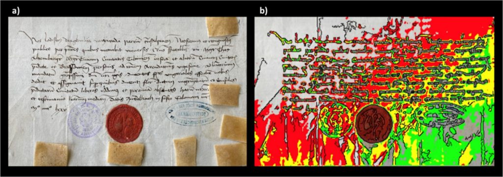
The researchers noted that while many mediaeval people may have handled these papers, it is also presumable that the most prominent ancient proteins can be attributed to the one who wrote and signed them – Prince Vlad the Impaler.

First, they discovered proteins pointing to ciliopathy, which affects the cellular cilia or the cilia anchoring structures, the basal bodies or ciliary function. This can manifest in a wide range of disorders, ranging from cerebral malformation to liver disease and intellectual disability.

They also uncovered signs of an undetermined inflammatory disease which likely involved his skin and respiratory tract.

Proteomics data also suggests that, according to some stories, he might also have suffered from a pathological condition called haemolacria – he could shed tears admixed with blood. This appears to confirm what some stories said about Vlad – that he sometimes cried tears of blood. While it is a known medical condition, it would have no doubt been terrifying for superstitious mediaeval people to behold when seen in someone with a reputation like Vlad the Impaler’s.

Non-human peptides also proved to be a window into the conditions of the time, hinting at common foods, pests and diseases. Database searches of the identified, as potential endogenous original components, 3 proteins from bacteria, 24 from viruses, 4 from fungi, 17 from insects (suggesting fruit flies), and 5 from plants (including rice, wheat and thale cress). Of the bacteria, they noted that some peptides related to Enterobacterales are specific to Yersinia pestis, the pathogenic bacterium causing plague, whereas another group is specific to E. coli.

Study Shows Intermittent Fasting Effective in Type 2 Diabetes

Photo by Photomix Company on Pexels

Time-restricted eating, also known as intermittent fasting, can help people with Type 2 diabetes lose weight and control their blood sugar levels, according to a new study published in JAMA Network Open from researchers at the University of Illinois Chicago.

Participants who ate only during an eight-hour window between noon and 8 pm each day actually lost more weight over six months than participants who were instructed to reduce their calorie intake by 25%. Both groups had similar reductions in long-term blood sugar levels, as measured by a test of haemoglobin A1C, which shows blood sugar levels over the past three months.

The study was conducted at UIC and enrolled 75 participants into three groups: those who followed the time-restricted eating rules, those who reduced calories and a control group. Participants’ weight, waist circumference, blood sugar levels and other health indicators were measured over the course of six months.

Senior author Krista Varady said that participants in the time-restricted eating group had an easier time following the regime than those in the calorie-reducing group. The researchers believe this is partly because patients with diabetes are generally told to cut back on calories by their doctors as a first line of defence, so many of these participants likely had already tried, and struggled with, that form of dieting. And while the participants in the time-restricted eating group were not instructed to reduce their calorie intake, they ended up doing so by eating within a fixed window.

“Our study shows that time-restricted eating might be an effective alternative to traditional dieting for people who can’t do the traditional diet or are burned out on it,” said Varady, a professor of kinesiology and nutrition. “For many people trying to lose weight, counting time is easier than counting calories.”

There were no serious adverse events reported during the six-month study. Occurrences of hypoglycaemia and hyperglycaemia did not differ between the diet groups and control groups.

Just over half the participants in the study were Black and another 40% were Hispanic. This is notable as diabetes is particularly prevalent among those groups, so having studies that document the success of time-restricted eating for them is particularly useful, the researchers said.

The study was small and should be followed up by larger ones, said Varady, who is also a member of the University of Illinois Cancer Center. While it acts as a proof of concept to show that time-restricted eating is safe for those with Type 2 diabetes, Varady said people with diabetes should consult their doctors before starting this sort of diet.

Source: University of Illinois Chicago

Which Entryway a Coronavirus Uses Affects its Infection Severity

Image by Fusion Medical on Unsplash

Until now, the reason why some coronaviruses such as SARS-CoV-2 affect humans more severely than other seasonal ones has eluded scientists. Now, results published in Nature have provided a piece of the puzzle by identifying a gateway used by the seasonal coronavirus HKU1 to enter human cells. HKU1 binds to a different receptor than SARS-CoV-2, which may partly explain the difference in severity between these two coronaviruses.

Receptors provide a useful means of figuring out coronavirus transmissibility and pathology as part of surveillance work on viral evolution. Seven coronaviruses are known for their ability to infect humans. Four of these are generally mild: HKU1, 229E, NL63 and OC43, while the other three are more pathogenic: SARS-CoV-1, Mers-CoV and SARS-CoV-2.

The HKU1 virus was first identified in an elderly patient with severe pneumonia in Hong Kong in 2005. Like SARS-CoV-2, HKU1 mainly infects upper respiratory tract cells. However, it rarely affects the bronchi and alveoli in the lungs. The HKU1 virus causes colds and other mild respiratory symptoms. Complications may also occur, including severe respiratory tract infections, particularly in young children, the elderly and immunocompromised individuals. It is estimated that 70% of children are infected before the age of 6. In total, 75 to 95% of the global population has been exposed to HKU1, which is comparable to other seasonal human coronaviruses.

At the cellular level, coronavirus spike proteins are cleaved after binding to their receptors. This cleavage phenomenon is vital for viral fusion, entry and multiplication. Some coronaviruses (SARS-CoV-2 and NL63) use the ACE2 receptor as a gateway for entering cells. Until now, HKU1 and OC43 were the only coronaviruses with unknown receptors.

Through collaboration between scientists at eight Institut Pasteur units, it was possible to identify the TMPRSS2 enzyme as the receptor to which HKU1 binds to enter cells. Once binding has occurred, TMPRSS2 triggers fusion of HKU1 with the cell, leading to viral infection. Through a combination of techniques performed in vitro and in cell culture, the scientists demonstrated that the TMPRSS2 receptor has high affinity with the HKU1 spike, which is not the case for SARS-CoV-2.

“Once a receptor has been identified for a virus, it is possible to characterise target cells more accurately, while also gaining insights on viral entry and multiplication mechanisms and infection pathophysiology,” comments, Olivier Schwartz, co-last author of the study and Head of the Institut Pasteur’s Virus and Immunity unit.

“Our findings also shed light on the various evolution strategies employed by coronaviruses, which use TMPRSS2 either to bind to target cells or trigger fusion and viral entry,” adds Julian Buchrieser, co-last author of the study and scientist in the Institut Pasteur’s Virus and Immunity unit.

These human-pathogenic viruses’ use of different receptors probably affects their degree of severity. Receptor levels vary among respiratory tract cells, thus influencing the sensitivity of cells to infection and viral spread. Once the route of viral entry into cells is known, it should also be possible to fight infection more effectively by developing targeted therapies and assess the risk of virulence posed by any future emerging coronaviruses.

In parallel with this work, Institut Pasteur teams led by Pierre Lafaye and Felix Rey have developed and characterised nano-antibodies (very small antibodies) that inhibit HKU1 infection by binding to the TMPRSS2 receptor. These reagents have been patented for potential therapeutic activities.

Source: Institut Pasteur

Life-saving TB Drug is Now Cheaper in South Africa – But Not as Cheap as It can be

Diagram by the United States-based National Institute of Allergy and Infectious Diseases showing the medicine options for drug-resistant tuberculosis. (Via Flickr, CC BY 2.0 Deed)

By Daniel Steyn for GroundUp

The South African government and pharmaceutical company Johnson & Johnson (J&J) have agreed to a lower price for bedaquiline, a medicine used to treat drug-resistant tuberculosis (DR-TB) in South Africa.

This comes off the back of mounting pressure from activists and amid an ongoing investigation by the Competition Commission, looking into J&J’s pricing of the drug.

An estimated 14 000 people in South Africa fell ill with DR-TB in 2019. Bedaquiline is one of the main drugs used to treat DR-TB. Before bedaquiline became available, treatment for DR-TB would consist of up to two years of injections with serious side effects. The bedaquiline-containing regimen has no injectables, far fewer side effects and is typically six months. 

Bedaquiline has been provided by the South African government since 2018.

In July, J&J agreed to sell bedaquiline to lower and middle-income countries through the Stop TB Partnership’s Global Drug Facility for $130 (R2470) per six-month regime, but South Africa does not make use of this facility due to national procurement policies.

Instead, about the same time that J&J made this announcement, the National Health Department agreed to pay J&J R5500 for the drug.

The Competition Commission announced in September that it will be investigating Johnson & Johnson’s pricing of the drug. The commission assisted the Department of Health in renegotiating the price, says department spokesperson Foster Mohale.

This week the department sent out a circular indicating that it will be paying R3,148 for bedaquiline.

Bedaquiline is prescribed to 7000 to 8000 people a year, Mohale told GroundUp. Mohale says the new price amounts to a 40% saving on bedaquiline for the next two years.

Candice Sehoma, Access Campaign Advocacy Advisor for Medicines Sans Frontiere (MSF), told GroundUp that the “momentous” cost saving is a “big achievement”. Sehoma says it is a sign that the global campaign to ensure accessible and affordable treatment for TB is yielding results.

MSF has estimated that bedaquiline could be manufactured and sold for profit for as little as $102 (R1940).

Fatima Hassan, director of the Health Justice Initiative, says that while the price drop is a victory, it is important to ensure that this does not happen again.

“The significant price reduction emphasises why price scrutiny is significant,” Hassan told GroundUp.

Alleged “evergreening”

J&J’s patent for bedaquiline expired in July 2023, but J&J had already applied for a new patent for a slightly different version of bedaquiline, which was granted. This meant their patent protection continued in South Africa after the original patent expired.

This amounts to “evergreening”, says Hassan. Evergreening, as explained in this article in The Conversation, “is achieved by seeking extra patents on variations of the original drug – new forms of release, new dosages, new combinations or variations, or new forms”.

The Competition Commission will be looking into J&J’s alleged “evergreening” as part of its investigation.

After making its agreement with the Global Drug Facility, J&J has announced it will not be enforcing the new patent – a move that will allow generic versions of the product to enter the market and further lower the price.

GroundUp sent questions to J&J but received no response.

Republished from GroundUp under a Creative Commons Attribution-NoDerivatives 4.0 International License.

Source: GroundUp

Study Reveals a Touchy Secret in Hair Follicles

Photo by Tobias Aeppli

Imperial researchers have discovered a hidden mechanism within hair follicles that allows the sensation of touch. Previously, touch was thought to be detected only by nerve endings present within the skin and surrounding hair follicles. This new research from Imperial College London has found that that cells within hair follicles (the structures surrounding the hair fibre) are also able to detect the sensation in cell cultures.

The researchers also found that these hair follicle cells release the neurotransmitters histamine and serotonin in response to touch. These findings, published in Science Advances, might help us in future to understand histamine’s role in inflammatory skin diseases like atopic dermatitis.

Lead author of the paper Dr Claire Higgins, from Imperial’s Department of Bioengineering, said: “This is a surprising finding as we don’t yet know why hair follicle cells have this role in processing light touch. Since the follicle contains many sensory nerve endings, we now want to determine if the hair follicle is activating specific types of sensory nerves for an unknown but unique mechanism.”

A touchy subject

We feel touch using several mechanisms: sensory nerve endings in the skin detect touch and send signals to the brain; richly innervated hair follicles detect the movement of hair fibres; and sensory nerves known as C-LTMRs, that are only found in hairy skin, process emotional, or ‘feel-good’ touch.

Now, researchers may have uncovered a new process in hair follicles. To carry out the study, the researchers analysed single cell RNA sequencing data of human skin and hair follicles and found that hair follicle cells contained a higher percentage of touch-sensitive receptors than equivalent cells in the skin.

They established co-cultures of human hair follicle cells and sensory nerves, then mechanically stimulated the hair follicle cells, finding that this led to activation of the adjacent sensory nerves.

They then decided to investigate how the hair follicle cells signalled to the sensory nerves. They adapted a technique known as fast scan cyclic voltammetry to analyse cells in culture and found that the hair follicle cells were releasing the neurotransmitters serotonin and histamine in response to touch.

When they blocked the receptor for these neurotransmitters on the sensory neurons, the neurons no longer responded to the hair follicle cell stimulation. Similarly, when they blocked synaptic vesicle production by hair follicle cells, they were no longer able to signal to the sensory nerves.

They therefore concluded that in response to touch, hair follicle cells release that activate nearby sensory neurons.

The researchers also conducted the same experiments with cells from the skin instead of the hair follicle. The cells responded to light touch by releasing histamine, but they didn’t release serotonin.

Dr Higgins said: “This is interesting as histamine in the skin contributes to inflammatory skin conditions such as eczema, and it has always been presumed that immune cells release all the histamine. Our work uncovers a new role for skin cells in the release of histamine, with potential applications for eczema research.”

The researchers note that the research was performed in vitro, and will need to be replicated in vivo The researchers also want to determine if the hair follicle is activating specific types of sensory nerves. Since C-LTMRs are only present within hairy skin, they are interested to see if the hair follicle has a unique mechanism to signal to these nerves that we have yet to uncover.

Source: Imperial College London

Terminally Ill Patients Need More than Prayer from Spiritual Leaders

Photo by Rodnae Productions on Pexels

A study conducted among advanced cancer patients in Soweto has found that most patients who received palliative care and are at the end of life, have spiritual needs beyond regular prayers from spiritual leaders. Furthermore, patients who received religious or spiritual care had less physical pain, used less morphine and had higher odds of dying where they wish than those who did not.

The study involving 233 participants was conducted by a team of local and international experts led by Wits researchers.

Lead researcher Dr Mpho Ratshikana-Moloko from the Centre for Palliative Care in the Faculty of Health Sciences at Wits University says that previous research has shown that religion and spirituality are important to most patients facing life-threatening illnesses. However, this study probed further.

Using the African Palliative Care Association Palliative Outcome Scale, the research confirmed previous international findings that nearly 98% of the participants had a religious or spiritual need.

The most common spiritual need expressed by patients in Soweto was “seeking a closer connection with their God” and “forgiveness for sins”, says Ratshikana-Moloko. This finding is of significance because it calls on faith leaders to provide relevant support that responds to the needs of patients. This research-led intervention empowers leaders to move beyond prayer, explains Ratshikana-Moloko.

“This is the first study to assess the spiritual and religious needs, and religious and spirituality care provided to advanced cancer patients who received palliative care in Soweto,” says Ratshikana-Moloko.

Since the study was concluded in 2018, Wits University has developed a course in Spiritual and Chaplaincy in Palliative Care. The first cohort of faith leaders from all religious backgrounds completed in September 2023.

Palliative care to increase

Palliative care is one of the key pillars in illness management among terminally ill patients who are judged by a specialist physician as unlikely to benefit from curative-intent therapy. Often, patients are unlikely to survive beyond six months.

The South African National Policy Framework and Strategy for Palliative Care (2017–2022) incorporates spirituality into health care. However, palliative care services in South and southern Africa and elsewhere, rarely address these needs, despite available policies, guidelines and evidence.

“We have to implement what we know. The integration of spiritual care within the clinical care setting is recommended,” Ratshikana-Moloko.

South Africa faces a heavy burden of communicable and non-communicable diseases. One in six deaths globally is due to cancer, and cancer diagnoses are expected to increase by 70% in the next two decades, especially in low- and middle-income countries.

“Failure to identify and address the religious and spiritual needs of terminally-ill patients may increase distress and suffering,” Ratshikana-Moloko.

A New Robotic ‘Hand’ that can Carry out Clinical Breast Examination

The device. Credit: George Jenkinson

University of Bristol researchers have created a robotic hand that could carry out Clinical Breast Examinations (CBE). The device is able to apply very specific forces over a range similar to forces used by human examiners and can detect lumps using sensor technology at larger depths than before.

This could revolutionise how women monitor their breast health by giving them access to safe electronic CBEs, located in easily accessible places, such as pharmacies and health centres, which provide accurate results. The technology is described in the journal Sensors.

Precision, repeatability and accuracy are of paramount importance in these tactile medical examinations to ensure favourable patient outcomes. A range of automatic and semi-automatic devices have been proposed to aid with optimising this task, particularly for difficult to detect and hard to reach situations such as during minimally invasive surgery.

The research team included a mix of postgraduate and undergraduate researchers, supervised by Dr Antonia Tzemanaki from Bristol Robotics Laboratory. Lead author George Jenkinson explained: “There are conflicting ideas about how useful carrying out Clinical Breast Examinations (CBE) are for the health outcomes of the population.

“It’s generally agreed upon that if it is well performed, then it can be a very useful and low risk diagnostic technique.

“There have been a few attempts in the past to use technology to improve the standard to which healthcare professionals can perform a CBE by having a robot or electronic device physically palpate breast tissue. But the last decade or so of technological advances in manipulation and sensor technology mean that we are now in a better position to do this.

“The first question that we want to answer as part of this is whether a specialised manipulator can be demonstrated to have the dexterity necessary to palpate a realistic breast size and shape.”

The team created their manipulator using 3D printing and other Computerised Numerical Control techniques and employed a combination of laboratory experiments and simulated experiments on a fake (silicone) breast and its digital twin, both modelled on a volunteer at the Simulation and Modelling in Medicine and Surgery research group at Imperial College London.

The simulations allowed the team to perform thousands of palpations and test lots of hypothetical scenarios such as calculating the difference in efficiency when using two, three, or four sensors at the same time. In the lab, they were able to carry out the experiments on the silicone breast to demonstrate the simulations were accurate and to experimentally discover the forces for the real equipment.

George added: “We hope that the research can contribute to and complement the arsenal of techniques used to diagnose breast cancer, and to generate a large amount of data associated with it that may be useful in trying to identify large scale trends that could help diagnose breast cancer early.

“One advantage that some doctors have mentioned anecdotally is that this could provide a low-risk way to objectively record health data. This could be used, for example, to compare successive examinations more easily, or as part of the information packet sent to a specialist if a patient is referred for further examination.”

As a next step, the team will combine CBE techniques learned from professionals with AI, and fully equip the manipulator with sensors to determine the effectiveness of the whole system at identifying potential cancer risks.

The ultimate goal is that the device and sensors will have the capability to detect lumps more accurately and deeper than it is possible only from applying human touch. It could also be combined with other existing techniques, such as ultrasound examination.

“So far we have laid all of the groundwork,” said George. “We have shown that our robotic system has the dexterity necessary to carry out a clinical breast examination – we hope that in the future this could be a real help in diagnosing cancers early.”

Source: The University of Bristol

The Seasons Affect Appetite in Unexpected Ways

Photo by Julian Jagtenberg on Pexels

Many people may feel that they are healthier in the summer: the sun is shining, they get plenty of vitamin D, and the days are long. However, recent research from the University of Copenhagen suggests that eating habits in winter may be better for metabolic health than eating habits in summer – at least in the case of mice. Researchers have examined the metabolism and weight of mice exposed to both ‘winter light’ and ‘summer light’.

“We found that even in non-seasonal animals, differences in light hours between summer and winter do cause differences in energy metabolism. In this case, body weight, fat mass and liver fat content,” says Lewin Small, who carried out the research while a postdoc at Novo Nordisk Foundation Center for Basic Metabolic Research at the University of Copenhagen. He adds:

“We found this mostly in mice exposed to winter light hours. These mice had less body weight gain and adiposity. They have more rhythmicity in the way they eat over a 24-hour period. And this then led to benefits in metabolic health.”

The study, published in Cell Metabolism, is the first of its kind to examine light hour’s influence on metabolism in mice, that are not considered seasonal animals as like humans they do not only breed in specific seasons. Animals breeding in specific seasons gain weight before the breeding season to save energy supplies.

Light hours impact metabolism

Lewin Small’s inspiration for initiating the study stemmed from the significant variation in daylight hours across various regions of the world.

“We study the influence of the time-of-day on aspects of metabolism such as exercise, obesity and diabetes. However, most studies that investigate this link do so assuming an equal length of day and night all year round,” says Lewin Small.

Therefore, they wanted to find out what the seasonal light differences meant for the metabolism. Most people in the world live with at least a two-hour difference in light between summer and winter.

“I come from Australia, and when I first moved to Denmark, I was not used to the huge difference in light between summer and winter and I was interested in how this might affect both circadian rhythms and metabolism,” says Lewin Small and adds:

“Therefore, we exposed laboratory mice to different light hours representing different seasons and measured markers of metabolic health and the circadian rhythms of these animals.”

Because the research was conducted using mice as the experimental subjects, it is not possible to assume that the same thing goes for humans.

“This is a proof of principle. Do differences in light hours affect energy metabolism? Yes, it does. Further studies in humans may find that altering our exposure to artificial light at night or natural light exposure over the year could be used to improve our metabolic health,” says Juleen Zierath, Professor at the Novo Nordisk Center for Basic Metabolism Research (CBMR) and senior author of the study.

Lewin Small adds that the findings are important to understand how eating patterns are affected by the light and seasons which might help us understand why some people gain more weight or if people gain more weight in a specific time of year.

“Differences in light between summer and winter could affect our hunger pathways and when we get hungry during the day,” he says.

Source: University of Copenhagen – The Faculty of Health and Medical Sciences

How Sleep Disruption Can Make Pain Feel Worse

Photo by Andrea Piacquadio on Pexels

People often experience headaches and body pain after a lack of sleep, but the mechanisms behind this phenomenon are unclear. A new study published in Nature Communications reveals that a certain endocannabinoid neurotransmitter plays a major role.

The animal-based study, led by investigators at Massachusetts General Hospital (MGH), a founding member of Mass General Brigham (MGB), found that the heightened pain sensitivity than can result from chronic sleep disruption (CSD) – or CSD-induced hyperalgaesia – involved signalling from a part of a brain known as the thalamic reticular nucleus (TRN).

Analyses of metabolites showed that the level of N-arachidonoyl dopamine (NADA), a type of neurotransmitter called an endocannabinoid, decreased in the TRN as a result of sleep deprivation.

Activity of the cannabinoid receptor 1, which is involved in controlling pain perception, also decreased in the thalamic reticular nucleus after CSD.

Administering NADA to the TRN reduced CSD-induced hyperalgaesia in mice.

This beneficial effect of administered NADA could be countered by blocking the cannabinoid receptor 1, suggesting that both the receptor and NADA play a role in pain sensitivity due to sleep deprivation.

“We provide a mechanism as to how sleep disruption leads to exaggerated pain, suggesting that harnessing the endocannabinoid system might break the vicious cycle between pain and sleep loss,” says co-senior author Shiqian Shen, MD, the clinical director of MGH’s Tele Pain Program.

Source: Massachusetts General Hospital

Science Finally Tackles the Question of How Warming up Improves Performance

Photo by Olga Guryanova on Unsplash

While top sports teams like the Springboks all know the importance of warming up their muscles before a game, it has not always been clear as to what is actually going on when muscles are warmed up, and whether all muscles are the same. Now, in a study recently published in the Journal of General Physiology, a Japanese research team has revealed how heating affects the contraction of different muscles, and how this might benefit populations in need of improved exercise performance as well as on the sports field.

Skeletal muscle contracts in response to electrical signals from the nervous system, which activate proteins in muscle cells, resulting in movement. The team previously explored how cardiac muscle contractions are affected by temperature, determining that the heart can contract efficiently within the body temperature range.

Next, using muscle proteins and advanced microscopy, the Osaka University-led team wanted to determine how temperature affects skeletal muscle: do skeletal muscles have similar temperature sensitivity, or are they different from cardiac muscle?

The research team found that some of the proteins in the muscle cells act as a temperature sensor, and that heating affects skeletal and cardiac contractile systems differently. “Our findings point to differences in the temperature sensitivity of proteins responsible for contraction in skeletal vs. cardiac muscles,” says co-lead author Kotaro Oyama. “Basically, the skeletal muscle that moves our body around is more sensitive to heating than the heart.”

The physiological significance of these findings will become clear when the functional difference between skeletal and cardiac muscle is considered. While skeletal muscle only generates a certain amount of force when required, the heart is meant to beat continuously.

“The higher temperature dependence of skeletal muscle may allow it to contract relatively quickly upon warming up, even from slight warming due to light movement or exercise. This means that the muscle can save energy and rest when not needed. In contrast, the lower temperature sensitivity of the heart may be beneficial for maintaining a continuous beat, regardless of temperature,” explains co-lead author Shuya Ishii.

This study provides new insights into how, at the protein level, warm-up before exercise enhances muscle performance. The discovery that some muscle proteins act as a temperature sensor may lead to a new hyperthermia strategy, in which skeletal muscle performance is improved by warming up the muscle. Incorporating appropriate warm-up routines into the daily lives of individuals, particularly the elderly population, could improve their muscle and exercise performance, thereby reducing the risk of injury and helping to maintain their independence.

Source: Osaka University