A woman with Systemic Lupus Erythematosus. Source: Wikimedia CC0
The autoimmune condition lupus occurs in women at a rate nine times higher than in men. Some of the factors that cause the disease’s high prevalence in women have eluded discovery, but in a new study published in the Journal of Clinical Investigation Insight, Johns Hopkins Medicine researcher investigated the immune system processes in lupus and the X chromosome, and uncovered answers about the disease’s frequency in females.
A number of dysregulated genetic and biological pathways contribute to the development of lupus and its varied symptoms of muscle and joint pain, skin rashes, kidney problems and other complications throughout the body. One such pathway involves a protein in the immune system called toll-like receptor 7 (TLR7), which, in lupus, reacts to the body’s own RNA, molecules that act as messengers of genetic information. TLR7’s reaction to RNA triggers an immune response that damages healthy tissue.
In the full article, researchers honed in on this TLR7 immune response in lupus, looking specifically at how a piece of genetic material only found in women, known as X-inactive specific transcript (XIST), could trigger TLR7’s immune system response. XIST is a type of RNA that plays a crucial role in inactivating one of the two X chromosomes found in female cells so that females do not have imbalanced gene expression.
“XIST has previously been implicated in autoimmunity, but more as something that could prevent autoimmune conditions like lupus, rather than drive the disease’s development,” says study author and lead researcher Erika Darrah, PhD. “Our findings show the opposite, that XIST actually plays a role in promoting autoimmunity – increasing the susceptibility to lupus and its severity in women.”
The research team first tested whether XIST could bind to TLR7 and initiate the receptor’s immune response using cellular experiments. They observed that XIST could strongly bind to TLR7 and trigger the production of molecules called interferons, an immune system protein seen at high levels in lupus that contributes to tissue damage in this disease. Rather than protect from TLR7 and interferon’s negative effects on the body, these tests illustrated that XIST drove the process of an overactive immune response and therefore contributed to lupus development.
“XIST has now taken on a different role, an alarm signal related to autoimmunity,” says study author Brendan Antiochos, MD. “The immune system activation through XIST and TLR7 is female-specific, helping explain the observation that lupus is so much more common in women compared to men.”
To further study XIST’s role in lupus, researchers also examined XIST levels in patients from two lupus cohorts. The team tested blood samples from patients at the Johns Hopkins Lupus Center for XIST levels, and also used publicly available data from another study that showed XIST and interferon levels in white blood cells taken from the kidneys of people with lupus. They assessed that not only did the levels of XIST in the kidney correlate with higher interferon levels, but also, those with more XIST in their blood cells experienced greater disease severity and worsened lupus symptoms.
Darrah and Antiochos say these findings may implicate XIST in other autoimmune conditions that are more often seen in women, and that more research should be conducted to investigate this female-specific process.
Researchers also say that understanding XIST’s role in lupus development may lead to creative therapies that target the XIST-TLR7 pathway, as well as offer an additional explanation for patients who may wonder about the origins of their disease.
New experimental evidence suggests that substances known as narrow-spectrum Wnt signaling inhibitors can suppressing breast cancer tumour growth in mice. These substances, which are derived from Clostridioides difficile bacteria, could have fewer side effects than existing treatments. Aina He of Shanghai Jiaotong University Affiliated Sixth People’s Hospital, China, and colleagues published these findings in the open access journal PLOS Biology.
While certain subtypes of breast cancer can be targeted with special medications, others can only be treated with standard chemotherapy. For some patients, chemotherapy may lead to the growth of stem cell-like cancer cells that are drug resistant. Previous studies suggest that medications that inhibit a specific biological process called Wnt signaling could potentially combat these cells, but so far, the potential benefits of Wnt signaling inhibitors have been hampered by their damaging side effects, particularly on bone density.
These side effects arise from the fact that humans have ten different versions of the Wnt signaling receptor, Frizzled, with distinct functions. Researchers have therefore recently developed new Wnt signaling inhibitors that could reduce side effects by targeting just three of these receptors. However, it has been unclear how effective these narrow-spectrum Wnt signaling inhibitors might be at treating cancer.
To shed new light, He and colleagues conducted a series of experiments with a specific narrow-spectrum Wnt signaling inhibitor known as TcdBFBD, which was derived from a toxin found naturally in the bacterial species Clostridioides difficile. They tested TcdBFBD in several different mouse models that mimic different types of breast cancer – basal-like and luminal-like – found in humans.
The researchers found evidence suggesting that TcdBFBD suppressed tumour growth and reduced the activity of stem cell-like cancer cells in the mice, without side effects on bone density. They also found evidence that TcdBFBD can synergise with the standard chemotherapy drug cisplatin to inhibit both basal-like and luminal-like breast cancer tumours in mice.
These findings provide preliminary evidence for the potential therapeutic promise of narrow-spectrum Wnt signaling inhibitors like TcdBFBD. However, more research will be needed to investigate their effectiveness in humans, examine how they might synergise with other cancer treatments beyond cisplatin, and explore their effects in additional types of cancer, such as serous ovarian cancer and oral squamous cell carcinoma.
The authors add, “A bacterial toxin fragment targets and suppresses breast cancer tumour-initiating and chemo-resistant cells.”
Although pets are generally perceived as having a positive impact on well-being, a new study has found that there was no association between well-being and owning a pet during the COVID pandemic. This finding, published in the Personality and Social Psychology Bulletin, was in spite of pets owners reporting that pet ownership improved their lives.
There is a general understanding that pets have a positive impact on one’s well-being. A new study by Michigan State University found that although pet owners reported pets improving their lives, there was not a reliable association between pet ownership and well-being during the COVID-19 pandemic.
The study assessed 767 people over three periods in May 2020. The researchers took a mixed-method approach that allowed them to look at several indicators of well-being while also asking people in an open-ended question to reflect on the role of pets from their point of view. Pet owners reported that pets made them happy. They claimed pets helped them feel more positive emotions and provided affection and companionship. They also reported negative aspects of pet ownership like being worried about their pet’s well-being and having their pets interfere with working remotely.
However, when their happiness was compared to nonpet owners, the data showed no difference in the well-being of pet owners and nonpet owners over time. The researchers found that it did not matter what type of pet was owned, how many pets were owned or how close they were with their pet. The personalities of the owners were not a factor.
“People say that pets make them happy, but when we actually measure happiness, that doesn’t appear to be the case,” said William Chopik, an associate professor in MSU’s Department of Psychology and co-author of the study. “People see friends as lonely or wanting companionship, and they recommend getting a pet. But it’s unlikely that it’ll be as transformative as people think.”
The researchers explored several reasons why there is not a difference between the well-being of pet owners and nonpet owners. One of them being that nonpet owners may have filled their lives with a variety of other things that make them happy.
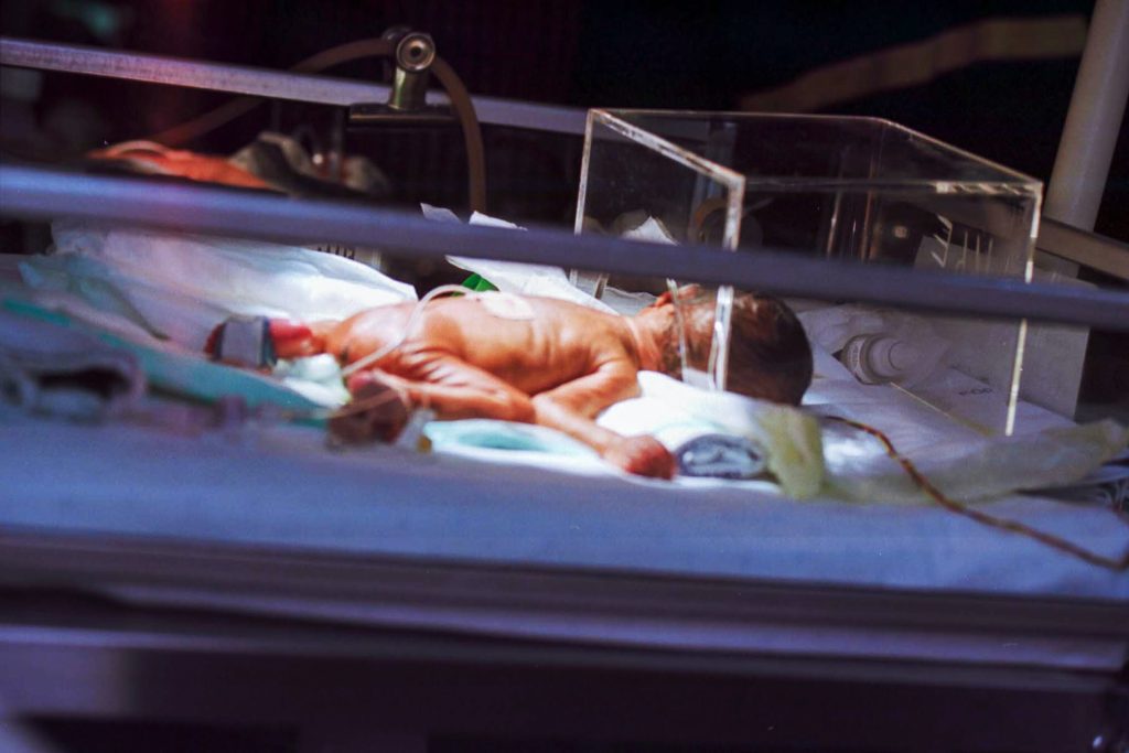
A treatment to move blood from the umbilical cord into an infant’s body may provide a safe option for preterm infants born after 28 weeks who need rapid support, suggests a study supported by the National Institutes of Health. The procedure, called umbilical cord milking, involves gently squeezing the cord between the thumb and forefinger and pushing the blood into the newborn’s abdomen.
The new findings suggest that concerns raised by a 2019 study of infants born before 28 weeks (which concluded that umbilical cord milking might increase the risk of bleeding inside the brain) do not apply to preterm infants born after 28 weeks. The current study appears in Pediatrics.
The standard procedure, delaying cord clamping while blood naturally flows into the infant’s body, takes 30 to 180 seconds. However, cord milking, takes about 20 seconds, reducing delay for infants who need immediate assistance, such as respiratory support. Both procedures allow for umbilical cord blood to reach the infant’s body before clamping, reducing the risk of anaemia and other complications seen among infants receiving immediate cord clamping and cutting.
The study was conducted by Anup Katheria, M.D., of the Sharp Mary Birch Hospital for Women & Newborns in San Diego, and colleagues in the United States, Canada and Europe. It was supported by NIH’s Eunice Kennedy Shriver National Institute of Child Health and Human Development.
More than 1000 infants were randomly assigned either to umbilical cord milking or delayed cord clamping. Rates of severe intraventricular haemorrhage and/or death did not differ significantly between the two groups (just over 1%). Moreover, the rates of overall intraventricular haemorrhage were also similar between the groups (approximately 12%). The researchers will follow all the infants in the study for two years to observe longer term outcomes.
A study of firefighters on a punishing training course has revealed clues as to why extreme exercise temporarily weakens the immune system – a phenomenon seen in elite athletes. The findings, published in Military Medical Research, may lead to better ways to support the health of people who undergo extreme exertion, such as firefighters tackling wildfires.
Thirteen firefighters volunteered for the study, average age 25 and male. They went through a rigorous training exercise, carrying 9 to 20kg of gear over hilly terrain during a 45-minute training exercise in the California sun. Gloves, helmets, flashlights, goggles, and more weighted them down as they sprinted through the countryside wearing fire-resistant clothing to show they were ready to serve as wildland firefighters.
After the training, they immediately gave samples of their blood, saliva, and urine for analysis. Two were excluded, one being unable to finish the course and the other arriving to late to provide a sample. The 11 participants who completed the course lost an average of 2.2% of their initial weight.
Then, the scientists from the Department of Energy’s Pacific Northwest National Laboratory (PNNL) analysed more than 4700 molecules, consisting of proteins, lipids, and metabolites, from each of the firefighters, looking to understand what happens when the body undergoes intense physical exercise. Measuring and interpreting the data from thousands of such measurements is a specialty of PNNL scientists who explore issues related to climate science and human health by analysing millions of sensitive measurements using mass spectrometry each year.
The researchers’ aim was to increase safety for first responders and others.
“Heat stress can be life threatening,” said Kristin Burnum-Johnson, a corresponding author of the study. “We wanted to take an in-depth look at what’s happening in the body and see if we’re able to detect danger from exhaustion in its earliest stages. Perhaps we can reduce the risk of strenuous exercise for first responders, athletes, and members of the military.”
As expected, the team detected hundreds of molecular changes in the firefighters. The differences before and after exercise underscored the body’s efforts at tissue damage and repair, maintenance of fluid balance, efforts to keep up with increased energy and oxygen demand, and the body’s attempts to repair and regenerate its proteins and other important substances.
But in the saliva, the team found some unexpected results. There was a change in the microbial mix of the mouth – the oral microbiome – showing that the body was increasingly on the lookout for bacterial invaders. Scientists also saw a decrease in signaling molecules important for inflammation and for fighting off viral infections.
A decrease in inflammation makes sense for people exercising vigorously; less inflammation allows people to breathe in air more quickly, meeting the body’s eager demand for more oxygen. Having fewer inflammatory signals in the respiratory system helps the body improve respiration and blood flow.
Less inflammation, more inhalation
But less inflammation leaves the body more vulnerable to viral respiratory infection, which other studies observed in elite athletes and others who exercise vigorously. Some studies have shown that a person is up to twice as likely to come down with a viral respiratory infection in the days after an especially energetic workout.
“People who are very fit might be more prone to viral respiratory infection immediately after vigorous exercise. Having less inflammatory activity to fight off an infection could be one cause,” said Ernesto Nakayasu, a corresponding author of the paper. He notes that the work provides a molecular basis for what clinicians have noticed in their patients who do strenuous workouts.
The team hopes that the findings will help explain why come people are more vulnerable to respiratory infection after a workout.
Dr Rodrigueze and Aaron James. Credit: NYU Langone Health
A surgical team from NYU Langone Health performed the world’s first whole-eye and partial-face transplant for a 46-year-old military veteran from Arkansas who survived a work-related high-voltage electrical accident. The surgery included transplanting the entire left eye and a portion of the face from a single donor, making this the first-ever human whole-eye transplant in medical history and the only successful combined transplant case of its kind.
While it is still unknown whether he will regain sight, since the May 2023 procedure, the transplanted left eye has shown remarkable signs of health, including direct blood flow to the retina. Although many questions remain in a case with no precedence, this groundbreaking achievement opens new possibilities for future advancements in vision therapies and related medical fields.
The recipient, Aaron James of Hot Springs, Arkansas, survived a deadly 7200-volt electric shock while working as a high-voltage lineman in June 2021, when his face accidentally touched a live wire. Despite multiple reconstructive surgeries, James had extensive injuries – including the loss of his left eye, his dominant left arm from above the elbow, his entire nose and lips, front teeth, left cheek area, and chin down to the bone.
“Aaron has been extremely motivated to regain the function and independence he lost after his injury. We couldn’t have asked for a more perfect patient,” said Dr Rodriguez. “We owe much of our success in this monumental endeavour to the exceptional institutional support we receive at NYU Langone and the unwavering dedication of our world-class team in delivering the highest level of care to our patient. This achievement demonstrates our capacity to embrace the most difficult challenges and drive continuous advancements in the field of transplantation and beyond.”
The NYU Langone team was introduced to James’ case just two months after his injury, allowing guidance during the early-phase reconstructions with specialists at a Texas medical centre where he was receiving care. The possibility of a face transplant was discussed over the next year, and an initial evaluation for the transplant took place one year following the initial injury in June 2022.
When Texas surgeons were forced to remove James’ left eye after injury due to severe pain, Dr. Rodriguez and his team recommended that the optic nerve be cut as close to the eyeball as possible, to preserve as much nerve length to maximize reconstructive options, including the hope of a potential transplant later. This began the discussion on the possibility of including an eye with the face, something that has never been attempted before.
NYU Langone’s multidisciplinary team, Dr Rodriguez, and the James family collectively made the decision to move ahead with a whole-eye transplant in combination with the face – understanding that at best it may only provide cosmetic benefits but leave many unknowns.
“Given Aaron needed a face transplant and will be taking immunosuppressive drugs regardless, the risk versus reward ratio of transplanting the eye was very low. Despite the eye being successfully transplanted, from a cosmetic standpoint, it would still be a remarkable achievement,” said Dr. Rodriguez.
This is the fifth face transplant performed under the leadership of Dr. Rodriguez and the first known whole-eye transplant in existence. “The mere fact that we’ve accomplished the first successful whole-eye transplant with a face is a tremendous feat many have long thought was not possible,” added Dr. Rodriguez. “We’ve made one major step forward and have paved the way for the next chapter to restore vision.”
One of the Shortest Wait Times for a Donor—Three Months
Once James’ case cleared all approvals within NYU Langone, he was officially listed as a potential recipient in February 2023 with the United Network for Organ Sharing (UNOS), the private, nonprofit organization that manages the nation’s organ transplant system.
Locally, the effort to find a donor for James was led by LiveOnNY, the organ procurement organization for the greater New York metropolitan area. In May 2023, just three months after James was listed for organ donation, coordinators from LiveOnNY identified a potential donor at another hospital in New York City. After a series of donor evaluations – including tests to determine if the eye was healthy and viable, led by Vaidehi S. Dedania, MD, retina specialist in the Department of Ophthalmology at NYU Langone – he was deemed an ideal donor. The total time from injury to transplant was a little under two years.
“The donor hero was a young man in his 30s who came from a family that strongly supports organ donation. He, in support by his family, generously donated tissues leading to this successful face and eye transplant, but also saved three other individuals between the ages of 20 and 70, donating his kidneys, liver, and pancreas,” said Leonard Achan, RN, MA, ANP, president and CEO of LiveOnNY. “LiveOnNY is proud to have collaborated with such a distinctive team of medical professionals at NYU Langone. This act of grace and innovative surgical procedure will have a multigenerational impact on all the recipients and their loved ones.”
A Question of Nerve
While corneal transplants have become relatively common, with thousands performed in the United States each year, successful whole-eye transplants to restore vision have remained elusive due to the complex nature of the eye and the challenges associated with nerve regeneration, immune rejection, and retinal blood flow.
The human eye is intricately connected to the brain through the optic nerve, part of the central nervous system and responsible for transmitting visual information to the brain. Reestablishing these nerve connections successfully is a fundamental requirement for a whole-eye transplant to restore vision and one of the biggest challenges.
Dr Rodriguez, in collaboration with the team at NYU Langone’s Transplantation and Cellular Therapy Center, part of the Blood and Marrow Transplant Program at Perlmutter Cancer Center, made the decision to combine the donor eye with donor bone marrow–derived adult stem cells. Bone marrow is a sponge-like tissue found inside the large bones in the body. Adult stem cells that are transplanted can work as a replacement therapy and natural repair crew, dividing again and again to create heathy cells that replace the damaged or dysfunctional elements.
“This is the first attempt of injecting adult stem cells into a human optic nerve during a transplant in the hopes of enhancing nerve regeneration,” said Samer Al-Homsi, MD, MBA, executive director of the Transplantation and Cellular Therapy Center and professor in the Department of Medicine at NYU Langone. “We chose to use CD34-positive stem cells which have been shown to harbor the potential to replace damaged cells and neuroprotective properties.”
During the transplant, bone marrow that was harvested from the donor’s vertebrae and processed preoperatively to isolate the CD34-positive stem cells was brought into the operating room (OR) and injected at the optic nerve connection of the recipient.
“We have now demonstrated that the procedure is safe and potentially efficacious, but we need time to determine if this step plays a role in enhancing the chance of sight restoration and if there’s anything further that can be done in the future to optimize the procedure,” added Dr Al-Homsi.
Innovations in Face Transplant Technology
Through collaboration with Depuy Synthes, the Orthopedics Company of Johnson & Johnson, and Materialise, state-of-the-art technology played a pivotal role in both presurgical planning and the actual surgery. Cutting-edge three-dimensional (3D) computer surgical planning, along with patient-specific 3D cutting guides, enabled precise alignment of bones and optimal placement of implantable plates and screws. This meticulous approach fit the grafted partial face and whole left eye onto James.
The successful surgery took place in NYU Langone’s Kimmel Pavilion, where expansive ORs enabled two highly skilled surgical teams to simultaneously operate in both the donor and recipient rooms. The surgical teams adhered to a carefully planned timetable – which was rehearsed many times over the last year – as the team proceeded with the transplantation and reconstruction process, seamlessly integrating the donor’s face and eye onto James as quickly as possible to ensure the optimal outcome. Dr. Rodriguez and his surgical team of 7 – and OR team of 80 – transplanted the following:
partial face, including the nose, left upper and lower eyelids, left eyebrow, upper and lower lips, and underlying skull, cheek, nasal and chin bone segments, with all of the tissues below the right eye including the underlying muscles, blood vessels, and nerves
left whole eye and socket including the orbital bones and all surrounding eye tissues including the optic nerve
As they do in all transplant operations, the surgeons sought to complete the procedure as quickly as possible to limit the ischemia time, or the amount of time the donated tissue is not receiving a blood supply.
“The progress we’ve seen with the eye is exceptional, especially considering that we have a viable cornea paired with a retina showing great blood flow five months after the procedure. This far exceeds our initial expectations, given our initial hope was that the eye would survive at least 90 days,” said Bruce E. Gelb, MD, a transplant surgeon at the NYU Langone Transplant Institute and vice chair of quality in the Department of Surgery. “We will continue to monitor, and I am excited to see what else we may learn over time.”
Dr. Rodriguez has since performed a less extensive follow-up surgery for James to optimise his functional and aesthetic outcome. James plans to have orthodontic treatment and dental rehabilitation in the coming months.
“Beyond the eye, the quality of Aaron’s results from the face transplant is special. You would never think he underwent such a procedure so recently. He looks great,” said Dr. Rodriguez.
The NYU Langone team has set the standard in the field of face transplants for eliminating and avoiding early rejection episodes as well as the frequency of rejections using a unique immunosuppression regimen.
As in previous face transplants, Dr. Rodriguez and his team collaborated with NYU’s advanced 3D media services center, LaGuardia Studio, to create a 3D-printed replacement of the donor’s face to restore the integrity of the donor’s identity after the organs were removed to return to his family. Traditionally, a molded, hand-painted silicone mask had been used. There are few printers in the world like the one at LaGuardia, which prints with 60,000 colors.
A Determined Patient Focused on Healing
Following the surgery, James spent just 17 days in the intensive care unit at NYU Langone, one of the shortest recoveries among Dr. Rodriguez’s face transplant recipients. He was discharged on July 6 to a nearby apartment. From there, he continued outpatient rehabilitation including physical, occupational, and speech therapy.
James continues to remain positive and eternally grateful to regain many elements of life he lost after the 2021 injury, especially the ability to taste, smell, and eat solid foods. On September 14, he returned home to Arkansas with his wife, Meagan, and daughter, Allie. James comes back to New York City monthly for follow-up appointments. He has the option to return to work as a safety manager for high-voltage line workers in the future.
“I’m grateful beyond words for the donor and his family, who have given me a second chance at life during their own time of great difficulty. I hope the family finds solace in knowing that part of the donor lives on with me,” said James. “I will also forever be thankful to Dr. Rodriguez and his team for changing my life. My family and I wouldn’t have been able to navigate this difficult journey without their expertise and support. Our hope is that my story can serve as inspiration for those facing severe facial and ocular injuries.”
James is looking forward to spending the upcoming Thanksgiving holiday with his family, getting to enjoy eating a holiday meal for the first time since his injury.
Seeing the Future
While James’ vision in his native right eye is intact, the transplanted left eye does not currently have any sight. However, over the last six months, his eye has shown remarkable signs of health in other regards based on various clinical tests that measure outcome.
Dr. Rodriguez, Dr. Dedania, and a multidisciplinary team of world-renowned researcher scientists and clinicians – including leaders in neurology, ophthalmology, radiology, and neuroradiology – continue to convene and discuss questions that remain related to the eye and ways to measure any indications toward sight restoration.
“What we’re witnessing now is not something we ever expected or thought we’d see,” said Dr. Dedania, who runs regular tests for James in relation to his eyes. “The first step is having an intact eyeball, a lot of things could come after that; this is a first in the world, so we are really learning as we go.”
James will continue to have various clinical tests on the left transplanted eye, including electroretinography, a test that measures the electrical response of the retina to light.
“This is certainly one extraordinary step in the right direction,” said Steven L. Galetta, MD, renowned neuro-ophthalmologist and the Philip J. Moskowitz, MD, Professor and Chair of Neurology at NYU Langone. “We’re now crossing into the frontier of the central nervous system. Whatever happens next allows the opportunity for various methods to try to enhance the remaining aspects of the retina, whether it be through growth factors, stem cells, or a device that can pick up the signals and then bypass things along that optic nerve pathway. I’m looking forward to further advancements from this case in collaboration with the very talented minds that made it happen here at NYU Langone.”
A multinational study of almost one million individuals confirms a strong and clear association between radiation exposure from CT scans in young people and an increased risk of blood cancers. These results, published in Nature Medicine, highlight the importance of continuing to apply strict radiological protection measures, particularly in paediatric populations.
The benefits of computed tomography (CT) for imaging in patient management (including diagnostic efficacy, treatment planning and disease follow-up) are undisputed. But in recent decades the extensive use of this procedure has raised concerns about the potential cancer risks associated with exposure to ionising radiation, particularly in young patients.
“The exposure associated with CT scans is considered low (less than 100mGy), but it is still higher than for other diagnostic procedures,” says Elisabeth Cardis, Head of the Radiation Group at ISGlobal and senior author of the study. Previous studies have suggested an increased risk of cancer in in children exposed to CT scans, but they had several methodological limitations.
To address these limitations, clinicians, epidemiologists and dosimetrists from nine European countries (Belgium, Denmark, France, Germany, Netherlands, Norway, Spain, Sweden, and UK) came together to conduct a multinational, European-funded study, EPI-CT, coordinated by the International Agency for Research on Cancer (IARC).
“Implementing this large, multinational study was challenging – it involved extracting data from radiology records of 276 hospitals and linking them to population-based registries in nine countries, all while maintaining the confidentiality of the individuals’ data,” says Cardis.
A dose-dependent association
The study analysed data from almost one million people, who underwent at least one CT scan before the age of 22. The dose of radiation delivered to the bone marrow, where blood cells are produced, was estimated for each person. By linking this information to national cancer registries, EPI-CT researchers were able to identify those who developed a blood cancer over time. Individuals were followed for an average of 7.8 years, although for those who had CT scans in the early years of the technology, researchers were able to monitor cancer incidence for more than 20 years after the first scan.
The results show a clear association between the total radiation doses to the bone marrow from CT scans and the risk of developing both myeloid and lymphoid malignancies. A dose of 100mGy approximately tripled the risk of developing a blood cancer. These results suggest that a typical scan today (with an average dose of about 8mGy) increases the risk of developing these malignancies by about 16%. “In terms of absolute risk, this means that, for every 10 000 children who have a CT scan, we can expect to see about 1–2 cases of cancer in the 12 years following the examination,” says first author Magda Bosch de Basea, ISGlobal researcher at the time of the study.
The authors point out that more work is needed to ensure that doses and technical parameters are systematically and adequately collected in the clinics in real time to further improve risk estimates in the future.
Public health implications
Today, more than one million children in Europe undergo CT scans every year. Although radiation doses from CT scans have decreased substantially in recent years, the findings of this study underline the need to raise awareness among the medical community and to continue to apply strict radiation protection measures, especially in the youngest patients. “The procedure must be properly justified – taking into account possible alternatives – and optimised to ensure that doses are kept as low as possible while maintaining good image quality for the diagnosis,” Cardis explains.
A panel of international experts representing the World Health Organization’s (WHO) Guideline Development Group has updated its guidance on treatments for patients with COVID.
The new recommendations published by The BMJ are part of a living guideline, developed by the WHO with the methodological support of MAGIC Evidence Ecosystem Foundation, to provide up to date, trustworthy guidance on the management of COVID and help doctors make better decisions with their patients.
The guidance incorporates the latest clinical trial evidence for existing and new COVID therapies and takes account of evidence relating to safety, prognosis, resources, access, and equity issues, as well as patient values and preferences.
The updates include:
Distinct risk categories to help doctors more accurately assess whether an individual is at high, moderate, or low risk of hospital admission and tailor treatment accordingly.
A new treatment benefit threshold of 1.5% (down from 6%) reduction in the risk of hospital admission. This reflects the lower baseline risk for most patients with non-severe COVID as well as more safety evidence and wider availability of therapies.
A recommendation to use the antiviral drug nirmatrelvir-ritonavir in patients with non-severe COVID at high and moderate risk of hospital admission.
A recommendation against use of the antiviral drugs remdesivir and molnupiravir for patients with non-severe COVID at moderate and low risk of hospital admission (treatment is suggested for patients at high risk of admission).
A recommendation against use of a new antiviral (VV116) for patients with COVIDexcept in clinical trials, regardless of illness severity.
A strong recommendation against the use of ivermectin for patients with non-severe COVID(advice against use of ivermectin in patients with severe or critical COVID, except in clinical trials, still exists).
The experts say the new recommendations reflect changes in the virulence and transmissibility of circulating SARS-CoV-2 variants and sub-variants, along with changes in immunity related to global vaccinations, which have led to lower baseline risks of severe illness and death for most patients with non-severe COVID.
They acknowledge that there are still uncertainties around COVID therapeutics and emerging evidence and say these recommendations need to be used in light of these uncertainties.
Medshield Medical Scheme, a prominent medical aid scheme, and Clicks, a leading retail pharmacy chain and Designated Service Provider on the Medshield Pharmacy and SmartCare Networks, are pleased to announce the expansion and enhancement of their partnership. This partnership aims to empower members with even greater access to quality care through Medshield’s SmartCare benefit, allowing access to a network of Clicks clinics for professional nurse and nurse-led virtual Family Practitioner (GP) consultations. This further cements their commitment to delivering healthcare excellence through technology.
Expanding the SmartCare Network
Medshield and Clicks have partnered to add 123 Clicks clinics to the existing 255 clinics in the SmartCare Network. This expansion guarantees that Medshield members can conveniently and efficiently access their SmartCare benefits at these selected Clicks clinics.
SmartCare: The Gateway to Modern Healthcare
Medshield’s flagship member benefit, SmartCare, is leading the charge in digital innovation in healthcare. By utilising the power of technology, SmartCare provides access to pharmacy clinics that offer a one-stop-shop for members to access professional nurse consultations, health risk assessments, sick notes, specialist referrals, medication and nurse-led virtual access to Family Practitioners (GP) when required. This benefit is redefining the way healthcare services are accessed and delivered, making it more convenient and efficient for both healthcare providers and members.
Medshield members have access to an unprecedented level of convenience through Clicks clinics, which are powered by the Udok telemedicine solution. These consultations cover prevention, diagnosis, and treatment, focusing on connecting patients, nurses, doctors, and medication for fast and convenient care.
Kevin Aron, Principal Officer at Medshield, explains, “When we introduced SmartCare, we aimed to offer a cutting-edge solution that would add more value for our members. Medshield was the pioneer medical scheme in South Africa to integrate this service as a new benefit for all members, without additional costs.”
The Medshield SmartCare Benefit
SmartCare offers a multitude of benefits to Medshield members, providing them with a holistic approach to healthcare:
Enhanced Access to Care: SmartCare provides Medshield members with easy access to quality care led by professional, licensed nurses at pharmacy clinics. The nurse will facilitate a virtual Family Practitioner (GP) consultation depending on the patient’s ailment. Once the patient has been diagnosed and treatment prescribed, the relevant medication is easily obtained from the pharmacy.
Stretch day-to-day medical aid benefits: Healthcare services offered by SmartCare pharmacy clinics such as Clicks are cost-effective, and enable members to receive quality care and their medication as a complete solution. Utilising the SmartCare benefits allows the member to receive quality care whilst minimising the use of their day-to-day benefit.
Improved Health Outcomes: SmartCare services implemented by pharmacy providers allow members to manage and receive preventative care through wellness checks and health risk assessments, providing access to early intervention services and ultimately leading to better health outcomes.
The Vision of Collaboration
“We are excited to announce our enhanced partnership with Clicks, a valued partner on the Medshield DSP Network. With the addition of 123 Clicks clinics to the SmartCare Network, we are reinforcing our commitment to provide Medshield members with access to high-quality healthcare services,” said Kevin Aron, Principal Officer at Medshield. “SmartCare is revolutionising healthcare delivery, and we are proud to offer this innovative solution to our members.”
The Medshield SmartCare way of adding value:
A Medshield member can visit any Clicks clinic on the SmartCare network for primary healthcare needs such as acute conditions, wellness checks, health risk assessments, vaccinations, or chronic medication prescriptions as prescribed by a Family practitioner (GP).
A registered nurse performs a thorough medical history and examination of the patient.
The nurse can advise the patient on over-the-counter medication available at the pharmacy.
A virtual consultation with a family practitioner is requested by the nurse through Clicks clinic’s Udok technology when further treatment is necessary. The doctor then completes the consultation with the assistance of the nurse.
The nurse can print the doctor’s written documentation, and the patient can fill their prescription at the pharmacy immediately.
Accessible Medications and Comprehensive Care
In addition to SmartCare consultations, Clicks pharmacies are available on all Medshield plans, making access to prescription medication convenient for members.
Rachel Wrigglesworth, Clicks’ Chief Healthcare Officer stated, “This partnership between Clicks and Medshield focuses on the wellbeing of our customers, which is our top priority. The collaboration has expanded to include Clicks clinics powered by Udok, a solution that offers real-time access to registered family practitioners through our Nurse-led consultations on the SmartCare benefit, funded by Medshield Medical Scheme. As a leader in the healthcare market, this partnership perfectly aligns with our commitment to increasing access to affordable primary healthcare for all South Africans. We are committed to the continued success of this collaboration.”
Embracing the Future of Healthcare
As the healthcare industry continues to evolve in the digital age, SmartCare stands as a shining example of how technology and innovation come together to provide added convenience and efficiency in healthcare. It empowers nurses to provide additional care for Medshield members through accessible technology. Unless it is a trauma situation, members can visit a Clicks clinic on the SmartCare network for acute and chronic conditions. By embracing the future of healthcare through the SmartCare benefit, Medshield members can expect to experience efficient and reliable medical consultations to enhance their wellbeing.
“Medshield is continuing to reinvent healthcare the smart way. The SmartCare benefit offers our members a new level of convenience, connecting members with nurses, doctors and medicine like never before,” concluded Aron.
A Strengthened Partnership
Expanding the Medshield and Clicks partnership demonstrates a solid commitment to providing excellent healthcare services and a shared vision of creating a more accessible and convenient healthcare experience for Medshield members. It is a testament to the excellent collaboration between Medshield and Clicks, ensuring that quality care is always easily accessible.
Photo by Towfiqu barbhuiya: https://www.pexels.com/photo/person-feeling-pain-in-the-knee-11349880/
New research reported in the journal Nature could lead to new targeted treatments for rheumatoid arthritis (RA). The findings showed that guesswork could be taken out of selecting treatments for each patient, and this might one day also be extended to other autoimmune conditions.
The study was led by University of Colorado School of Medicine faculty members Fan Zhang, PhD, and Anna Helena Jonsson, MD, PhD. The Accelerating Medicines Partnership: Rheumatoid Arthritis and Systemic Lupus Erythematosus (AMP: RA/SLE) Network collected inflamed tissue from 70 patients with RA from across the country and the United Kingdom. Jonsson supervised the team of scientists who processed these samples for analysis, and Zhang led the computation analysis of the data. These efforts yielded a cell atlas encompassing more than 300 000 cells from synovial tissue. Further analysis revealed that there are six different subgroups of RA based on their cellular makeup.
“We hope the data will help us discover new treatment targets,” says Jonsson, assistant professor of rheumatology. “We wanted to make it public so that researchers across the country and across the world can continue working on new treatment ideas for rheumatoid arthritis going forward.”
No more guess-and-check
Jonsson, a practicing rheumatologist as well as a researcher, knows that RA patients respond differently to different treatments. Until now, she says, rheumatologists used a “guess and check” method to find a treatment that works for an individual patient.
With the new data and powerful computational classification methods developed by Zhang and the computational analysis team, the researchers were able to quantitatively classify RA types into what they call ‘cell-type abundance phenotypes’, or CTAPs. Developed methods, together with the new cell atlas, can start to identify which patients will respond to which treatments.
“Even when you classify rheumatoid arthritis inflammation using these simple markers – T cell markers, B cells, macrophages and other myeloid cells, fibroblasts, endothelial cells – what we found is that each of those categories is associated with very specific kinds of pathogenic cell types we’ve already discovered,” Jonsson says. “Previous rheumatoid arthritis research found that T cell populations called peripheral helper T cells are relevant in rheumatoid arthritis, as are B cells called antibody-producing B cells, and other specific cell types. What we found is that they’re usually not found all together.
“For example, the peripheral helper cells are found with the B cells in only one category of RA, and the pathogenic macrophage populations tend to exist in a different category. Because of this, we can start asking questions about how these specific partners work together.”