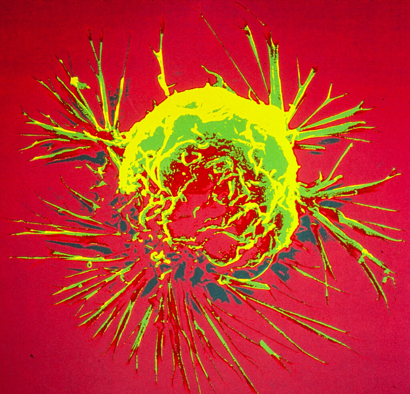
Colourised scanning electron micrograph of a breast cancer cell. Credit: NIH
A novel therapeutic approach that combines human epidermal growth receptor factor 2 (HER2)-targeted therapies with the cholesterol-lowering drug lovastatin can reduce the number of cancer treatments required to prevent tumour growth. Monitored by immuno-PET scans, this combination therapy has the potential to personalise treatment for cancer patients and spare them from harmful side effects. This research was published in The Journal of Nuclear Medicine.
Antibody-drug conjugates (ADCs) have become an eminent cancer treatment because of their ability to precisely target tumours with potent efficacy. HER2-ADC therapies have been effective in treating breast, lung, bladder, and stomach cancers. Although usually well-tolerated, multiple doses of the drugs can result in severe side effects, including low blood counts, liver damage, and lung damage. Strategies that reduce toxic side effects caused by ADCs and predictive biomarkers of ADC toxicity are currently an unmet clinical need.
“In this study, we sought to determine whether a single dose of HER2-ADCs could be administered in combination with lovastatin (which temporarily elevates cell-surface HER2) to achieve therapeutic efficacy similar to that of a multiple dose regime,” said Patricia Pereira, PhD, assistant professor at the Washington University School of Medicine. “We also used HER2-targeted immuno-PET to monitor changes in HER2 expression after ADC therapy.”
Researchers injected mice with cultured gastric cancer cells and patient-derived gastric cancer cells. When tumours grew sufficiently, the mice were divided into groups and received various treatment schedules (no treatment, multiple doses of ADC, multiple doses of ADC with lovastatin, single dose of ADC, or single dose of ADC with lovastatin). Immuno-PET was used to investigate the dosing regimen and the efficacy of the treatment schedules.
A single dose of ADC therapy combined with lovastatin was found to reduce tumour volume at rates similar to those resulting from multiple doses of ADC in a preclinical setting. The study results showed that immuno-PET can noninvasively monitor HER2 tumour levels after treatment with HER2-targeted ADC therapies.
“This preclinical work is significant because it has the potential to improve therapy for patients with HER2-positive cancers,” noted Pereira. “It not only simplifies treatment by exploring single-dose schedules of antibody-drug conjugates but can also reduce side effects by minimizing the number of doses required. Additionally, it personalises therapy using molecular imaging, enhancing treatment efficacy.”
She continued, “The findings suggest a future where molecular imaging techniques play a critical role in guiding drug development and cancer treatment decisions, particularly as various ADCs are being tested and approved for cancer treatment. Currently, there is no perfect way to select tumours or monitor their response to ADCs. This research indicates that molecular imaging can bridge this gap by providing real-time insights into therapy response.”
Research on long-term memories has largely focused on the role of neurons but in recent years, research is revealing that other cell types are also vital in memory formation and storage. A new study reveals the crucial role of vascular system cells (pericytes) in the formation of long-term memories of life events – memories that are lost in diseases such as Alzheimer’s. The research, published in the journal Neuron, shows that pericytes, which wrap around the capillaries work in concert with neurons to help ensure that long-term memories are formed.
Pericytes help maintain the structural integrity of the capillaries. Specifically, they control the amount of blood flowing in the brain and play a key role in maintaining the barrier that stops pathogens and toxic substances from leaking out of the capillaries and into brain tissue.
“We now have a firmer understanding of the cellular mechanisms that allow memories to be both formed and stored,” says Cristina Alberini, a professor in New York University’s Center for Neural Science and the paper’s senior author. “It’s important because understanding the cooperation among different cell types will help us advance therapeutics aimed at addressing memory-related afflictions.”
“This work connects important dots between the newly discovered function of pericytes in memory and previous studies showing that pericytes are either lost or malfunction in several neurodegenerative diseases, including Alzheimer’s disease and other dementia,” explains author Benjamin Bessières, a postdoctoral researcher in NYU’s Center for Neural Science.
The discovery, reported in the new Neuron article, of the pericytes’ significance in long-term memory emerged because Alberini, Bessières, Kiran Pandey, and their colleagues examined the role of insulin-like growth factor 2 (IGF2) – a protein that was known to increase following learning in brain regions, such as the hippocampus, and to play a critical role in the formation and storage of memories.
They found that IGF2’s highest levels in the brain cells of the hippocampus do not come from neurons or glial cells, or other vascular cells, but, rather, from pericytes.
A cheap and widely available prescription drug can improve symptoms of irritable bowel syndrome in patients seen in GP surgeries, according to research findings published in The Lancet and presented today at UEG Week 2023.
Amitriptyline, a tricyclic which is commonly used at low doses for a range of health concerns, has been found to improve irritable bowel syndrome (IBS) symptoms too, according to the results of the ATLANTIS trial.
The study was conducted in primary care, with GPs prescribing the drug and patients managing their own dose based on the severity of their symptoms, using an adjustment document designed for the trial. Most people with IBS are seen and managed in primary care by their GP, which means that the results of this trial are likely to be applicable to many people with the condition.
Led by researchers at the Universities of Leeds, Southampton, and Bristol and funded by the National Institute for Health and Care Research (NIHR), the study showed that patients taking amitriptyline were almost twice as likely to report an overall improvement in symptoms as those taking a placebo.
Now the trial team is recommending that GPs support their patients with IBS to use amitriptyline to manage their symptoms – and has made the dose adjustment document available for clinicians and patients.
Co-chief Investigator Alexander Ford, Professor of Gastroenterology in the University of Leeds’s School of Medicine, said: “Amitriptyline is an effective treatment for IBS and is safe and well tolerated. This new rigorously conducted research indicates that general practitioners should support patients in primary care to try low-dose amitriptyline if their IBS symptoms haven’t improved with recommended first-line treatments.”
Most treatments for IBS, which affects around 1 in 20 people, only have a modest effect and people often have ongoing troublesome symptoms.
Amitriptyline was originally used at high doses to treat depression, but is now superseded by newer and better antidepressants.
Previous small trials of low-dose tricyclic antidepressants for IBS suggested a possible benefit in patients seen in hospital clinics, who often have more difficult to treat symptoms, but this new study is the first randomised controlled trial of low-dose amitriptyline versus a placebo tablet for IBS in primary care. It is also the largest trial of amitriptyline for IBS undertaken worldwide.
GPs already prescribe low-dose amitriptyline to treat chronic nerve and back pain, and to help prevent migraine attacks. NICE guidelines currently state that GPs could consider using a low dose tricyclic, like amitriptyline, for IBS but, until now, the evidence for a benefit has been uncertain.
Based on the results of the trial, which showed a clear benefit of amitriptyline, GPs can offer low-dose amitriptyline to people with IBS as part of shared decision making if symptoms don’t improve with first-line treatments.
Co-chief Investigator Hazel Everitt, Professor of Primary Care Research at the Primary Care Research Centre, University of Southampton, said: “Prior to ATLANTIS, GPs haven’t often prescribed amitriptyline for IBS as the research evidence was uncertain, but our new research provides good evidence of benefit.
“GPs already prescribe low-dose amitriptyline for other conditions, such as chronic pain and poor sleep, and when we interviewed GPs as part of this research, they were willing to prescribe it for IBS if the research evidence supported this. Participants were also keen to have another option to try to help their IBS symptoms and most were happy to self-adjust their dose depending on symptoms and side effects.’’
Some 463 people with IBS took part, recruited from 55 general practices across the UK.
Participants were put at random into two groups – those receiving amitriptyline and those receiving a placebo. Participants controlled how many tablets of the trial medication they took, receiving support via the patient dose adjustment document that was developed with patient representatives especially for this trial. This enabled participants to increase or decrease the number of tablets based on their IBS symptoms and any side effects experienced.
IBS scores were measured using the IBS-SSS scale. Amitriptyline participants scored a 99-point improvement compared with a 69-point improvement among placebo participants.
Participants taking amitriptyline reported a bigger improvement in their symptom scores after six months compared with those taking a placebo. Those taking amitriptyline were almost twice as likely as those taking a placebo to report an overall improvement in IBS symptoms, with amitriptyline performing better across a wide range of IBS symptom measures.
Researchers monitored participants’ anxiety or depression scores and found that they were not altered – suggesting that the beneficial effects of the medication were via the gut, not because of any effect as an antidepressant.
No safety concerns were identified and side effects in people on amitriptyline were mostly mild, such as a dry mouth in the morning.
Matthew Ridd, GP and Professor of Primary Health Care at the Centre for Academic Primary Care, University of Bristol, said: “Pragmatic trials like this are always challenging to do in primary care and the team worked hard to overcome the additional challenges of the Covid-19 pandemic. It’s fantastic that we’ve found that amitriptyline is an effective and safe option for patients with IBS to try.”
Amanda Farrin, Professor of Clinical Trials and Evaluation of Complex Interventions, who leads the Complex Intervention Division of the Leeds Clinical Trials Research Unit, said: “The participants in the ATLANTIS trial had moderate to severe symptoms and an average duration of IBS of 10 years. The fact that amitriptyline had such a big effect over a placebo is significant because it can help improve the quality of life of patients with this condition.”
Professor Andrew Farmer, Director NIHR’s Health Technology Assessment (HTA) Programme, said: “The results of this study are hugely encouraging. It shows that a drug already widely available to treat a number of other conditions appears to be safe and effective for people with IBS. The findings the research team have shared around the adjustment of dosages can be tremendously helpful to GPs in guiding them when treating patients.
“IBS affects a significant number of people in the UK and can have a debilitating effect on their day-to-day lives. This is another excellent example of how high-quality research can lead to positive changes in health and social care practice and treatments for the benefit of patients and healthcare professionals.”
A three-year clinical trial has shown that the sublingual immunotherapy (SLIT) is safe in peanut-allergic children ages one to four, with a greater likelihood of desensitisation and remission the earlier the treatment began. SLIT approach where the treatment is given as a small amount of liquid under the tongue, instead of peanut flour that is mixed with other food and then eaten like it is during oral immunotherapy, or OIT.
Published in the Journal of Allergy and Clinical Immunology, this is the first randomised, controlled trial to investigate – in this young age group – the efficacy and feasibility of SLIT.
The study included 50 peanut-allergic children between the ages of one and four, randomised to receive 4mg peanut SLIT versus placebo. Participants were randomised 1:1 to receive either peanut SLIT or placebo. Desensitization to peanut was assessed by double-blind, placebo-controlled food challenge (DBPCFC) after three years of treatment.
Findings showed that peanut SLIT can be highly effective in treating peanut-allergic toddlers with almost 80% tolerating 15 peanuts without allergic symptoms after completing the treatment. With most typical peanut-allergic reactions being caused by one peanut or less, these results would translate into strong protection against exposures to peanut. In addition, researchers showed that remission of the peanut allergy may be possible after peanut SLIT with 63% of the toddlers maintaining their protection three months after stopping the treatment. These new findings show that early intervention with peanut SLIT is promising and warrants further development.
Led by Edwin Kim, MD, associate professor of paediatrics at the UNC School of Medicine, said: “From our prior studies in older children, we were optimistic that peanut SLIT could have a similar treatment effect in toddlers.
“However, what we found was even better. The desensitisation levels we saw were higher than expected and on par with levels we normally would only expect with oral immunotherapy. Just as important, rather than wearing off quickly, we were excited to see that over 60% stayed protected three months after stopping the treatment.”
One of the presumed strengths of the SLIT approach when compared to OIT has been its overall safety and simple administration. While most treatment side effects with OIT are mild to moderate, severe reactions requiring emergency treatment do occur and there remains a critical need to develop treatments with more manageable side effects.
“Peanut OIT is currently available and being offered by increasing numbers of allergists, however we are quickly learning that in addition to its known risk of allergic reactions, the actual doing of OIT can be very difficult for many families,” said Kim. “Peanut SLIT could be a good option to consider as it may be able to provide comparable levels of protection while being safe and easier to administer.”
Compared to OIT, the SLIT approach is likely to be a safer option, Kim said, with the most common side effect consisting of oral itching. Treatments that can protect children from allergic reactions while still being safe and practical for busy families can be life-changing, and researchers are hopeful that peanut SLIT can be one of those options.
“Even with the push to introduce peanut in early childhood in order to prevent the allergy, peanut allergy remains one of the most common food allergies,” said Kim. “A result of early peanut introduction is that we are diagnosing peanut allergy at younger and younger ages making it vitally important to develop treatments that can be safe and effective at preventing allergic reactions in these young children.”
Talk to your patients about safe, effective use of medicines
Photo by Cottonbro on Pexels
Approximately one in ten patients experience an adverse drug reaction during their care1. This can lead to serious harm or even death. Sanofi is committed to reducing these numbers by working with healthcare practitioners to create a culture of patient safety.
“Patient safety is a top priority for Sanofi,” says Yusuf Dawood, Multi-Country Safety Head for Sanofi Southern Africa. “We believe that patients should be essential partners in their healthcare journeys, and we are committed to working with healthcare professionals alongside their patients to ensure optimal therapeutic outcomes. We call on all healthcare practitioners to join us in raising awareness of patient safety. By working together, we can advocate for improved communication and reduce patient harm.”
Here are some key tips for healthcare practitioners on how to improve patient safety:
Ask patients about their concerns and listen to their feedback. They can provide valuable insights into their own health and well-being and by engaging them, healthcare practitioners can ensure that potential issues are detected as soon as possible and handled appropriately.
Provide patients with clear and concise information about their care. Patients need to understand what their diagnosis is, what treatment options are available, and what the benefits and risks of each option are. They also need to know what to expect during and after their treatment, and how to manage any side effects or complications. By giving patients accurate and easy-to-understand information, healthcare practitioners can empower them to make informed choices about their care.
Communicate with patients and other members of the healthcare team. Use simple and unambiguous language, avoid jargon and acronyms, and confirm that the patient has understood the information they have been given. Use tools such as checklists, handovers, and feedback loops to ensure that the information they share is complete and accurate.
Follow safety protocols and procedures. Healthcare practitioners need to adhere to guidelines, policies, protocols, best practices and standards of care established by professional bodies and regulatory authorities, which have been designed to prevent or minimise harm to patients.
Report issues immediately. Report any patient safety issues to the appropriate authorities in the interest of public safety. Report any medication-related patient safety issues to the relevant pharmaceutical companies. This enables companies to continuously monitor the benefit-risk profile of their products and ensure the safe use of medicines.
“Patient safety should be a top priority for healthcare professionals and pharmaceutical companies because the goal of both sectors is to improve and protect the well-being of individuals,” says Dawood. “When safety is compromised, it not only jeopardises the health and trust of patients but also undermines the credibility and integrity of the entire healthcare system. By working with pharmaceutical companies like Sanofi, healthcare professionals can provide real-world feedback on drug efficacy and side effects. This collaborative approach ensures that treatments are both safe and effective.
Join Sanofi in championing patient care. Let’s collaborate, communicate, and make every patient’s journey safer.
Reference 1. Ribeiro, M. et al. (2018) ‘Increase of 10% in the rate of adverse drug reactions for each drug administered in hospitalized patients’, Clinics, 73, pp. 1–6. doi:10.6061/clinics/2018/e185.
Relief could be on the way for people with painful hand osteoarthritis after a new study found an affordable existing drug can help. Until now there has been no effective treatment.
Published in The Lancet, the paper investigated methotrexate, a low-cost, effective treatment for inflammatory joint conditions such as rheumatoid arthritis and psoriatic arthritis. It has been widely used in Australia and globally since the early 1980s.
Researchers led by Monash University and Alfred Health found that methotrexate reduced symptoms in those with hand osteoarthritis (OA). A 20mg weekly oral dose over six months had a moderate effect in reducing pain and stiffness in patients with symptomatic hand OA.
Hand OA is a disabling condition that causes pain and affects function, impeding daily activities such as dressing and eating. It can significantly reduce quality of life. About one in two women and one in four men will experience symptoms from hand OA by the time they turn 85.
About half will have inflamed joints, which cause pain and are associated with significant joint damage. Despite the high prevalence and disease burden, there are no effective medications.
Senior author Professor Flavia Cicuttini said that the study identified the role of inflammation in hand OA and the potential benefit of targeting patients who experience painful hand OA.
“In our study, as with most studies of osteoarthritis, both the placebo group and methotrexate groups’ pain improved in the first month or so,” Professor Cicuttini said.
“However, pain levels stayed the same in the placebo group but continued to decrease in the methotrexate group at three and six months, when they were still decreasing. The pain improvement in the methotrexate group was twice as much as in the placebo group.
“Based on these results, use of methotrexate can be considered in the management of hand osteoarthritis with an inflammatory pattern. This provides clinicians with a treatment option for this group, which tends to get more joint damage.”
Professor Cicuttini said in patients with hand OA and inflammation, the effects of methotrexate were present at about three months and by six months it was very clear if it worked.
“At that time patients and their doctors can decide whether to continue or stop it,” she said. “This is very similar to what we currently do with other forms of inflammatory arthritis.”
The randomised, double-blind, placebo-controlled trial of 97 people with hand OA and MRI-detected inflammation assessed whether 20mg of methotrexate weekly reduced pain and improved function compared to placebo in patients with symptomatic hand OA and synovitis (inflammation) over six months.
Professor Cicuttini said the results could provide relief for people with hand OA inflammation, which was particularly common in women as they experienced menopause.
“Further trials are needed to establish whether the effect of methotrexate extends beyond six months, for how long we need to treat patients, and whether methotrexate reduces joint damage in patients with hand osteoarthritis and associated inflammation,” she said.
Professor Cicuttini now plans to conduct an extension trial to address these questions, in particular whether women who develop hand OA around menopause and often have severe pain and joint damage may benefit.
Reducing overall calorie intake may rejuvenate muscles and activate biological pathways important for good health, according new study, published in the journal Aging Cell. Calorie restriction, which cuts intake of calories but not essential nutrients, has long been known to delay the progression of age-related diseases in animal models. This finding, by researchers at the National Institutes of Health and their colleagues, suggests the same biological mechanisms may also apply to humans.
Researchers analysed data from participants in the Comprehensive Assessment of Long-Term Effects of Reducing Intake of Energy (CALERIE), a study supported by the National Institute on Aging (NIA) that examined whether moderate calorie restriction conveys the same health benefits seen in animal studies. They found that during a two-year span, the goal for participants was to reduce their daily caloric intake by 25%, but the highest the group was able to reach was a 12% reduction. Even so, this slight reduction in calories was enough to activate most of the biological pathways that are important in healthy aging.
“A 12% reduction in calorie intake is very modest,” said corresponding author and NIA Scientific Director Luigi Ferrucci, MD, PhD. “This kind of small reduction in calorie intake is doable and may make a big difference in your health.”
The research team next sought to understand the molecular underpinnings of the benefits seen in limited, previous research of calorie restriction in humans. One study showed that individuals on calorie restriction lost muscle mass and an average of 20 pounds of weight over the first year and maintained their weight for the second year. However, despite losing muscle mass, calorie restriction participants did not lose muscle strength, indicating calorie restriction improved the amount of force generated by each unit of muscle mass, called muscle specific force.
For the current study, scientists used thigh muscle biopsies from CALERIE participants that were collected when individuals joined the study and at one-year and two-year follow ups.
To figure out which human genes were impacted during calorie restriction, the scientists isolated messenger RNA (mRNA), a molecule that contains the code for proteins, from muscle samples. The team determined the protein sequence of each mRNA and used the information to identify which genes originated specific mRNAs. Further analysis helped the scientists establish which genes during calorie restriction were upregulated, meaning the cells made more mRNA; and which were downregulated, meaning the cells produced less mRNA. The researchers confirmed calorie restriction affected the same gene pathways in humans as in mice and non-human primates. For example, a lower caloric intake upregulated genes responsible for energy generation and metabolism, and downregulated inflammatory genes leading to lower inflammation.
“Since inflammation and aging are strongly coupled, calorie restriction represents a powerful approach to preventing the pro-inflammatory state that is developed by many older people,” said Ferrucci.
A groundbreaking, easy-to-use 3D printable finger prosthesis created by a recent University of Houston graduate could offer amputees a low-cost solution to restore finger functionality. David Edquilang first designed Lunet, which doesn’t need metal fasteners, adhesives or special tools to assemble, as an undergraduate student at the Gerald D. Hines College of Architecture and Design. While standard prostheses can cost thousands of dollars, Edquilang aims to make his design open access on the internet, instead of selling it.
Edquilang explains: “Lunet began when I decided to design and 3D print prototype finger mechanisms for a prosthetic hand for fun in my free time. 2 weeks and 18 prototypes later, I created a mechanism and finger structure that closely replicated the range of motion of real fingers.”
Edquilang’s mentor at UH was Associate Professor Jeff Feng, co-director of UH’s Industrial Design program. Through a partnership with Harris Health System, Feng learned of a patient who had her fingers amputated due to frostbite. Inspired by working on an upper limb prosthesis Edquilang previously developed with student Niell Gorman, working closely with Professor Feng, Edquilang created prosthetic fingers that returned mobility to the patient, allowing her to pick up objects again.
Edquilang continues: “My professor and I were then referred to a finger amputee who lost 3 of her fingers. I applied the mechanism I created to design a finger prosthesis for her. Nearly 40 design iterations and multiple rounds of patient testing were performed to ultimately create a functional prosthesis that fit her.
His “breakthrough” came from a literal break in his design.
“After we finished working with this amputee patient, I continued to tinker with my finger designs. I intentionally broke one of my finger prototypes to see where its structural weakpoint is. It broke at the distal knuckle. This led to me having a breakthrough in the design. I added a linkage that replaces the previously rigid distal knuckle, and I stumbled upon inventing a novel finger mechanism that was more flexible and nearly unbreakable. I then set on refining the design to be more functional, easily 3D printable, and more visually appealing. Inspiration from cyberpunk art and fighter jets influenced the design. 28 design iterations and a myriad of prototypes later resulted in Lunet.”
“It feels great knowing you have the capability to positively impact people’s lives and give them help they otherwise wouldn’t be able to get,” said Edquilang.
“Not every good idea needs to be turned into a business. Sometimes, the best ideas just need to be put out there ,” said Edquilang, who graduated with a Bachelor of Science in Industrial Design last year. “Medical insurance will often not cover the cost of a finger prosthesis, since it is not considered vital enough compared to an arm or leg. Making Lunet available online for free will allow it to help the greatest number of people.”
Lunet wins awards
The prosthetic design garnered Edquilang a 2023 Red Dot: Luminary award, the highest level of recognition accorded at the Red Dot Award: Design Concept. He and Feng took home the coveted accolade at Red Dot’s ceremony last month in Singapore.
“Good results come from dedication. Extraordinary results come from experimentation. Incredible results come from a combination of both,” he said upon winning the award. He has also received a number of other accolades, including iFDesign, and national runner up for the James Dyson Award.
“David’s recent success in winning the most prestigious design awards across the world is the best manifestation of the unparalleled education and training students experience in our Industrial Design program,” Feng said. “Built upon a belief that every student is a creative individual, the program pedagogy focuses on methods of cultivating innovative minds, which is enforced with rigorous professional training.”
Lunet’s geometry inspired its name
Lunet is made up of two common types of 3D printed plastics: polylactic acid and thermoplastic polyurethane. Each finger is made up of four parts held together by plastic pins. Edquilang describes arcs and circular orbits as the foundation for the motion of the finger mechanism. The geometric basis of the design evoked the idea that the prosthesis orbits around the user’s joints like a moon, or lunet, hence the name.
Another element of Lunet’s uniqueness is that it is nearly impossible to break; other finger prosthetics can be complicated and require many parts.
“The problem with higher mechanical complexity is that these designs are less durable,” Edquilang said. “The more parts you have, the more points of failure. You need to make prosthetic fingers robust and as strong as possible, so it doesn’t break under normal use, yet you want the design to be simple. This was one of the greatest challenges in making Lunet.”
He encourages other design students not to be afraid to experiment and fail because that is often how one can learn to improve the most.
“Where the world has an abundance of problems, designers have an abundance of talent, and we should not be selfish with it,” Edquilang said.
Researchers at the University of Oxford have produced an engineered tissue representing a simplified cerebral cortex by 3D printing human stem cells. The results, published in the journal Nature Communications, showed that, when implanted into mouse brain slices, the structures became integrated with the host tissue.
The breakthrough technique could lead to tailored repairs for brain injuries. The researchers demonstrated for the first time that neural cells can be 3D-printed to mimic the architecture of the cerebral cortex.
Brain injuries, including those caused by trauma, stroke and surgery for brain tumours, typically result in significant damage to the cerebral cortex. For example, each year, around 70 million people globally suffer from traumatic brain injury (TBI), with 5 million of these cases being severe or fatal. Currently, there are no effective treatments for severe brain injuries, leading to serious impacts on quality of life.
Tissue regenerative therapies, especially those in which patients are given implants derived from their own stem cells, could be a promising route to treat brain injuries in the future. Up to now, however, there has been no method to ensure that implanted stem cells mimic the architecture of the brain.
In this new study, the University of Oxford researchers fabricated a two-layered brain tissue by 3D printing human neural stem cells. When implanted into mouse brain slices, the cells showed convincing structural and functional integration with the host tissue.
Lead author Dr Yongcheng Jin (Department of Chemistry, University of Oxford) said: ‘This advance marks a significant step towards the fabrication of materials with the full structure and function of natural brain tissues. The work will provide a unique opportunity to explore the workings of the human cortex and, in the long term, it will offer hope to individuals who sustain brain injuries.’
The cortical structure was made from human induced pluripotent stem cells (hiPSCs), which have the potential to produce the cell types found in most human tissues. A key advantage of using hiPSCs for tissue repair is that they can be easily derived from cells harvested from patients themselves, and therefore would not trigger an immune response.
The hiPSCs were differentiated into neural progenitor cells for two different layers of the cerebral cortex, by using specific combinations of growth factors and chemicals. The cells were then suspended in solution to generate two ‘bioinks’, which were then printed to produce a two-layered structure. In culture, the printed tissues maintained their layered cellular architecture for weeks, as indicated by the expression of layer-specific biomarkers.
When the printed tissues were implanted into mouse brain slices, they showed strong integration, as demonstrated by the projection of neural processes and the migration of neurons across the implant-host boundary. The implanted cells also showed signalling activity, which correlated with that of the host cells. This indicates that the human and mouse cells were communicating with each other, demonstrating functional as well as structural integration.
The researchers now intend to further refine the droplet printing technique to create complex multi-layered cerebral cortex tissues that more realistically mimic the human brain’s architecture. Besides their potential for repairing brain injuries, these engineered tissues might be used in drug evaluation, studies of brain development, and to improve our understanding of the basis of cognition.
The new advance builds on the team’s decade-long track record in inventing and patenting 3D printing technologies for synthetic tissues and cultured cells.
Senior author Dr Linna Zhou (Department of Chemistry, University of Oxford) said: “Our droplet printing technique provides a means to engineer living 3D tissues with desired architectures, which brings us closer to the creation of personalised implantation treatments for brain injury.”
Senior author Associate Professor Francis Szele (Department of Physiology, Anatomy and Genetics, University of Oxford) added: “The use of living brain slices creates a powerful platform for interrogating the utility of 3D printing in brain repair. It is a natural bridge between studying 3D printed cortical column development in vitro and their integration into brains in animal models of injury.”
Senior author Professor Zoltán Molnár (Department of Physiology, Anatomy and Genetics, University of Oxford) said: “Human brain development is a delicate and elaborate process with a complex choreography. It would be naïve to think that we can recreate the entire cellular progression in the laboratory. Nonetheless, our 3D printing project demonstrates substantial progress in controlling the fates and arrangements of human iPSCs to form the basic functional units of the cerebral cortex.”
Dr Bukiwe Spondo recently received the Rural Doctor of the Year award at the Rural Health Conference held in Chintsa in the Eastern Cape. PHOTO: Supplied
By Biénne Huisman for Spotlight
Describing the rutted gravel road between Butterworth and Tafalofefe District Hospital in the Eastern Cape, Dr Bukiwe Spondo uses the word “terrible” at least eighteen times. Dipping through the Amatole District, the 55-kilometre journey can take several hours. With heavy rain, tractors may be required to dislodge ambulances and often even staff have difficulty getting to work because of the mud.
Since 2007, Spondo and her colleagues have offered a multitude of services at Tafalofefe in the lush but impoverished Centane village. First off, she moved the hospital’s ARV clinic from an out-building to inside the premises – reducing stigma – “because if patients went into that building on the outside, automatically everyone knew,” she says.
In 2012, having observed how patients stopped taking treatment due to travel costs, she started driving up to 40 kilometres a day twice weekly to nine clinics in the area, where up to fifty patients would be queuing to see her. To make life easier for patients, she started pre-packing medication to take to them at the clinics. Later she opened a CHAMP (Clinical HIV /AIDS Management Programme) site at Tafalofefe to see complicated cases referred from the clinics, and a multi-drug-resistant TB (MDR-TB) review clinic in conjunction with Butterworth Provincial Hospital.
“As a rural doctor, you become a social worker, a pharmacist, a priest – you do everything,” she says, laughing.
Rural doctor of the year
Spondo’s efforts have not gone unnoticed. Last month at the Rural Doctor’s Association of South Africa (RuDASA’s) annual Rural Health Conference, she received the Rural Doctor of the Year award. RuDASA chairperson Dr Lungile Hobe conferred the award at the event hosted near Chintsa. Spondo is quick to point out that she also won an Amatole District leadership award last year.
Speaking to Spotlight over Zoom, she says, “So the roads here at Centane are terrible. It becomes a challenge to get ambulances through and the chopper cannot fly either when it’s raining. I mean, the other day a truck was stuck, crossing the road so the ambulance couldn’t pass. We had to take a private car from the hospital to go meet the ambulance halfway.”
She adds that the community hoped that roads would be improved after a devastating accident five kilometres from Tafalofefe in 2020 when an overloaded 65-seater bus plunged into a gorge, causing 25 deaths and 62 injuries. But, she says, the improvements never come.
At Tafalofefe, the two nearest referral hospitals are Cecilia Makiwane and Frere Provincial in East London, situated an additional 110 kilometres or 90-minute drive from Butterworth along the N2 highway. Housed in a pale building, Tafalofefe has 160 beds served by 41 professional nurses and seven doctors – including three community service doctors who joined last year. The additions have increased capacity, for example, emergency caesareans are now available around the clock.
Taking healthcare to the people
The hospital has three 4×4 bakkies [pick-ups] for visiting or transporting patients. It is in one of these that Spondo travels to see patients in remote corners between the Kobonqaba and Kei Rivers on Tuesdays and Thursdays.
“Clinics are part of decentralised primary healthcare goals,” she says. “But the problem was that if there were complicated cases – like if a patient is taking ARVs and then develop side effects, the sisters are not equipped to handle that. For example, if there is a kidney problem, they [cannot] do anything about that.
“And in time, I realised that for these people traveling to the hospital costs too much money. Let’s say, for example, the clinic at Qolora – for a person to travel from Qolora to Tafalofefe is R100. A return ticket is R200. And you know, most people here are unemployed. They can’t afford this. By the time they have saved up enough money to travel to the hospital, it’s too late. Like it would be the end stage of their kidney problem. You could not send this patient for dialysis, nothing could be done to help them. This is why I started my outreach trips.”
In motivating for Spondo to receive the RuDASA award, Tafalofefe’s CEO Masizakhe Madlebe pointed out how her work days start at 7am, only finishing once all patients had been seen, whether at the hospital or at one of the local clinics. In addition, he notes how, over the years, Spondo has mentored youth in the area, including children whose parents had succumbed to AIDS, and school girls on topics like life goals and contraceptives. He adds that Spondo even reached into her own pocket to pay school fees for children without parents.
Spondo relays how she noticed girls as young as twelve years old in their maternity ward, giving birth. “Myself and some nurses we went to two schools in the area to educate them, to discuss goals and contraceptives,” she says. “We started with grade 12 pupils. No teachers were present. It was just us and them. And I was surprised at how free they were talking. I said to them education is more important. I said to them – You see me? I am a doctor. One day you can be a doctor too, but you need to be educated. I told them they could come to Tafalofefe any time if they needed to talk, that I could help them apply for tertiary degrees, to college or to university.”
Spondo has kept a close eye on children orphaned by AIDS in the area. “I tell them to bring me their June, September, and December school reports, so I can see how they’re doing, so I can motivate them,” she says.
“These kids, I’ve seen them grow up. Some of them I saw angry – with everyone, with their own deceased parents. And I explained to them, don’t be angry. It’s not your mother’s fault. It’s not your father’s fault. It was the government’s fault for not giving your parents access to ARVs. But now, take your own ARVs and you will be fine. Some of them have passed high school with distinction, some even now have access to universities.”
Bringing her skills back home
Alongside two brothers whom she describes as “wonderful”, Spondo grew up in the village of Nqamakwe, on the opposite side of Butterworth. Her parents have passed away, but she still considers Nqamakwe her home. Here her family’s farming interests include cattle, goats, and sheep.
She attended Blythswood Secondary School in Nqamakwe – excelling at biology and physics, even though maths was hard work. “Becoming a doctor was just something I always wanted,” she says, relaying how in her formative years she had been a sickly child who often required medical care. This changed, she says, as she cannot remember ever being sick as an adult.
Spondo graduated from medical school at the University of KwaZulu-Natal in 2002, completing her internship at Cecilia Makiwane and her community service at Tafalofefe and Frere in 2004.
Speaking with rapid enthusiasm, she says how happy she is to bring her healthcare skills back home to serve the community that shaped her own humanity.
“I mean, I know these people inside out. I was born in front of them, raised in front of them,” she says. “These are our relatives, our aunts, our grannies. It’s giving back to them, to the community that raised you, that has done everything for you. Who supported you through all these years.”
She adds that Tafalofefe’s clinical manager, Sambona Ntamo, grew up near Butterworth too.
“Who would look after these people if we didn’t?” she asks.
Where does she find the resilience that drives her passion to care for sick people, often queuing at the end of long rutted roads?
“Lots of exercise,” she says, smiling.
At Tafalofefe there is a staff gym with a treadmill, a bicycle, weight lifts, and pilates balls.
“I tell the guys after work it’s gym time, it’s gym time, it’s gym time!” she says. “We’ve got a key and everyone knows that even if they want to go to the gym after midnight, they may get the key and go.”
Photographs capture an air of camaraderie at Tafalofefe. Staff sharing a meal of tripe and creamed spinach on heritage day, a farewell gathering for a retiring nurse with balloons and huge gifts in silver wrapping, [and] women knitting countless bright beanies for babies delivered in the maternity ward. A picture inside the hospital’s paediatric room shows youngsters on plastic motorbikes and mothers holding toddlers wrapped in blankets.
Spondo and her own eight-year-old son, Lutho desperately – which means the greatest one – live in a doctor’s house on the hospital’s premises. They travel to their family home in Nqamakwe over weekends.
For Spondo, being a doctor does not feel like a job. “When you do something you love, it doesn’t feel like a job,” she says. “Being a doctor is something I look forward to every morning. When patients return to me, saying they feel better with a smile on their faces, saying thank you for the treatment – that just makes my day.”