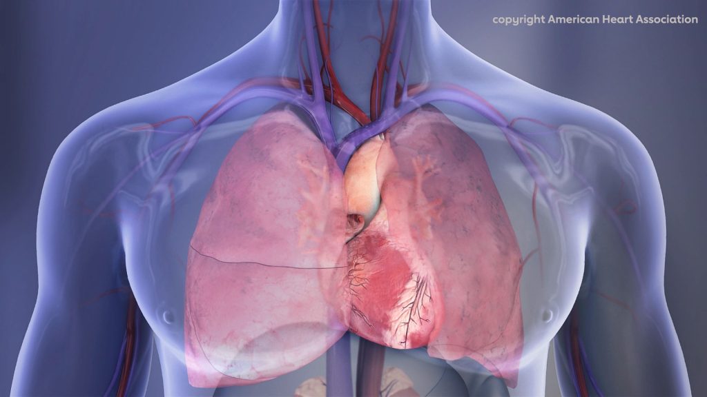
A new American Heart Association scientific statement suggests surgery be considered for more people with high-risk pulmonary embolism (PE). The statement, published in the journal Circulation, also calls for data quality registries for high-risk patients with pulmonary embolism and more research to better understand the disease process and effective treatments.
Nearly 45% of patients experiencing PE will progress to severe symptoms, where the clot causes high pressure in the lungs and subsequent damage to the right heart chamber, with a high risk of death. Even therapy following current guideline-directed treatment has a high rate of death, estimated at approximately 40% of cases in some groups.
Treatment options for patients with severe pulmonary embolism include anticoagulation therapy or thrombolysis (either intravenously or via catheter procedure), or advanced surgical interventions such as surgical embolectomy and mechanical circulatory support. Often, surgical techniques are a last resort after other treatments are unsuccessful. The statement suggests that considering surgery earlier may help improve survival for patients with severe PE.
“This statement demonstrates that modern surgical management strategies and mechanical circulatory support results in excellent survival (97%) even among the sickest patients, including those who present with cardiac arrest and have had CPR,” said Joshua B. Goldberg, MD, chair of the statement writing group.
“Modern surgical strategies and mechanical circulatory support are drastically underutilised,” he said. “It is the hope of the multidisciplinary group of authors that this scientific statement will provide a greater awareness of the safety and efficacy of modern surgical management and mechanical circulatory support in treating the most unstable patients so that lives may be saved. In addition, we hope this statement will facilitate improved understanding of the disease process and effective treatments and encourage future research to improve the survival of patients with this common and deadly disease.”
The writing group proposes strategies to determine risk more accurately and identify earlier which PE patients may benefit from surgical intervention. They also suggest increased education for clinicians to encourage the use and integration of surgical strategies earlier in PE treatment. Additionally, the statement supports the development of patient registries, particularly focused on data that provides useful context for clinicians and surgeons to understand the progression from intermediate to high-risk pulmonary embolism and treatment outcomes across patients at various risk levels.
New research published in Dermatologic Surgery indicates that simple laser treatments to the skin may help to prevent the development of basal cell carcinoma and squamous cell carcinoma, collectively known as keratinocyte carcinoma.
Conducted by a team of researchers from Massachusetts General Hospital, a founding member of Mass General Brigham, the work describes an easy-to-implement strategy to protect skin health.
Nonablative fractional lasers (NAFL) deliver heat in a fractional manner that leaves it fully intact after treatment, unlike ablative fractional lasers that remove the top layer of skin. Currently they are used to treat things such as scars, sun-damaged skin and age spots, but their effectiveness for preventing skin damage is unknown.
To investigate, Mathew Avram, MD, JD, director of the Mass General Dermatology Laser & Cosmetic Center, and his colleagues studied patients who had been successfully treated for facial keratinocyte carcinoma in the past. Such patients have a 35% risk of experiencing a subsequent keratinocyte carcinoma within 3 years and a 50% risk within 5 years.
In the study, 43 patients received NAFL therapy and 52 served as controls and did not receive NAFL therapy.
The rate of subsequent facial keratinocyte carcinoma development over an average follow-up of more than 6 years was 20.9% in NAFL-treated patients and 40.4% in controls, indicating that patients treated with NAFL had about half the risk.
Controlling for age, gender, and skin type, control patients were 2.65-times more likely to develop a new facial keratinocyte carcinoma than NAFL-treated patients.
Also, among patients who developed a facial keratinocyte carcinoma, the time to development was significantly longer in patients treated with NAFL compared with untreated patients.
“These findings suggest that NAFL treatment may have an important role in protecting against subsequent keratinocyte carcinomas,” says Avram.
“While the mechanism of NAFL’s protective effect is not completely understood, it is suspected that NAFL treatment reduces the overall burden of photo damaged keratinocytes and may promote a wound healing response, which gives healthy skin cells a selective advantage.”
Avram noted that additional studies are warranted to more critically assess the role of NAFL in skin cancer prevention, to reveal the duration of its protective effects, and to determine optimal treatment parameters.
“Based on this research, it’s encouraged for patients to have nonablative laser treatments to help prevent skin cancer if they are at risk or notice abnormalities,” says Avram.
As COVID cases rise again around the world and the more infectious XBB.1.5 variant spreads rapidly, health minister Joe Paahla has emphasised the importance of getting vaccinated and boosted.
About 19 million people in South Africa (just over 30% of the population) are fully vaccinated and four million booster shots have been administered. The country is administering just over 40 000 jabs a week.
At the moment only people over 50 are eligible for a second booster. But according to Dr Nicholas Crisp, Deputy Director-General for the National Department of Health, all adults will be eligible in February. “As soon as the systems are all in place and staff orientated, the department will announce,” Crisp told GroundUp.
But finding a booster shot has become difficult. Privately-owned facilities have mostly discontinued their rollout of the vaccine, although a handful of Dis-Chem pharmacies still do vaccinations. Public sector health facilities are the only alternative.
Active vaccination sites can be found on the government’s Find My Jab website. Some are “visiting” sites only, open once or twice a week, and others are permanently open, but it is advised to call ahead to confirm availability.
“The department is trying to find a more efficient way of updating which vaccination sites are active and those are being reflected and changed weekly on Find My Jab,” says Crisp.
The Western Cape Health Department makes weekly updates to this list of vaccination sites in the province.
One concerned reader from Pennington in KwaZulu-Natal, who is over the age of 50 and HIV-positive (meaning COVID poses a higher risk for him) told GroundUp that his local clinic no longer offered vaccines. It had been ten months since his previous booster. He went to the nearest hospital but was refused a jab and told to wait for an SMS.
He called the vaccination hotline and was told to send a copy of his ID and vaccination card to be registered on the system and receive an SMS, despite already having received jabs in the past.
Without a device to send the documents, and 60km of flood-damaged road between him and and his nearest PostNet, he has still not received his booster shot.
Contrary to popular belief, rest may not always be the best treatment after a concussion, according to the results of a large multi-centre study published in JAMA Network Open. In fact, an early return to school may be associated with a lower symptom burden after suffering a concussion and, ultimately, faster recovery.
“We know that absence from school can be detrimental to youth in many ways and for many reasons,” says study lead author Christopher Vaughan, PsyD, neuropsychologist at Children’s National Hospital. “The results of this study found that, in general, an earlier return to school after a concussion was associated with better outcomes. This helps us feel reassured that returning to some normal activities after a concussion – like going to school – is ultimately beneficial.”
In this cohort study, data from over 1600 youths aged five to 18 were collected across nine paediatric emergency departments in Canada. Because of the large sample size, many factors associated with greater symptom burden and prolonged recovery were first accounted for through the complex statistical approach used to examine the data. The authors found that an early return to school was associated with a lower symptom burden 14 days post-injury in the 8 to 12 and 13 to 18-year-old age groups.
“Clinicians can now confidently inform families that missing at least some school after a concussion is common, often between 2 and 5 days, with older kids typically missing more school,” Dr Vaughan says. “But the earlier a child can return to school with good symptom management strategies and with appropriate academic supports, the better that we think that their recovery will be.”
The results suggest a possible mechanism of therapeutic benefit to the early return to school. This could be due to:
Socialisation (or avoiding the deleterious effects of isolation).
Reduced stress from not missing too much school.
Maintaining or returning to a normal sleep/wake schedule.
Returning to light-to-moderate physical activity sooner (also consistent with previous literature).
Scientists have worked out why selective serotonin reuptake inhibitors (SSRIs), a common antidepressant class, cause around a half of users to feel emotionally ‘blunted’. In a study published in Neuropsychopharmacology, they show that the drugs interfere with reinforcement learning, which allows humans to adapt to their environment.
As their name implies, SSRIs target the neurotransmitter serotonin, and are commonly used to treat more resistant depression and anxiety. One of their widely-reported side effects is ‘blunting’, where patients report feeling emotionally dull and no longer finding things as pleasurable as they used to. Between 40–60% of patients taking SSRIs are believed to experience this side effect.
To date, most studies of SSRIs have only examined their short term use, but, for clinical use in depression these drugs are taken chronically, over a longer period of time. Researchers sought to address this by recruiting healthy volunteers and administering one of the best tolerated SSRIs, escitalopram, over several weeks and assessing the impact the drug had on their performance on a suite of cognitive tests.
In total, 66 volunteers took part in the experiment, 32 of whom were given escitalopram while the other 34 were given a placebo. Volunteers took the drug or placebo for at least 21 days and completed a comprehensive set of self-report questionnaires and were given a series of tests to assess cognitive functions including learning, inhibition, executive function, reinforcement behaviour, and decision-making.
No differences were found in ‘cold’ cognition – such as attention and memory, nor any differences found in most tests of ‘hot’ cognition – cognitive functions that involve our emotions.
However, the key novel finding was that there was reduced reinforcement sensitivity on two tasks for the escitalopram group compared to those on placebo. Reinforcement learning is how we learn from feedback from our actions and environment.
In order to assess reinforcement sensitivity, the researchers used a ‘probabilistic reversal test’. In this task, a participant would typically be shown two stimuli, A and B. If they chose A, then four out of five times, they would receive a reward; if they chose B, they would only receive a reward one time out of five. Volunteers would not be told this rule, but would have to learn it themselves, and at some point in the experiment, the probabilities would switch and participants would need to learn the new rule.
The team found that the escitalopram group was less likely to use the positive and negative feedback to guide their learning of the task compared to the placebo group. This suggests that the drug affected their sensitivity to the rewards and their ability to respond accordingly.
The finding may also explain the one difference the team found in the self-reported questionnaires, that volunteers taking escitalopram had more trouble reaching orgasm when having sex, a side effect often reported by patients.
Professor Barbara Sahakian, senior author, from the Department of Psychiatry at the University of Cambridge and a Fellow at Clare Hall, said: “Emotional blunting is a common side effect of SSRI antidepressants. In a way, this may be in part how they work – they take away some of the emotional pain that people who experience depression feel, but, unfortunately, it seems that they also take away some of the enjoyment. From our study, we can now see that this is because they become less sensitive to rewards, which provide important feedback.”
Dr Christelle Langley, joint first author also from the Department of Psychiatry, added: “Our findings provide important evidence for the role of serotonin in reinforcement learning. We are following this work up with a study examining neuroimaging data to understand how escitalopram affects the brain during reward learning.”
To date, it has been assumed that bones lack lymphatic vessels, but new research published in the journal Cell not only mapped them within bone tissue, but demonstrated their role in bone and blood cell regeneration and reveals changes associated with ageing.
The network of vessels that form the lymphatic system plays an important role in draining excess fluid from tissues, clearing waste products and supporting immune responses.
The fine network of lymph vessels extends throughout the body, but a small number of sites such as the brain, eye and bone were previously assumed to lack lymph tissue. The hard tissue of bone in particular has traditionally made studying the distribution and role of blood and lymph more difficult.
Researchers used light-sheet imaging to identify and visualise the lymphatic vessels of bone in high-resolution 3D, revealing an active network of lymph vessels within bone. The researchers further identified some of the key signals happening between lymph vessels, blood stem cells and bone stem cells.
Dr Lincoln Biswas, co-first author of this study, said: ‘Interestingly after injury, lymphatic vessels in bone show dynamic crosstalk with blood stem cells and with specialised perivascular cells in order to accelerate bone healing. Such interactions between lymphatics and bone stem cells can harnessed to promote bone healing such as in fracture repair.’
The researchers found that lymphatic vessels in bone increase during injury via a signalling molecule called IL6, and trigger expansion of bone progenitor cells by secreting a different signal, called CXCL12. Dr Junyu Chen, a co-first author of the study now based at Sichuan University said: “Ageing is associated with diminished capacity for bone repair, and our findings show that lymphatic signalling is impaired in aged bones. Remarkably, the administration of young lymphatic endothelial cells restores healing of aged bones, thus providing a future direction to promote bone healing in elderly.”
Dr Anjali Kusumbe, who led the research said: “I am very excited as these findings not only demonstrate that lymphatic vessels do exist in bone but also reveal their critical interactions with blood stem cells and perivascular bone stem cells after injury to promote healing, thereby presenting lymphatics as a therapeutic avenue to stimulate bone and blood regeneration. Further, these findings are very fundamental, opening doors for understanding the impact of bone lymphatics on the immune system and their role in bone and blood diseases.”
Central nervous system (CNS) injuries often result in a catastrophic loss of sensory, motor and visual functions, and poses one of the most difficult medical challenges today. Neuroscientists report in PNAS that they recently identified a small molecule that can effectively stimulate nerve regeneration and restore visual functions after optic nerve injury.
“There is currently no effective treatment available for traumatic injuries to the CNS, so there is an immediate need for potential drug to promote CNS repair and ultimately achieve full function recovery, such as visual function, in patients,” said research leader Dr Eddie Ma Chi-him at City University of Hong Kong.
Enhancing mitochondrial dynamics and motility is key for successful axon regeneration
Axons are responsible for transmitting signals between neurons and from the brain to muscles and glands. The first step for successful axon regeneration is to form active growth cones and the activation of a regrowth programme, involving the synthesis and transport of materials to regrow axons. These are all energy-demanding processes, which require the active transport of mitochondria (the powerhouse of the cell) to injured axons at the distal end.
Injured neurons therefore face special challenges that require long-distance transport of mitochondria from the soma to distal regenerating axons, where axonal mitochondria in adults are mostly stationary and local energy consumption is critical for axon regeneration.
A research team led by Dr Ma identified a therapeutic small molecule, M1, which can increase the fusion and motility of mitochondria, resulting in sustained, long-distance axon regeneration. Regenerated axons elicited neural activities in target brain regions and restored visual functions within four to six weeks after optic nerve injury in M1-treated mice.
Small molecule M1 promotes mitochondrial dynamics and sustains long-distance axon regeneration
“Photoreceptors in the eyes [retina] forward visual information to neurons in the retina. To facilitate the recovery of visual function after injury, the axons of the neurons must regenerate through the optic nerve and relay nerve impulses to visual targets in the brain via the optic nerve for image processing and formation,” explained Dr Ma.
To investigate whether M1 could promote long-distance axon regeneration after CNS injuries, the research team assessed the extent of axon regeneration in M1-treated mice four weeks after injury. Strikingly, most of the regenerating axons of M1-treated mice reached 4mm distal to the crush site (ie near optic chiasm), while no regenerating axons were found in vehicle-treated control mice. In M1-treated mice, the survival of retinal ganglion cells (RGCs, neurons that transmit visual stimuli from the eye to the brain) was significantly increased from 19% to 33% four weeks after optic nerve injury.
“This indicates that the M1 treatment sustains long-distance axon regeneration from the optic chiasm, i.e. midway between the eyes and target brain region, to multiple subcortical visual targets in the brain. Regenerated axons elicit neural activities in target brain regions and restore visual functions after M1 treatment,” Dr Ma added.
M1 treatment restores visual function
To further explore whether M1 treatment can restore visual function, the research team gave the M1-treated mice a pupillary light reflex test six weeks after the optic nerve injury. They found that the lesioned eyes of M1-treated mice restored the pupil constriction response upon blue light illumination to a level similar to that of non-lesioned eyes, suggesting that M1 treatment can restore the pupil constriction response after optic nerve injuries.
In addition, the research team assessed the response of the mice to a looming stimulus — a visually induced innate defensive response to avoid predators. The mice were placed into an open chamber with a triangular prism-shaped shelter and a rapidly expanding overhead-black circle as a looming stimulus, and their freeze and escape behaviours were observed. Half of the M1-treated mice responded to the stimulus by hiding in a shelter, showing that M1 induced robust axon regeneration to reinnervate subcortical visual target brain regions for complete recovery of their visual function.
Potential clinical application of M1 for repairing nervous system injury
The seven-year-long study highlights the potential of a readily available, non-viral therapy for CNS repair, which builds on the team’s previous research on peripheral nerve regeneration using gene therapy.
“This time we used the small molecule, M1, to repair the CNS simply by intravitreal injection into the eyes, which is an established medical procedure for patients, eg for macular degeneration treatment. Successful restoration of visual functions, such as pupillary light reflex and response to looming visual stimuli was observed in M1-treated mice four to six weeks after the optic nerve had been damaged,” said Dr Au Ngan-pan, Research Associate in the Department of Neuroscience.
The team is also developing an animal model for treating glaucoma-related vision loss using M1 and possibly other common eye diseases and vision impairments such as diabetes-related retinopathy, macular degeneration and traumatic optic neuropathy. Thus, further investigation is warranted to evaluate the potential clinical application of M1. “This research breakthrough heralds a new approach that could address unmet medical needs in accelerating functional recovery within a limited therapeutic time window after CNS injuries,” said Dr Ma.
People with irritable bowel syndrome (IBS) have lower bacterial diversity in the intestine than do healthy people, according to research appearing in Microbiology Spectrum. The investigators believe that theirs is the first analysis to find a clear association between IBS and reduced diversity in the microbiota of the gut. The an open-access journal of the American Society for Microbiology.
Normally, “More than 10 000 species of microorganism live in the human intestine,” said corresponding author Jung Ok Shim, MD, PhD, a professor at Korea University College of Medicine. Disruption of the microbiome of the human gastrointestinal tract can trigger IBS. Typically, IBS causes bloating, diarrhoea, and stomach pain or cramps.
Previous studies of gut bacteria in patients with IBS have been controversial, with inconsistent results, due to small sample size and lack of consistent analytical methods used among these studies, said Shim. The investigators combined their own dataset with 9 published, shared datasets, encompassing 576 IBS patients and 487 healthy controls, analysing them with a “unified data processing and analytical method.”
The researchers found that the gut bacterial community is less diverse in IBS patients than in healthy people, said Shim. Additionally, the abundance of 21 bacterial species differed between IBS patients and healthy controls. However, the findings were not statistically significant in the paediatric cohort due to small sample size.
The investigators proved that the disturbed gut bacterial community “is associated with IBS, though this does not mean that the relationship is causal,” said Shim. “Functional studies are needed to prove whether the change in gut micro-organisms contributes to development of IBS.”
Even though IBS is a common disorder, its pathogenesis remains unknown, and as yet there is no effective treatment strategy. “Based on the epidemiological studies of IBS patients, altered gut microbiota was proposed as one of the possible causes of IBS,” the researchers write. “Acute bacterial gastroenteritis can cause chronic, asymptomatic, low-grade intestinal wall inflammation sufficient to alter neuromuscular and epithelial cell function.”
An international study demonstrates for the first time that degradation in epigenetic information can drive ageing in an organism, independently of changes to the genetic code itself. Published in the journal Cell, the work shows that a breakdown in epigenetic information causes mice to age and that restoring the integrity of the epigenome reverses those signs of ageing.
“We believe ours is the first study to show epigenetic change as a primary driver of ageing in mammals,” said the paper’s senior author, David Sinclair, professor of genetics at Harvard Medical School.
The team’s extensive series of experiments provide long-awaited confirmation that DNA changes are not the only, or even the main, cause of ageing. Rather, the findings show, chemical and structural changes to chromatin contribute ageing without changing the genome.
“We expect the findings will transform the way we view the process of ageing and the way we approach the treatment of diseases associated with ageing,” said co-first author Jae-Hyun Yang, research fellow in genetics in the Sinclair lab.
Since it is easier to manipulate epigenetics than DNA, this could lead to a whole new avenue of research. Studies in nonhuman primates are currently underway.
“We hope these results are seen as a turning point in our ability to control aging,” said Sinclair. “This is the first study showing that we can have precise control of the biological age of a complex animal; that we can drive it forwards and backwards at will.”
Beyond mutations
A reigning, decades-old theory of ageing was that it arises from an accumulation of changes to DNA, primarily genetic mutations, which over time prevent more and more genes from functioning properly. Over time, researchers began finding contradictory evidence: in some human and mice, high mutation rates was not accompanied by premature ageing, while many types of aged cells lacked mutations. Some researchers believed that epigenetics could be the true culprit.
A component of epigenetics is the physical structures such as histones that bundle DNA into tightly compacted chromatin and unspool portions of that DNA when needed. Bundled up, genes are inaccessible when but are available to be copied and used to produce proteins when they’re unspooled. Thus, epigenetic factors regulate which genes are active or inactive in any given cell at any given time.
By acting as a toggle for gene activity, these epigenetic molecules help define cell type and function. Since each cell in an organism has basically the same DNA, it’s the on-off switching of particular genes that differentiates a nerve cell from a muscle cell from a lung cell.
“Epigenetics is like a cell’s operating system, telling it how to use the same genetic material differently,” said Yang, who is co-first author with Motoshi Hayano, a former postdoctoral fellow in the Sinclair lab who is now at Keio University School of Medicine in Tokyo.
In the late 1990s and early 2000s, Sinclair’s lab and others showed in yeast and mammals that epigenetic changes were associated with ageing but could not determine whether they caused it or were caused by it. At least, this new study let the scientists disentangle epigenetic causes from genetics.
ICE mice
The team’s main experiment involved creating temporary, fast-healing cuts in the DNA of lab mice, which mimicked those breaks chromosomes that mammalian cells receive on a daily basis from things like breathing, exposure to sunlight and cosmic rays, and contact with certain chemicals. This let the researchers simulate a sped-up life.
Most of the breaks did not happen in the DNA’s coding regions, so did not cause mutations. Rather, the breaks altered the way DNA is folded.
Sinclair and colleagues called their system ICE, short for inducible changes to the epigenome.
At first, epigenetic factors paused their normal job of regulating genes and moved to the DNA breaks to coordinate repairs. Afterward, the factors returned to their original locations.
But as time passed, things changed. The researchers noticed that these factors got ‘distracted’ and did not return home after repairing breaks. The epigenome grew disorganised and began to lose its original information. Chromatin got condensed and unspooled in the wrong patterns, a hallmark of epigenetic malfunction.
As the mice lost their youthful epigenetic function, they began to look and act old. The researchers saw a rise in biomarkers that indicate ageing. Cells lost their identities as, for example, muscle or skin cells. Tissue function faltered. Organs failed.
The team used a tool recently developed by Sinclair’s lab to measure how biologically old the mice were, based on how many sites across the genome lost the methyl groupsnormally attached to them. Compared to untreated mice born at the same time, the ICE mice had aged significantly more.
Young again
Next, the researchers gave the mice a gene therapy that reversed the epigenetic changes they’d caused, which Sinclair likened to rebooting a malfunctioning computer.
The therapy delivered a trio of genes (Oct4, Sox2, and Klf4, together named OSK) that are active in stem cells and can help rewind mature cells to an earlier state. (Sinclair’s lab used OSK to restore sight in blind mice in 2020.)
The ICE mice’s organs and tissues resumed a youthful state.
The therapy “set in motion an epigenetic program that led cells to restore the epigenetic information they had when they were young,” said Sinclair. “It’s a permanent reset.”
How exactly OSK treatment achieved that remains unclear.
At this stage, Sinclair says the discovery supports the hypothesis that mammalian cells maintain a kind of backup copy of epigenetic software that, when accessed, can allow an aged, epigenetically scrambled cell to reboot into a youthful, healthy state.
For now, the extensive experiments led the team to conclude that “by manipulating the epigenome, aging can be driven forwards and backwards,” said Yang.
From here
The ICE method offers researchers a new way to explore the role of epigenetics in ageing and other biological processes.
Because signs of ageing developed in the ICE mice after only six months rather than toward the end of the average mouse life span of two and a half years, the approach also saves time and money for researchers studying aging.
Yang said that researchers can also look beyond OSK gene therapy to other methods such as drugs, to determine how lost epigenetic information might be restored in aged organisms.
Watch the team describe their research in the video below.
Scanning Electron Micrograph of Pseudomonas aeruginosa. Credit: CDC/Janice Carr
Using a modified version of the bacterium Mycoplasma pneumoniae, researchers have designed the first ‘living medicine’ to treat lung infections. Their method is reported in the journal Nature Biotechnology. The treatment targets Pseudomonas aeruginosa, a common source of hospital-acquired infections and which is naturally multi-drug resistant.
Researchers removed the M. pneumoniae‘s ability to cause disease and repurposing it to attack P. aeruginosa instead. The modified bacterium is used in combination with low doses of antibiotics that would otherwise not work on their own.
Researchers tested the efficacy of the treatment in mice, finding that it significantly reduced lung infections. The ‘living medicine’ doubled mouse survival rate compared to not using any treatment. Administering a single, high dose of the treatment showed no signs of toxicity in the lungs. Once the treatment had finished its course, the innate immune system cleared the modified bacteria in a period of four days.
P. aeruginosa infections are difficult to treat because the bacteria lives in communities that form biofilms. Biofilms can attach themselves to various surfaces in the body, forming impenetrable structures that escape the reach of antibiotics.
P. aeruginosa biofilms can grow on the surface of endotracheal tubes used by critically-ill patients who require mechanical ventilators to breathe. This causes ventilator-associated pneumonia (VAP), a condition affecting 9–27% of patients who require intubation. The incidence exceeds 50% for patients intubated because of severe COVID. VAP can extend the duration in intensive care unit for up to 13 days and kills 9–13% of patients.
The authors of the study engineered M. pneumoniae to dissolve biofilms by equipping it with the ability to produce various molecules including pyocins, toxins naturally produced by bacteria to kill or inhibit the growth of Pseudomonas bacterial strains. To test its efficacy, they collected P. aeruginosa biofilms from the endotracheal tubes of patients in intensive care units. They found the treatment penetrated the barrier and successfully dissolved the biofilms.
“We have developed a battering ram that lays siege to antibiotic-resistant bacteria. The treatment punches holes in their cell walls, providing crucial entry points for antibiotics to invade and clear infections at their source. We believe this is a promising new strategy to address the leading cause of mortality in hospitals,” says Dr María Lluch, co-corresponding author of the study.
With the aim of using the ‘living medicine’ to treat VAP, the researchers will carry out further tests before reaching the clinical trial phase. The treatment is expected to be administered using a nebuliser.
M. pneumoniae is one of the smallest known species of bacteria. Dr Luis Serrano first had the idea to modify the bacteria and use it as a ‘living medicine’ two decades ago. Dr Serrano is a specialist in synthetic biology, a field that involves repurposing organisms and engineering them to have new, useful abilities. With just 684 genes and no cell wall, the relative simplicity of M. pneumoniae makes it ideal for engineering biology for specific applications.
One of the advantages of using M. pneumoniae to treat respiratory diseases is that it is naturally adapted to lung tissue. After administering the modified bacterium, it travels straight to the source of a respiratory infection, where it sets up shop like a temporary factory and produces a variety of therapeutic molecules.
By showing that M. pneumoniae can tackle infections in the lung, the study opens the door for researchers creating new strains of the bacteria to tackle other types of respiratory diseases such as lung cancer or asthma. “The bacterium can be modified with a variety of different payloads – whether these are cytokines, nanobodies or defensins. The aim is to diversify the modified bacterium’s arsenal and unlock its full potential in treating a variety of complex diseases,” says ICREA Research Professor Dr. Luis Serrano.
In addition to designing the ‘living medicine’, Dr. Serrano’s research team are also using their expertise in synthetic biology to design new proteins that can be delivered by M. pneumoniae. The team are using these proteins to target inflammation caused by P. aeruginosa infections.
Though inflammation is the body’s natural response to an infection, excessive or prolonged inflammation can damage lung tissue. The inflammatory response is orchestrated by the immune system, which release mediator proteins such as cytokines. One type of cytokine, IL-10, has well-known anti-inflammatory properties and is of growing therapeutic interest.
Dr Serrano’s research group used protein-design software to engineer new versions of IL-10 purposefully optimised to treat inflammation. The cytokines were designed to be created more efficiently and to have higher affinity, meaning less cytokines are needed to have the same effect.
The researchers engineered strains of M. pneumoniae that expressed the new cytokines and tested its efficacy in the lungs of mice with acute P. aeruginosa infections. They found that engineered versions of IL-10 were significantly more effective at reducing inflammation compared to the wild type IL-10 cytokine.
According to Dr Ariadna Montero Blay, co-corresponding author of that study, “live biotherapeutics such as M. pneumoniae provide ideal vehicles to help overcome the traditional limitations of cytokines and unlock their huge potential in treating a variety of human diseases. Engineering cytokines as therapeutic molecules was critical to tackle inflammation. Other lung diseases such as asthma or pulmonary fibrosis could also stand to benefit from this approach.”