Foetuses yawn in the womb, with more yawns associated with a lower weight at birth
Photo by Mart Production on Pexels
Even in the womb, where all oxygen is provided by the parental placenta, foetuses can – and do – yawn. More yawns during observation were associated with a lower weight at birth – potentially indicating mild foetal stress in the womb, according to a study published February 25, 2026 in the open-access journal PLOS One by Damiano Menin, of the Università degli Studi di Ferrara in Italy, and colleagues.
Yawning is a behaviour found across vertebrates – and no one quite knows why. In humans, foetuses yawn in the womb from about 11 weeks. Even though there is no air to breathe, they slowly open their mouths, make motions similar to inhalation and exhalation, and close their mouths again. To understand more about foetal yawns, the authors of this study used ultrasound to observe 32 healthy foetuses (56% female, 44% male) between 23 and 31 weeks. Each foetus was observed for 22.5 minutes.
The authors found that the foetuses yawned between zero and six times during the observation period, with an average of 3.63 yawns per hour. They also showed that foetuses that yawned more during their observations were more likely to have a low weight at birth, which is considered as an indicator of mild distress – though all foetuses in the study were born healthy.
The researchers did not perform any manipulations to see if they could affect foetal yawning and also did not record measures such as foetal heart rate or maternal temperature which might potentially be associated with the behaviour. Additionally, no high-risk pregnancies were observed. Based on their research, the authors suggest that frequent foetal yawning might be a sign of mild distress in the healthy foetus.
The authors add: “We found that yawning frequencies in the womb are negatively related to birth weight, potentially indicating a stress-related response in healthy fetuses. This suggests that even before birth, yawning may serve as an indicator of a foetus’s well-being.”
Right side heart failure. Credit: Scientific Animations CC4.0
A simple neck scan can identify men with double the risk of heart failure, according to a new study led by UCL researchers and funded by the British Heart Foundation and the National Institute for Health and Care Research.
A carotid ultrasound, like the ultrasound for pregnant women, is quick and painless, using a small handheld device moved gently over the neck to scan the arteries underneath.
When around 1600 men over the age of 70 received the scan, it showed the ‘flexibility’ of their carotid arteries – how much they stretch and expand with each heartbeat.
Researchers found that the quarter of men with the least flexible carotid arteries were 2.5 times more likely to develop heart failure than those with the most flexible carotid arteries.
These people could be encouraged by doctors to eat more healthily, do more exercise and take medications, if needed, to help reduce their risk of developing heart failure.
GPs do not currently routinely carry out the cheap and easy scan on healthy patients without symptoms. But, where GP surgeries have the capacity, offering a neck scan to older people to measure the flexibility of their arteries could help them better understand their risk of future heart failure, according to the researchers.
Having relied upon data from the British Regional Heart Study, which began in the 1970s and only involved men, researchers highlight that these findings next need to be looked at in women.
Dr Atinuke Akinmolayan (UCL Primary Care & Population Health), who is now a GP, said: “The carotid ultrasound is a safe, cheap and painless investigation, and our findings suggest it may be able to provide an early warning sign for heart failure.
“More research is needed, especially to see if this works for women, but this is something GPs could look at offering to people over the age of 60, where possible and believed needed.
“A patient who gets an ultrasound result indicating they may be at higher risk of future heart failure could have an important conversation with their doctor about lifestyle changes they could make to lower that risk.”
Doctors tend to scan the two carotid arteries, which run up either side of the neck, when someone has had a stroke or is at risk of a stroke following a transient ischaemic attack, known as a ‘mini-stroke’. A scan can identify carotid artery disease – a build-up of fatty material which can cause a stroke by breaking off and travelling into the brain or by narrowing the arteries and stopping blood reaching the brain.
However, the carotid arteries may be a red flag for heart failure also. This is because, when the carotid arteries become less flexible, they do not expand properly to let blood through. This can raise blood pressure, which forces the heart muscle to work harder. Over time, this can lead to heart failure.
The study, published in the Journal of the American Heart Association, looked at 1631 British men, aged 71 to 92, who had a carotid artery ultrasound between 2010 and 2012 as part of the British Regional Heart Study.
A carotid ultrasound, sometimes called a Doppler scan, takes an average of 15 to 30 minutes for most people, although this can vary.
A small handheld sensor is moved back and forth over the neck, generating sound waves which bounce off the arteries. That provides an echo which changes in frequency when blood flow is reduced in the blood vessels because they are narrowed by built-up fatty material.
The narrowing identified by a carotid scan can then be used to calculate the arteries’ flexibility, after factoring in other measures, including blood pressure. Researchers were able to identify the quarter of men with the least flexible carotid arteries, and the quarter of men whose carotid arteries were most flexible.
They then compared the rates of heart failure in each group over an average of six years after their neck scans.
Even after considering other causes of heart failure, like age, weight, smoking and whether people had previously suffered a heart attack, the quarter of men with the least flexible carotid arteries had 2.5 times the risk of developing heart failure, compared to the quarter with the most flexible carotid arteries.
In a separate finding, looking at the thickness of people’s carotid arteries rather than their flexibility, the study found that men with thicker carotid arteries were more likely to have a heart attack or die from one.
For every ‘unit’ increase in the thickness of the carotid artery wall – with a unit equalling 0.16 millimetres – the risk of having a heart attack increased by about 29 per cent, even after considering other relevant factors like age and weight.
However the thickness of the carotid arteries was not found to be significantly linked to future heart failure in the study.
There are around 200 000 new cases of heart failure diagnosed every year in the UK.
It occurs when the heart is not pumping blood around the body as well as it should, most commonly when the heart muscle has been damaged – for example, after a heart attack.
Heart failure can cause extreme fatigue, shortness of breath and fainting.
Professor Bryan Williams, chief scientific and medical officer at the British Heart Foundation (BHF), who is also Chair of Medicine at UCL Institute of Cardiovascular Science, said: “The findings of this study are interesting and show that stiffening of arteries is associated with increased risk of heart failure, most likely due to the heart having to work harder against the resistance caused by these stiffer arteries.
“It is an important signal that whenever we detect such changes in the carotid arteries, we should also be thinking of the potential impact on the heart and an increased risk of heart failure – which we have treatment strategies to prevent.”
Prof Llewellyn Padayachy is pioneering work in non-invasive techniques to assess and measure raised pressure inside the skull.
Paediatric neurosurgeon Professor Llewellyn Padayachy, Head of the Department of Neurosurgery at the University of Pretoria’s (UP) Steve Biko Academic Hospital, is redefining how brain-related diseases are diagnosed and treated, especially in low-resource settings. He’s at the forefront of pioneering work in non-invasive techniques to assess and measure raised pressure inside the skull, known as intracranial pressure (ICP).
As part of his PhD 15 years ago, Prof Padayachy set out to find safer methods for earlier diagnosis of brain tumours in children, a patient group that often presented far too late, with tumours already dangerously large. This trend of delayed diagnosis shifted his research focus to detecting raised ICP, pressure within the skull – a critical marker when diagnosing life-threatening neurological conditions. Traditionally, assessing this pressure involves invasive procedures and highly specialised equipment, resources that are often unavailable in rural or primary care settings.
“Ultimately, this non-invasive system offers a ‘thermometer for the brain’ – a simple yet powerful diagnostic tool that enables earlier treatment, better outcomes and more equitable healthcare access,” Prof Padayachy explains. “This research provides a lifesaving bridge between innovation and accessibility, especially on a continent where neurosurgery is severely under-resourced.”
At the heart of this innovation is the concept of the eye as a window to the brain. Initially using ultrasound imaging to measure the optic nerve sheath – along with technologies like optical coherence tomography (which uses light waves to take cross-sectional images of eye tissue), intraocular tonometry (to measure pressure inside the eye) and retinal scanning – his team has refined methods for non-invasively assessing ICP, without radiation or surgical intervention. This offers a faster, safer and more portable method for diagnosing neurological diseases.
Prof Padayachy’s initial work has since expanded to include adult patients, and now plays a crucial role in identifying a range of central nervous system disorders, including brain tumours, hydrocephalus, infections and intracranial bleeding, conditions where early detection is essential for effective treatment. This non-invasive approach has major benefits for both patients and health systems.
Early detection of conditions like brain tumours and hydrocephalus allows for intervention when symptoms are still mild and treatment is most effective. Detecting tumours earlier is the best modifier of outcome.
This eye-based technique is designed for point-of-care diagnosis. It is a simple, rapid method that can be employed in GP practices, rural clinics or by assistant nurse, with minimal training. By analysing high volumes of data using machine-learning algorithms, a “traffic light” system has been developed to streamline diagnosis: green for normal, orange for uncertain and red for urgent intervention.
The reduced risk and cost of this approach eliminates the dangers of invasive testing and reliance on expensive imaging tools like magnetic resonance imaging (MRI) and computed tomography (CT) scans, which are often unavailable in rural areas.
It can support broader disease management by aiding in the diagnosis of not just tumours but various central nervous system disorders, including bleeds, infection, strokes and traumatic brain injuries. This technology is also being tested in countries like Norway and Germany, and is applicable to astronauts who experience raised intracranial pressure in microgravity.
A solution for Africa, with global impact
According to the World Health Organization (WHO), more than two billion people around the world lack access to safe surgical care, with low- and middle-income countries carrying the greatest burden. Africa faces immense challenges in neurosurgery, such as severe underfunding, a lack of training positions and a high burden of disease.
There is one neurosurgeon per four million people, far below the WHO’s recommendation of one per 200 000. This shortage, compounded by the lack of a central brain tumour registry and limited access to diagnostics, severely impacts patient outcomes. In South Africa alone, limited infrastructure and only a handful of neurosurgical training posts mean that even the brightest medical talent can be lost in the system.
“We have more than 70 applicants for a single registrar training post,” Prof Padayachy says. “This is completely inadequate. This research demonstrates how innovation born out of necessity can help us overcome these hurdles.”
This non-invasive technique isn’t just capable of transforming care in Africa; its application in diagnosing visual impairment due to raised intracranial pressure in astronauts, where a conventional tool like lumbar puncture is difficult to use, highlights its versatility. Ultrasound, which is portable and radiation-free, is the only imaging modality suitable for space. The same “thermometer for the brain” now being tested in orbit began in the clinics of South Africa.
“With the right support, we can create a self-sustaining model for research in Africa, by Africans,” Prof Padayachy says. “We certainly have the talent, and we can develop the tools to lead the world in non-invasive brain diagnostics.”
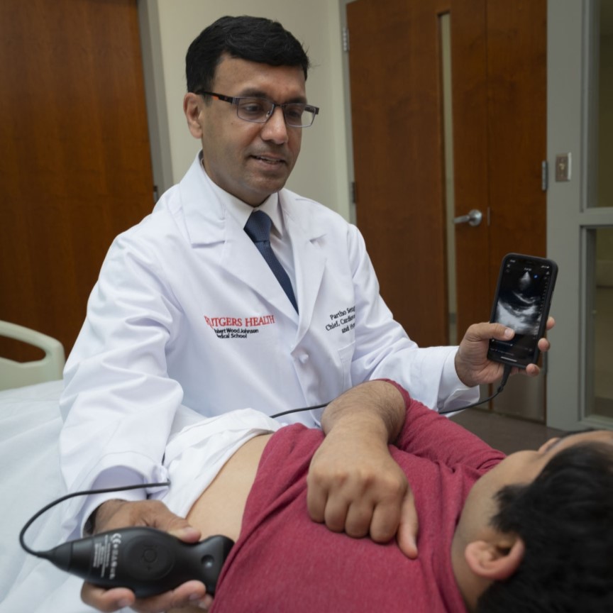
When hospitalised patients struggle to breathe, doctors typically reach for their stethoscopes, but results from a clinical study in JAMA Network Opensuggest they should diagnose the problem with portable ultrasounds instead.
The study, by Rutgers Universitty and RWJBarnabas Health, found initial exams with portable ultrasounds led to better diagnoses, shorter hospital stays and big cost savings. However, the findings revealed a need for additional training and workflow integration to help clinicians transition from traditional tools to this promising new technology.
“The study clearly shows that ultrasound is the superior diagnostic technology, even for long-time stethoscope users who get a few hours of ultrasound training,” said senior study author Partho Sengupta, Henry Rutgers professor and chief of cardiology at Rutgers Robert Wood Johnson Medical School (RWJMS), chief of cardiology at Robert Wood Johnson University Hospital (RWJUH) and member of the RWJBarnabas Health Medical Group.
The explanation here is simple. Ultrasound gives you more information, and more concrete information, about what’s going on
Partho Sengupta, Henry Rutgers professor
The study enrolled 208 patients admitted with shortness of breath to Robert Wood Johnson University Hospital in New Brunswick. About half of them underwent diagnosis via point-of-care ultrasound devices that attach to smartphones. The rest underwent diagnosis via existing standards of care.
Initial diagnosis with ultrasound trimmed a patient’s average length of hospital stay from 11.9 days to 8.3 days. In all, initial diagnosis with ultrasound saved 246 bed-days and about $751 000 in direct costs across the cohort, while 30-day readmissions were similar between groups.
“The explanation here is simple. Ultrasound gives you more information, and more concrete information, about what’s going on,” Sengupta said. “When clinicians can see fluid in the lungs, a failing heart or a stiff inferior vena cava in minutes, they can target therapy sooner or rule out a cardiopulmonary cause and look elsewhere.”
To keep things simple and encourage buy-in, scans focused on a handful of cardiac views and a six-zone lung sweep. The exam was designed to be quick and binary: signs of congestion or not, systolic function reduced or not.
The study provided several hours of ultrasound training to participating hospitalists (doctors who oversee and coordinate hospital patient care) so that each could perform and interpret the ultrasound exam in 10 to 15 minutes.
Nevertheless, most hospitalists who underwent the training let sonographers perform the exams and cardiologists interpret them. Only 20% of patients received an ultrasound diagnosis from one of the hospitalists.
Sengupta said time pressure on rounds and a lack of incentives make it hard to add a 10-minute procedure, even if it changes care.
“This is consistent with what we observe in day-to-day clinical practice,” Sengupta said. “Although the ultrasound probe fits in your pocket and attaches to the back of a smartphone, its use in clinical settings remains inconsistent. This study overcame those barriers by leveraging a multidisciplinary framework.”
Kameswari Maganti, professor of cardiology at RWJMS and section chief for non-invasive cardiology at RWJUH, led the image interpretation with the sonography team that worked closely with the RWJUH hospitalist team, led by RWJMS faculty Catherine Chen and Payal Parikh, as well as the engineering and data science team, headed by Naveena Yanamala.
“This coordinated team effort was key to developing and delivering a streamlined protocol that significantly reduced hospital stays and healthcare costs,” Sengupta said.
The researchers reported that ultrasound findings altered medical decisions in roughly a third of cases, including new diagnoses and changes in therapy. They also noted that longer-stay patients appeared to benefit the most, a hint that ultrasound-guided triage and treatment may pay particular dividends when cases are complex.
As with most single-center implementations, caveats apply. The model relied on trained sonographers and cardiology reads, which may not be available everywhere. Broader studies across multiple hospitals will be needed to confirm the cost and length-of-stay benefits, and to test strategies that make adoption stick.
Still, the argument for seeing more and guessing less is gaining ground. A bedside view of the heart and lungs, delivered early in admission, may help the right treatments arrive sooner.
Illustration of new ultrasound device. Credit: Morgan Roberts.
Scientists have long been looking for a way to modulate brain function, which could improve our understanding of how the brain works and help to treat neurological diseases, using non-invasive methods that don’t involve surgery.
One technology that could help is transcranial ultrasound stimulation (TUS), which was recently discovered to be able to modulate the activity of neurons (the brain’s key communication cells) by delivering gentle mechanical pulses that influence how these cells send signals.
But to date current systems have struggled to reach deeper areas of the brain with sufficient precision to target specific brain structures. Conventional TUS systems often affect broader regions than intended, limiting their utility for targeted neuromodulation.
The study, published in Nature Communications, introduces a new ultrasound device capable of influencing deep brain regions without surgery for the first time, targeting areas around 1000 times smaller than conventional ultrasound devices can pinpoint and 30 times smaller than previous deep brain ultrasound devices.
The new technology features 256 elements configured within a special helmet to send focused beams of ultrasound to specific parts of the brain in order to turn neuronal activity up or down. It also includes a soft plastic face mask which helps to target the ultrasound waves more precisely by keeping the head still.
The research team demonstrated the system’s capabilities on seven human volunteers by targeting a part of the thalamus, a small structure in the centre of the brain that helps to relay sensory and motor information, called the lateral geniculate nucleus (LGN). The LGN is involved in processing visual information.
In the first experiment, participants looked at a flashing checkerboard, which sent signals to the brain through the eyes. During stimulation with the ultrasound device, a functional magnetic resonance imaging (fMRI) scan showed significantly increased activity in the participants’ visual cortex, confirming precise targeting of the LGN.
A second experiment revealed sustained decreases in visual cortex activity for at least 40 minutes after ultrasound stimulation, highlighting the system’s potential for inducing lasting changes in brain function.
Though participants did not consciously perceive any changes in what they were seeing during the experiments, the brain scans revealed significant changes in neural activity. The ultimate goal is to harness these effects to produce clinically beneficial outcomes, such as stopping hand tremors.
Professor Bradley Treeby, senior author of the study from UCL Medical Physics and Biomedical Engineering, said: “This advance opens up opportunities for both neuroscience research and clinical treatment. For the first time, scientists can non-invasively study causal relationships in deep brain circuits that were previously only accessible through surgery.
“Clinically, this new technology could transform treatment of neurological and psychiatric disorders like Parkinson’s disease, depression, and essential tremor, offering unprecedented precision in targeting specific brain circuits that play key roles in these conditions.
“The ability to precisely modulate deep brain structures without surgery represents a paradigm shift in neuroscience, offering a safe, reversible, and repeatable method for both understanding brain function and developing targeted therapies.”
In addition to its research applications, the system could pave the way for new clinical interventions. Deep brain stimulation (DBS), currently used to treat conditions like Parkinson’s disease, requires invasive surgery and carries associated risks. The new ultrasound system offers a non-invasive alternative with comparable precision, potentially allowing clinicians to test areas of the brain that could be used to treat disease before surgery or even replace surgical approaches altogether.
Recognising this clinical potential, several members of the research team have recently founded NeuroHarmonics, a UCL spinout company developing a portable, wearable version of the system. The company aims to make precise, non-invasive deep brain therapy accessible for both clinical treatment and broader therapeutic applications.
Dr Eleanor Martin, first author of the study from UCL Medical Physics and Biomedical Engineering, said: “We designed the system to be compatible with simultaneous fMRI, enabling us to monitor the effects of stimulation in real time. This opens up exciting possibilities for closed-loop neuromodulation and personalised therapies.”
The researchers emphasise that further studies are needed to fully understand the mechanisms underlying TUS-induced neuromodulation. However, the results mark a significant milestone in the development of safe, effective, and targeted brain stimulation technologies.
A surgical lesion site is highlighted in orange following MR-guided focused ultrasound treatment. Structural brain connections associated with optimal tremor response or side effects, as identified in the present study, are depicted in various colors. The background features an ultra-high resolution MRI image acquired at Massachusetts General Hospital. Image courtesy of Andreas Horn, Mass General Brigham.
Essential tremor, a common neurological movement disorder, causes uncontrollable shaking, most often in the hands, but it can also occur in the arms, legs, head, voice, or torso. Essential tremor impacts an estimated 1% of the worldwide population and around 5% of people over 60.
Investigators from Mass General Brigham identified a specific subregion of the brain’s thalamus that, when included during magnetic resonance-guided focused ultrasound (MRgFUS) treatment, can result in optimal and significant tremor improvements while reducing side effects. Their results are published in Science Advances.
“This one-time, noninvasive treatment can have immediate, long-lasting and lifechanging effects for patients and was pioneered here at Brigham and Women’s Hospital 30 years ago,” said co-senior author G. Rees Cosgrove, MD, FRCSC, director of functional neurosurgery at Brigham and Women’s Hospital. “The results of this study will help make the procedure even more safe and effective than it already is and will help other centres around the world improve their outcomes.”
MRgFUS treatment of essential tremor creates a small, permanent lesion in a specific nucleus in the thalamus that is thought to be part of the brain circuit mediating the disorder and disrupts the tremor-causing activity. The research team analysed data from 351 thalamotomy patients that were treated across three international hospitals, the largest cohort assessed to date, to identify the optimal location for this procedure and better understand its impacts on clinical improvements and side effects.
The study identified a set of optimal sites and brain connections to target, as well as locations and connections to avoid that lead to side effects. The team then tested whether this ‘sweet spot’ could be used as a model to predict the outcomes in a cohort of patients treated with the same procedure at another centre, which proved true. The more the ‘sweet spot’ was lesioned, the better the outcome was in all patients’ one-year, post-procedure comparison data. According to the researchers, when thalamotomy patients have good tremor control at one year, it is typically sustained over multiple years.
“Seeing how this procedure can make such a huge impact on patients’ lives is what motivated me to pursue this research,” said lead author Melissa Chua, MD, a senior resident in the Brigham’s Department of Neurosurgery. “It is very exciting to have such robust validation and to be moving toward this treatment becoming even more precise and personalized in the future.”
Next, the team plans to further analyse patient data for a more detailed picture of the evolution of this technology and how patient outcomes have improved, to fully understand the parameters that go into achieving long-term tremor control and minimise side effects.
“It is incredible when you can provide a patient with relief from these tremors,” Cosgrove said. “It is like a gift when patients who have not been able to sing, speak in public, write, or even drink from a cup for years can once again do so – we see it in case after case.”
Pancreatic cancer. Credit: Scientific Animations CC BY-SA 4.0
Pancreatic cysts are fluid-filled sacs that can form in the pancreas. Some remain benign, while others have the potential to develop into pancreatic cancer. A recent study, which followed 257 patients in Japan for an average of five years, showed that the presence or absence of invasive nodules in pancreatic cysts is key to assessing whether these cysts are benign or cancerous.
The findings, published in the journal Annals of Surgery, may help patients diagnosed with a high risk of pancreatic cancer to avoid unnecessary surgery.
Pancreatic cancer is one of the most life-threatening and rapidly growing cancers. Pancreatic cysts, known as pancreatic intraductal papillary mucinous neoplasms (IPMNs), are gaining attention as one of the precursors of the cancer that can be identified by radiological imaging. In this context, patients diagnosed with pancreatic cysts are referred for further evaluation, and if they meet the criteria for being at particularly high risk of developing cancer, called high-risk stigmata, they are often recommended for surgery.
However, it was not clear whether all patients who met the criteria would need to undergo surgery. “In fact, among patients who underwent surgery, there were a number of cases where pathological examination results showed that their IPMNs were still benign and had not progressed to cancer,” explained Ryohei Kumano from Nagoya University, the first author of the study. “Pancreatic surgery is a significant burden for patients, so we wanted to find a more accurate way to diagnose whether their IPMNs are benign or cancerous in order to avoid unnecessary surgery.”
A research group consisting of Professor Hiroki Kawashima and Dr Kumano from Nagoya University Graduate School of Medicine, Professor Eizaburo Ohno from Fujita Health University, and their colleagues focused on the presence or absence of invasive nodules in 257 IPMN patients with high-risk stigmata. The researchers evaluated the prognosis of the patients with and without these nodules.
Invasive nodules, solid growths within cysts that have begun to invade surrounding tissues, are difficult to detect with a conventional method that uses a CAT scan. Therefore, the researchers instead used contrast-enhanced endoscopic ultrasound, which is thought to detect invasive nodules more accurately.
To track the prognosis of patients with and without invasive nodules between surgical and non-surgical groups, the researchers followed them for an average of about five years (ranging from 6 months to 24 years, depending on the patient).
The results showed that the presence or absence of invasive nodules had a significant impact on their survival. For patients with invasive nodules, undergoing surgery had a positive effect on improving their survival. On the other hand, most patients without invasive nodules had a favorable outcome even without surgery.
Endoscopic ultrasound (EUS) enables differentiation between non-invasive and invasive nodules within IPMN, providing crucial information for surgical decision making. (Credit: Ryohei Kumano)
In this study, a total of 21 patients who did not have invasive nodules opted for clinical monitoring instead of surgery. Notably, their five-year survival rates were 84.7% for overall survival and 100% for disease-specific survival.
In addition, in patients at higher risk for surgery, such as the elderly, there was little difference in survival rates between patients who underwent surgery and those who did not, if they had no invasive nodules. “Avoiding surgery, especially in such patients, seems to be a reasonable treatment strategy, given the fact that pancreatic surgery is highly invasive, carries a high risk of complications, and requires a long recovery period,” Kumano said.
“We expect that our findings will contribute to future clinical guidelines for IPMNs, leading to more accurate cancer diagnosis and optimised treatment selection.”
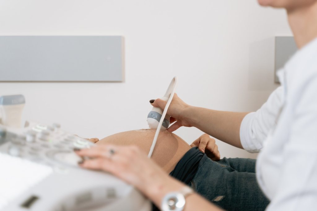
Published in Annals of Family Medicine, a University of Minnesota Medical School research team found that implementing point-of-care ultrasounds (POCUS) to assess the viability and gestational age of pregnancies in the first trimester enhanced care for pregnant patients and cut emergency visits by 81% for non-miscarrying patients.
Previously, early pregnancy care was provided through separate appointments for ultrasound, risk assessment and patient education. This new integrated approach allows patients who are under 14 weeks pregnant to receive comprehensive care during a single visit. This includes ultrasound-based pregnancy dating, immediate assessment of pregnancy viability, risk evaluation and on-site counselling – all based on real-time ultrasound results.
“Our study demonstrates that the use of point-of-care ultrasound provides meaningful benefit to the patients we serve by addressing early pregnancy problems at the time they are identified,” said Allison Newman, MD, an assistant professor at the U of M Medical School and family medicine physician at M Health Fairview Clinic. “POCUS in early pregnancy helps clinicians more efficiently and accurately diagnose problems without compromising the quality of needed first trimester assessments – saving time, money and stress for patients.”
The research team introduced this integrated approach at M Health Fairview Clinic – Bethesda in autumn 2022, allowing the clinic to quickly identify high-risk cases and offer timely intervention for issues such as miscarriage or abnormal pregnancies. They found:
The clinic saw an 81% reduction in emergency visits, urgent clinic appointments and first-trimester phone inquiries for non-miscarrying patients.
Clinic implementation led to more timely diagnosis of abnormal pregnancies and improved education and support for all patients, including those who experience miscarriage.
For miscarriage cases, the time from initial concern to diagnosis decreased from an average of 5.8 days to 1.7 days.
Suggested next steps include rolling out the process more widely within other family medicine practices and performing a wider study across multiple sites.
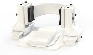
Chronic pain is often caused by faulty signals emerging deep within the brain, giving false alarms about a wound that has since healed, a limb that has since been amputated, or other intricate, hard-to-explain scenarios. Effective treatment options are sorely needed; now, a new device from the University of Utah may represent a practical long-sought solution, using ultrasound to target pain centres deep inside the brain.
Researchers at the University of Utah’s John and Marcia Price College of Engineering and Spencer Fox Eccles School of Medicine have published promising findings about an experimental therapy that has given many participants relief after a single treatment session.
At the core of this research is Diadem, a new biomedical device that uses ultrasound to noninvasively stimulate deep brain regions, potentially disrupting the faulty signals that lead to chronic pain.
The Diadem Device
The findings of a recent clinical trial are published in the journal Pain. This study constitutes a translation of two previous studies, published in Nature Communications Engineering and IEEE Transactions on Biomedical Engineering, which describe the unique features and characteristics of the device.
The randomised sham-controlled study recruited 20 participants with chronic pain, who each experienced two 40-minute sessions with Diadem, receiving either real or sham ultrasound stimulation. Patients described their pain a day and a week after their sessions, with 60% of the experimental group receiving real treatment reporting a clinical meaningful reduction in symptoms at both points.
“We were not expecting such strong and immediate effects from only one treatment,” says Riis.
“The rapid onset of the pain symptom improvements as well as their sustained nature are intriguing, and open doors for applying these noninvasive treatments to the many patients who are resistant to current treatments,” Kubanek says.
Diadem’s approach is based on neuromodulation, a therapeutic technique that seeks to directly regulate the activity of certain brain circuits. Other neuromodulation approaches are based on electric currents and magnetic fields, but those methods cannot selectively reach the brain structure investigated in the researchers’ recent trial: the anterior cingulate cortex.
After an initial functional MRI scan to map the target region, the researchers adjust Diadem’s ultrasound emitters to correct for the way the waves deflect off of the skull and other brain structures. This procedure was published in Nature Communications Engineering.
The team is now preparing for a Phase 3 clinical, trial which is the final step before FDA approval to use Diadem as a treatment for the general public.
An ultrasound test that detected 96% of ovarian cancers in postmenopausal women should replace current standard of care test in the UK according to a new study.
In a paper published in Lancet Oncology, research led by Professor Sudha Sundar from the University of Birmingham compared all currently available tests to diagnose ovarian cancer in postmenopausal women head-to-head in a high-quality diagnostic test accuracy study.
Of the six diagnostic tests investigated, the IOTA ADNEX model which looks at ultrasound features (how the lump looked like on ultrasound) had the best accuracy of all and could detect up to 96% of women with ovarian cancer.
The ultrasound test outperforms the current standard of care in the UK significantly and so the researchers recommend that the IOTA ultrasound ADNEX model should replace the current standard of care test called risk of malignancy (RMI1) test in the UK which identifies 83% of ovarian cancers.
Sudha Sundar, Professor of Gynaecological Cancer at the University of Birmingham and consultant in gynaecological cancer surgery at Sandwell and West Birmingham NHS Trust said:
“This is the first time that a head-to-head study of all available ovarian cancer tests have been done in the same population. Here we studied their use with symptomatic, postmenopausal women who are most at risk of this cancer. Our trial found that the IOTA ADNEX ultrasound protocol had highest sensitivity for detecting ovarian cancer compared to the standard of care and other test.
“The ultrasound test also performs well when delivered by a trained sonographer who have received specific training and certification and quality assurance, and as the vast majority of ultrasound scans are performed by sonographers it is important that a new standard is able to be delivered by as many clinical professionals as possible.
“We found that the higher sensitivity of the IOTA ADNEX model is likely to lead to some women who don’t have cancer also being flagged up as having a higher risk of cancer. We however did discuss this extensively with patients, cancer charity Target ovarian cancer and NHS experts who all agreed that in postmenopausal women who are at higher risk of ovarian cancer, picking up more women with cancer would benefit women overall.”
The research team note that the IOTA ADNEX model achieved 96% accuracy when delivered by NHS sonographers who were appropriately trained and received quality assurance. As most scans worldwide are carried out by sonographers rather than gynaecologists, introductory free online resources have been created by the researchers for NHS staff to undergo the specialist ultrasound training and get certification and quality assurance.