Tag: ultrasound

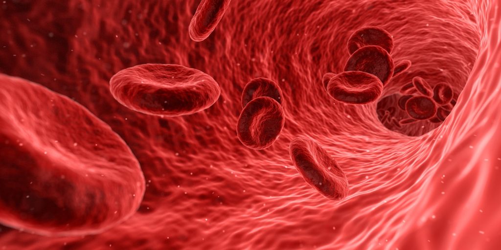
Pushing Kidney-stone Fragments Reduces Stones’ Recurrence

Kidney and ureteral stones. Credit: Scientific Animations CC4.0

Sometimes all it takes is a little push. That is the conclusion of a study, published recently in the Journal of Urology, in which doctors used a handheld ultrasound device to nudge patients’ kidney-stone fragments.

As many as 50% of patients who have kidney stones removed surgically still have small fragments remaining in the kidneys afterward. Of those patients, about 25% find themselves returning for another operation within five years to remove the now-larger fragments.

UW Medicine researchers found, however, that patients who underwent the stone-moving ultrasound procedure had a 70% lower risk of such a recurrence.

“I think the main takeaways of this study are removing fragments reduces relapse and using a noninvasive, hand-held ultrasound device to help clear these kidney stone fragments,” said UW Medicine urologist Dr Jonathan Harper, the study’s senior author.

The multisite, randomised and controlled trial was conducted from May 2015 to April 2024. Almost all of the 82 participants were from the UW Medicine or the VA Puget Sound health systems. All had stone fragments that had persisted in their kidneys for months, and their ureters were free of stones and fragments.

In the study, 40 underwent ultrasound treatment to encourage fragments to clear from the kidneys, while 42 control-group members received no such treatment.

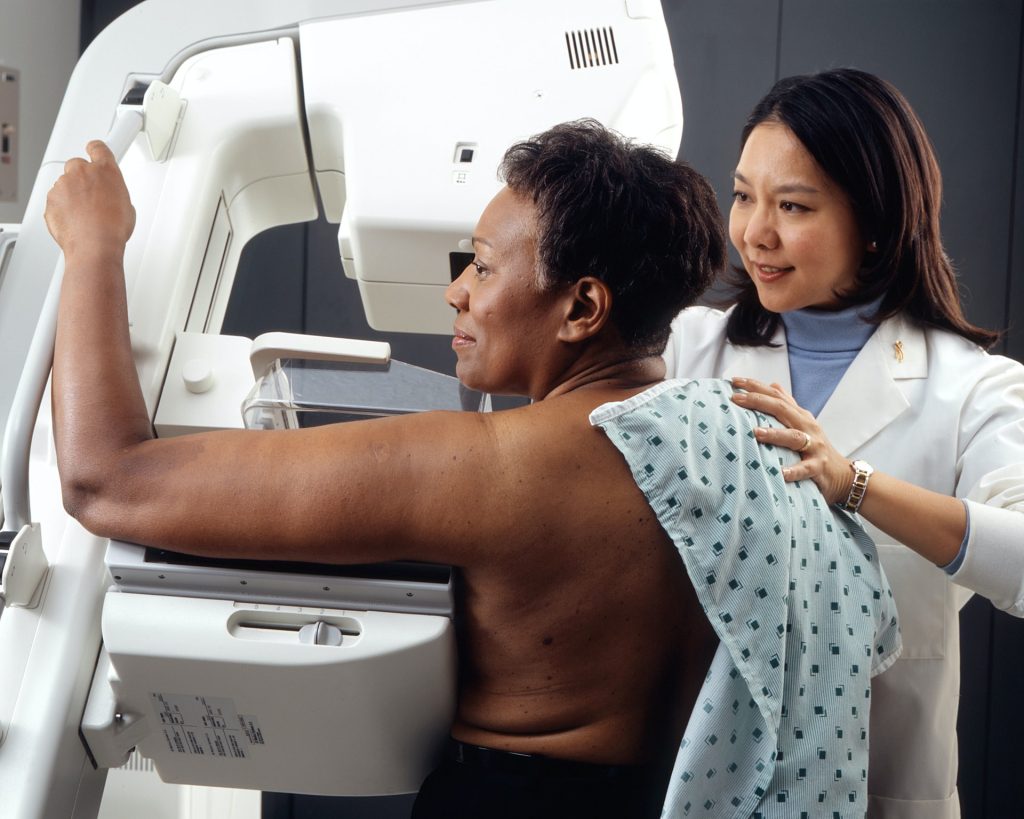
With patients awake in a clinic office setting, doctors used a wand that generated ultrasonic pulses through the skin to move the fragments closer to the ureter, where they could be naturally expelled, sometimes with the next urination, Harper noted.

Harper and his co-lead author on the paper, urologist Dr Mathew Sorensen, have worked on this technology and treatment for 15 years. They also use this technology, called burst wave lithotripsy, to blast larger stones into smaller pieces; those successes were published in 2022.

The pushing and breaking technologies are used with the same ultrasound platform.

Harper expressed hope that both clinical uses of the technology would become commonplace. A company, SonoMotion, is commercialising the technology, which was developed at the University of Washington, he added.

“I see a lot of potential in this It could become as common as getting your teeth cleaned. If you have a couple of small stones which could cause future problems, you make an office appointment and in 30 minutes you’re done.

“This could really revolutionise kidney stone treatment,” Harper said.

Source: University of Washington School of Medicine

Sonic ‘Tweezers’ can Manipulate Objects inside the Body

Photo by Pawel Czerwinski on Unsplash

In 2018, Arthur Ashkin won the Nobel Prize in Physics for inventing optical tweezers: laser beams that can be used to manipulate microscopic particles. While useful for many biological applications, optical tweezers require extremely controlled, static conditions to work properly.

“Optical tweezers work by creating a light ‘hotspot’ to trap particles, like a ball falling into a hole. But if there are other objects in the vicinity, this hole is difficult to create and move around,” says Romain Fleury, head of the Laboratory of Wave Engineering in EPFL’s School of Engineering.

Fleury and postdoctoral researchers Bakhtiyar Orazbayev and Matthieu Malléjac have spent the last four years trying to move objects in uncontrolled, dynamic environments using soundwaves. In fact, the team’s method – wave momentum shaping – is entirely indifferent to an object’s environment or even its physical properties. All the information that’s required is the object’s position, and the soundwaves do the rest.

“In our experiments, instead of trapping objects, we gently pushed them around, as you might guide a puck with a hockey stick,” Fleury explains.

The unconventional method, funded by the Swiss National Science Foundation (SNSF) Spark program, has been published in Nature Physics in collaboration with researchers from the University of Bordeaux in France, Nazarbayev University in Kazakhstan, and the Vienna University of Technology in Austria.

Very simple, very promising

If soundwaves are the hockey stick in Fleury’s analogy, then a floating object like a ping-pong ball is the puck. In the lab’s experiments, the ball was floating on the surface of a large tank of water, and its position was captured by an overhead camera. Audible soundwaves emitted from a speaker array at either end of the tank directed the ball along a pre-determined path, while a second array of microphones ‘listened’ to the feedback, called a scattering matrix, as it bounced off of the moving ball. This scattering matrix, combined with the camera’s positional data, allowed the researchers to calculate in real time the optimal momentum of the soundwaves as they nudged the ball along its path.

“The method is rooted in momentum conservation, which makes it extremely simple and general, and that’s why it’s so promising,” Fleury says.

He adds that wave momentum shaping is inspired by the optical technique of wavefront shaping, which is used to focus scattered light, but this is the first application of the concept to moving an object. What’s more, the team’s method is not limited to moving spherical objects along a path: they also used it to control rotations, and to move more complex floaters like an origami lotus.

Mimicking conditions inside the body

Once the scientists succeeded in guiding a ping-pong ball, they performed additional experiments with both stationary and moving obstacles designed to add inhomogeneity to the system. Successfully navigating the ball around these scattering objects demonstrated that wave momentum shaping could perform well even in dynamic, uncontrolled environments like a human body. Fleury adds that sound is a particularly promising tool for biomedical applications, as it is harmless and noninvasive.

“Some drug delivery methods already use soundwaves to release encapsulated drugs, so this technique is especially attractive for pushing a drug directly toward tumour cells, for example.”

Source: Ecole Polytechnique Fédérale de Lausanne

New Ultrasound and Genetics Combination Precisely Targets Neurons in Diseased Regions

McKelvey School of Engineering researchers have developed a noninvasive technology combining a holographic acoustic device with genetic engineering that allows them to precisely target affected neurons in the brain, creating the potential to precisely modulate selected cell types in multiple diseased brain regions. (Credit: Yaoheng Yang)

Brain diseases such as Parkinson’s disease involve damage in more than one region of the brain, requiring technology that could precisely and flexibly address all affected regions simultaneously. Researchers have developed a noninvasive technology combining a holographic acoustic device with genetic engineering that allows them to precisely target affected neurons in the brain. This has the potential to precisely modulate selected cell types in multiple diseased brain regions. 

Hong Chen, associate professor of biomedical engineering and neurosurgery at Washington University in St. Louis and her team created AhSonogenetics, or Airy-beam holographic sonogenetics, a technique that uses a noninvasive wearable ultrasound device to alter genetically selected neurons in the brains of mice. Results of the proof-of-concept study were published in Proceedings of the National Academy of Sciences

AhSonogenetics brings together several of Chen’s group’s recent advances into one technology. In 2021, she and her team launched Sonogenetics, a method that uses focused ultrasound to deliver a viral construct containing ultrasound-sensitive ion channels to genetically selected neurons in the brain. They use low-intensity focused ultrasound to deliver a small burst of warmth, which opens the ion channels and activates the neurons. Chen’s team was the first to show that sonogenetics could modulate the behaviour of freely moving mice.

In 2022, she and members of her lab designed and 3D-printed a flexible and versatile tool known as an Airy beam-enabled binary acoustic metasurface that allowed them to manipulate ultrasound beams. She also is developing Sonogenetics 2.0, which combines the advantage of ultrasound and genetic engineering to modulate defined neurons noninvasively and precisely in the brains of humans and animals. AhSonogenetics brings them together as a potential method to intervene in neurodegenerative diseases. 

“By enabling precise and flexible cell-type-specific neuromodulation without invasive procedures, AhSonogenetics provides a powerful tool for investigating intact neural circuits and offers promising interventions for neurological disorders,” Chen said. 

Sonogenetics gives researchers a way to precisely control the brains, while airy-beam technology allows researchers to bend or steer the sound waves to generate arbitrary beam patterns inside the brain with a high spatial resolution. Yaoheng (Mack) Yang, a postdoctoral research associate who earned a doctorate in biomedical engineering from McKelvey Engineering in 2022, said the technology gives the researchers three unique advantages.

“Airy beam is the technology that can give us precise targeting of a smaller region than conventional technology, the flexibility to steer to the targeted brain regions, and to target multiple brain regions simultaneously,” Yang said.

Chen and her team, including first authors Zhongtao Hu, a former postdoctoral research associate, and Yang, designed each Airy-beam metasurface individually as the foundation for wearable ultrasound devices that were tailored for different applications and for precise locations in the brain.

Chen’s team tested the technique on a mouse model of Parkinson’s disease. With AhSonogenetics, they were able to stimulate two brain regions simultaneously in a single mouse, eliminating the need for multiple implants or interventions. This stimulation alleviated Parkinson’s-related motor deficits in the mouse model, including slow movements, difficulty walking and freezing behaviours.

The team’s Airy-beam device overcomes some of the limits of sonogenetics, including tailoring the design of the device to target specific brain locations, as well as incorporating the flexibility to adjust target locations in a single brain.

Hu said the device, which costs roughly $50 to make, can be tailored in size to fit various brain sizes, expanding its potential applications. 

“This technology can be used as a research platform to speed neuroscience research because of the capability to flexibly target different brain regions,” Hu said. “The affordability and ease of fabrication lower the barriers to the widespread adoption of our proposed devices by the research community for neuromodulation applications.”

Source: Washington University in St. Louis

Spinal Surgeons can Now Monitor their Procedure’s Effects Mid-surgery

Photo by Natanael Melchor on Unsplash

With technology developed at UC Riverside, scientists can, for the first time, make high resolution images of the human spinal cord during surgery. The advancement could help bring real relief to millions suffering chronic back pain.

The technology, known as fUSI or functional ultrasound imaging, not only enables clinicians to see the spinal cord, but also enables them to map the cord’s response to various treatments in real time. A paper published today in the journal Neuron details how fUSI worked for six people undergoing electrical stimulation for chronic back pain treatment.

“The fUSI scanner is freely mobile across various settings and eliminates the requirement for the extensive infrastructure associated with classical neuroimaging techniques, such as functional magnetic resonance imaging (fMRI),” said Vasileios Christopoulos, assistant professor of bioengineering at UCR who helped develop the technology. “Additionally, it offers ten times the sensitivity for detecting neuroactivation compared to fMRI.”

Until now, it has been difficult to evaluate whether a back pain treatment is working since patients are under general anaesthesia, unable provide verbal feedback on their pain levels during treatment. “With ultrasound, we can monitor blood flow changes in the spinal cord induced by the electrical stimulation. This can be an indication that the treatment is working,” Christopoulos said.

The spinal cord is an “unfriendly area” for traditional imaging techniques due to significant motion artifacts, such as heart pulsation and breathing. “These movements introduce unwanted noise into the signal, making the spinal cord an unfavorable target for traditional neuroimaging techniques,” Christopoulos said.

By contrast, fUSI is less sensitive to motion artifacts, using echoes from red blood cells in the area of interest to generate a clear image. “It’s like submarine sonar, which uses sound to navigate and detect objects underwater,” Christopoulos said. “Based on the strength and speed of the echo, they can learn a lot about the objects nearby.”

Christopoulos partnered with the USC Neurorestoration Center at Keck Hospital to test the technology on six patients with chronic low back pain. These patients were already scheduled for the last-ditch pain surgery, as no other treatments, including drugs, had helped to ease their suffering. For this surgery, clinicians stimulated the spinal cord with electrodes, in the hopes that the voltage would alleviate the patient’s discomfort and improve their quality of life.

“If you bump your hand, instinctively, you rub it. Rubbing increases blood flow, stimulates sensory nerves, and sends a signal to your brain that masks the pain,” Christopoulos said. “We believe spinal cord stimulation may work the same way, but we needed a way to view the activation of the spinal cord induced by the stimulation.”

The Neuron paper details how fUSI can detect blood flow changes at unprecedented levels of less than 1mm/s. For comparison, fMRI is only able to detect changes of 2cm/s.

“We have big arteries and smaller branches, the capillaries. They are extremely thin, penetrating your brain and spinal cord, and bringing oxygen places so they can survive,” Christopoulos said. “With fUSI, we can measure these tiny but critical changes in blood flow.”

Generally, this type of surgery has a 50% success rate, which Christopoulos hopes will be dramatically increased with improved monitoring of the blood flow changes. “We needed to know how fast the blood is flowing, how strong, and how long it takes for blood flow to get back to baseline after spinal stimulation. Now, we will have these answers,” Christopoulos said.

Moving forward, the researchers are also hoping to show that fUSI can help optimise treatments for patients who have lost bladder control due to spinal cord injury or age. “We may be able to modulate the spinal cord neurons to improve bladder control,” Christopoulos said.

“With less risk of damage than older methods, fUSI will enable more effective pain treatments that are optimised for individual patients,” Christopoulos said. “It is a very exciting development.”

Source: University of California Riverside

Focused Ultrasound can Shut Down Pain Centre in Brain

Source: CC0

A new method has been developed that could non-invasively ease pain, avoiding the side effects of pain medication and the addiction problems associated with current opioid pain relievers.

This new study by Wynn Legon, assistant professor at the Fralin Biomedical Research Institute at Virginia Tech, and his team targets the insula, the location for pain reception deep within the brain. Their study, published in the journal PAIN, found that soundwaves from low-intensity focused ultrasound aimed at this spot can reduce both the perception of pain and other effects of pain, such as heart rate changes.

“This is a proof-of-principle study,” Legon said. “Can we get the focused ultrasound energy to that part of the brain, and does it do anything? Does it change the body’s reaction to a painful stimulus to reduce your perception of pain?”

Unlike ultrasound scans, focused ultrasound delivers a narrow band of sound waves to a tiny point. At high intensity, ultrasound can ablate tissue. At low-intensity, it can cause gentler, transient biological effects, such as altering nerve cell electrical activity

Neuroscientists have long studied how non-surgical techniques, such as transcranial magnetic stimulation, might be used to treat depression and other issues. Legon’s study, however, is the first to target the insula and show that focused ultrasound can reach deep into the brain to ease pain.

The study involved 23 healthy human participants. Heat was applied to the backs of their hands to induce pain. At the same time, they wore a device that delivered focused ultrasound waves to a spot in their brain guided by magnetic resonance imaging (MRI).

Participants rated their pain perception in each application on a scale of zero to nine. Participants reported an average reduction in pain of three-fourths of a point.

“That might seem like a small amount, but once you get to a full point, it verges on being clinically meaningful,” said Legon, also an assistant professor in the School of Neuroscience in Virginia Tech’s College of Science.

“It could make a significant difference in quality of life, or being able to manage chronic pain with over-the-counter medicines instead of prescription opioids.”

Researchers also monitored each participant’s heart rate and heart rate variability as a means to discern how ultrasound to the brain also affects the body’s reaction to a painful stimulus.

The study also found the ultrasound application reduced physical responses to the stress of pain – heart rate and heart rate variability, which are associated with better overall health.

“Your heart is not a metronome. The time between your heart beats is irregular, and that’s a good thing,” Legon said.

“Increasing the body’s ability to deal with and respond to pain may be an important means of reducing disease burden.”

The effect of focused ultrasound on those factors suggests a future direction for the Legon lab’s research – to explore the heart-brain axis, or how the heart and brain influence each other, and whether pain can be mitigated by reducing its cardiovascular stress effects.

Source: Virginia Tech

Low-frequency Ultrasound Improves Blood Oxygenation

Source: CC0

Research conducted by a team of scientists from Kaunas universities, Lithuania, revealed that low-frequency ultrasound influences blood parameters. The findings suggest that ultrasound’s effect on haemoglobin can improve oxygen’s transfer from the lungs to bodily tissues.

The research was undertaken on 300 blood samples collected from 42 pulmonary patients.

The samples were exposed to six different low-frequency ultrasound modes at the Institute of Mechatronics of Kaunas University of Technology (KTU). The calculations were made at the KTU Artificial Intelligence Centre.

Improved oxygen circulation and reduced blood pressure

KTU professors Vytautas Ostasevicius and Vytautas Jurenas say that the ongoing research papers are related to blood platelet aggregation.

The research of the KTU team revealed that the ultrasound affects not only platelet count but also red blood cells (RBC), which can result in better oxygen circulation and lowered blood pressure.

“During exposure to low-frequency ultrasound, aggregated RBCs are dissociated into single RBCs, whose haemoglobin molecules interact with oxygen over the entire surface area of RBCs, which is larger than that of aggregated RBCs and improves oxygen saturation in blood. The number of dissociated single RBCs per unit volume of blood decreases due to the spaces between them, compared to aggregates, which reduces blood viscosity and affects blood pressure,” explains Prof Ostasevicius, the Head of KTU Institute of Mechatronics.

The scientists claim that the effect of ultrasound on the haemoglobin in RBCs was higher than its impact on platelet aggregation, which is responsible for blood clotting.

Their findings have been supported by an additional analysis made at the LSMU Laboratory of Molecular Cardiology.

“This means that low-frequency ultrasound can be potentially used for improving oxygen saturation in lungs for pulmonary hypertension patients. Keeping in mind the recent COVID-19 pandemic, we see a huge potential in exploring the possibilities of our technology further,” says Prof Ostasevicius.

Partnership between medical and engineering scientists

In medicine, high-frequency ultrasound from 2 to 12MHz is used for both diagnostic and therapeutic purposes.

“Acoustic waves emitted by high-frequency ultrasound have a limited penetration depth into the body, so external tissues are more affected by high-frequency ultrasound than internal organs. Low-frequency ultrasound acoustic waves, penetrate deeper into the internal organs with a more uniform sound pressure distribution,” explains Prof Jurenas.

There are numerous applications for ultrasound in medical settings.

“For example, focused ultrasonic waves are used to break kidney stones, and to kill cancer cells. Maybe ultrasound can be used to activate certain medications. Or, to alleviate the delivery of antibiotics to the inflamed areas?” says Prof Jurenas.

The technology used in the above-described study is only one illustration of many successful working partnerships between engineers and physicians.

For example, just recently, the researchers of KTU Institute of Mechatronics have created the frame for immobilising the Gamma Knife radiosurgery patients at the Clinics of the Lithuanian University of Health Sciences.

“We believe, that using the know-how of different areas one can achieve greater results,” say KTU researchers about interinstitutional and interdisciplinary cooperation.

Source: Kaunas University of Technology

New Technique Enhances Clarity of Photoacoustic Imaging in Dark Skin

Photo by Nsey Benajah on Unsplash

In photoacoustic imaging, laser light is pulsed through the skin into tissues, which release ultrasound signals with which the internal structure can be imaged. This works well for people with light skin but has trouble getting clear pictures from patients with darker skin. A Johns Hopkins University-led team found a way to deliver clear pictures of internal anatomy, regardless of skin tone. Their technique is described in the journal Photoacoustics.

In experiments the new imaging technique produced significantly sharper images for all people – and excelled with darker skin tones. It produced much clearer images of arteries running through the forearms of all participants, compared to standard imaging methods where it was nearly impossible to distinguish the arteries in darker-skinned individuals.

“When you’re imaging through skin with light, it’s kind of like the elephant in the room that there are important biases and challenges for people with darker skin compared to those with lighter skin tones,” said co-senior author Muyinatu “Bisi” Bell, Associate Professor at Johns Hopkins. “Our work demonstrates that equitable imaging technology is possible.”

“We show not only there is a problem with current methods but, more importantly, what we can do to reduce this bias,” Bell said.

The findings advance a 2020 report that showed pulse oximeters, which measure oxygen rates in the blood, have higher error rates in Black patients.

“There were patients with darker skin tones who were basically being sent home to die because the sensor wasn’t calibrated toward their skin tone,” Bell said.

Bell’s team created a new algorithm to process information from photoacoustic imaging, a method that combines ultrasound and light waves to render medical images. Body tissue absorbing this light expands, producing subtle sound waves that ultrasound devices turn into images of blood vessels, tumours, and other internal structures. But in people with darker skin tones, melanin absorbs more of this light, which yields cluttered or noisy signals for ultrasound machines.

The team was able to filter the unwanted signals from images of darker skin, in the way a camera filter sharpens a blurry picture, to provide more accurate details about the location and presence of internal biological structures.

The researchers are now working to apply the new findings to breast cancer imaging, since blood vessels can accumulate in and around tumours. Bell believes the work will improve surgical navigation as well as medical diagnostics.

“We’re aiming to mitigate, and ideally eliminate, bias in imaging technologies by considering a wider diversity of people, whether it’s skin tones, breast densities, body mass indexes – these are currently outliers for standard imaging techniques,” Bell said. “Our goal is to maximise the capabilities of our imaging systems for a wider range of our patient population.”

Source: John Hopkins University

How Accurate is Supplemental Ultrasound in Breast Cancer Screening Failures?

Photo by National Cancer Institute on Unsplash

Dense breast tissue, which contains a higher proportion of fibrous tissue than fat, is a risk factor for breast cancer and also makes it more difficult to identify cancer on a mammogram. Many US states have enacted laws that require women with dense breasts to be notified after a mammogram, so that they can choose to undergo supplemental ultrasound screening to improve cancer detection. A recent study published by Wiley online in CANCER, a peer-reviewed journal of the American Cancer Society, evaluated the results of such additional screening to determine its benefits and harms to patients.

Although supplemental ultrasound screening may detect breast cancers missed by mammography, it requires additional imaging and may lead to unnecessary breast biopsies among women who do not have breast cancer. Therefore, it is important to use supplemental ultrasound only in women at high risk of mammography screening failure – in other words, women who develop breast cancer after a mammogram shows no signs of malignancy.

Brian Sprague, PhD, of the University of Vermont Cancer Center, and his colleagues evaluated 38 166 supplemental ultrasounds and 825 360 screening mammograms without supplemental ultrasounds during 2014–2020 at 32 US imaging facilities within three regional registries of the Breast Cancer Surveillance Consortium.

The team found that 95.3% of supplemental ultrasounds were performed in women with dense breasts. In comparison, 41.8% of mammograms without additional screening were performed in women with dense breasts.

Among women with dense breasts, a high risk of interval invasive breast cancer was present in 23.7% of women who underwent ultrasounds, compared with 18.5% of women who had mammograms without additional imaging.

The findings indicate that ultrasound screening was highly targeted to women with dense breasts, but only a modest proportion of these women were at high risk of mammography screening failure. A similar proportion of women who received only mammograms were at high risk of mammography screening failure.

“Among women with dense breasts, there was very little targeting of ultrasound screening to women who were at the highest risk of a mammography screening failure. Rather, women with dense breasts undergoing ultrasound screening had similar risk profiles to women undergoing mammography screening alone,” said Dr Sprague. “In other words, many women at low risk of breast cancer despite having dense breasts underwent ultrasound screening, while many other women at high risk of breast cancer underwent mammography alone with no supplemental screening.”

Clinicians can consider other breast cancer risk factors beyond breast density to identify women who may be appropriate for supplemental ultrasound screening. Publicly available risk calculators from the Breast Cancer Surveillance Consortium are available that also consider age, family history, and other factors (https://www.bcsc-research.org/tools).

Source: Wiley

Portable Ultrasound Works Just as Well in Diagnosing Forearm Fractures in Kids

Photo by cottonbro studio

Portable ultrasound devices could provide an alternative to x-ray machines for diagnosing forearm fractures in children, which could alleviate waiting times for families in hospital emergency departments (ED).

Griffith University researchers Professor Robert Ware and Senior Lecturer Peter Snelling compared functional outcomes in children given an ultrasound and those who received an x-ray on a suspected distal forearm fracture. Dr Snelling said the ultrasounds were performed by nurses, physiotherapists and emergency physicians at four south-east Queensland hospitals.

“They treated 270 children, aged between five and 15 years, during the randomised trial, which included a check-up 28 days later and another check-in at eight weeks,” Dr Snelling said. “The findings show the majority of children had similar recoveries and returned to full physical function.”

Less than one-third of children who were given an ultrasound needed a follow-up x-ray and care at an orthopaedic clinic. Those who didn’t have a buckle fracture or fractured arm were discharged from hospital without the need for further imaging.

Professor Ware said children who had an ultrasound initially had fewer x-rays, and shorter stays in the ED. “Families were also more satisfied with the treatment they received,” he said. “The results are promising and have wider implications beyond in hospital diagnosis and follow up care.

“By using a bedside ultrasound, this frees up the x-ray machine for patients who really need it and can potentially be a cost-cutting measure for hospitals as they reduce the number of x-rays without comprising the safety of patients.

“It also would be extremely beneficial in rural or remote areas eliminating the need for children and their families to travel to a larger hospital for an x-ray.”

Source: Griffith University

Measuring Tissue Stiffness with Ultrasound Yields Sharper Images

Researchers have developed a new ultrasound method that for the first time can measure the level of tension in human tissue – a key indicator of disease. The breakthrough, published in the journal Science Advances, could be used to build new ultrasound machines that are able to better discriminate between abnormal tissue, scarring, and cancer.

Images produced by the current techniques ultrasound used in healthcare aren’t usually enough to diagnose whether tissues are abnormal. To improve diagnosis, the researchers developed a way to measure forces such as tension by using an ultrasound machine. Tension is generated in all living tissue, so measuring it can indicate whether tissue is functioning properly or if it’s affected by disease.

The researchers harnessed a technique from a rail project at the University of Sheffield, which uses sound waves to measure tension along railway lines. The technique, used both for rail and medical ultrasound, relies on a simple principle: the greater the tension, the faster sound waves propagate. Using this principle, the researchers developed a method that sends two sound waves in different directions. The tension is then related to the speed of the waves by using mathematical theories developed by the researchers.

Previous ultrasound methods have struggled to show the difference between stiff tissue or tissue under tension. The developed technique is the first capable of measuring tension for any type of soft tissue, and without knowing anything about it. In this new paper, the researchers describe the new method and demonstrate how they used it to measure tension inside a muscle.

Study leader Dr Artur Gower, Lecturer in Dynamics at the University of Sheffield, said: “When you go to the hospital, a doctor might use an ultrasound device to create an image of an organ, such as your liver, or another part of your body, such as the gut, to help them explore what the cause of a problem might be. One of the limitations of ultrasounds used in healthcare now is that the image alone is not enough to diagnose whether any of your tissues are abnormal.

“What we’ve done in our research is develop a new way of using ultrasound to measure the level of tension in tissue. This level of detail can tell us whether tissues are abnormal or if they are affected by scarring or disease. This technique is the first time that ultrasound can be used to measure forces inside tissue, and it could now be used to build new ultrasound machines capable of diagnosing abnormal tissue and disease earlier.”

Source: University of Sheffield