Mechanoreceptors Set off Cascade of Damage Milliseconds After Spinal Trauma

Mechanoreceptors are present in the spinal cord from birth, are sensitive to mechanical stimuli, and play an important role in triggering the pathological events that follow trauma. What happens if they are blocked? The extent of the damage decreases. This is the finding of a new study published in The Journal of Physiology and conducted by a team at Scuola Internazionale Superiore di Studi Avanzati (SISSA), led by Professor Giuliano Taccola, with Atiyeh Mohammadshirazi as first author.
Everything happens within the very first milliseconds after the trauma, the scientists explained. It is during this brief time window that these spinal mechanoreceptors become active, triggering an impairment of the electrical signals that underlies normal neural communication. This initial event sets off a cascade of neurotoxic factors known as secondary damage, which amplifies and spreads the original traumatic lesion over the following hours and days.
Understanding the role of these receptors, according to the authors, is important not only for clarifying what happens during spinal shock. As demonstrated in the experiments, when their activity is blocked, the functional damage is also reduced. For this reason, spinal mechanoreceptors may represent a potential target for strategies aimed at reducing the disabling consequences of spinal cord injury.
Physical trauma disrupts electrical signaling
“It is well known that physical trauma to the spinal cord disrupts the flow of electrical signals that underlie the functioning of our nerve fibers. This phenomenon is known as DIP (Depolarizing Injury Potential). It begins almost immediately after trauma and continues propagating the primary damage over the following weeks, progressively worsening the lesion,” explain Atiyeh Mohammadshirazi and Giuliano Taccola. “However, the origins of this phenomenon are not yet fully understood.”
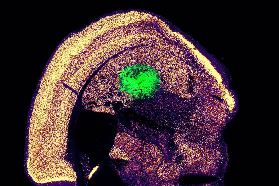
Yet, thanks to experiments carried out using highly sophisticated instrumentation invented by Professor Taccola and John Fischetti, the two scientists say, “we were able to discover something truly new.”
Mechanoreceptors and their role in the spread of damage
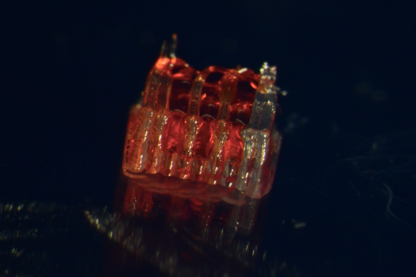
Receptors are cellular structures that respond to specific signals. Among them are mechanoreceptors, specialized proteins located on the cell membrane of sensitive cells that act as sensors for mechanical forces such as compression. Mechanoreceptors are found throughout the body, including around the spinal cord and within its central canal. In this environment, according to the SISSA research, they appear to play an important role in the propagation of injury.
The two authors explain, “In the progression of damage, depolarization precedes other well-known events such as the release of neurotoxic agents, and the inflammatory response that ultimately leads to cell death, the transient spinal hypoxia, and the rapid cell neuronal loss in the area of the primary lesion.” In this context, mechanoreceptors seem to contribute to initiating the depolarization process.
Mohammadshirazi and Taccola confirm: “When we blocked their activity in our experiments, we observed that the functional damage was significantly contained and limited.”
A possible avenue for reducing trauma-induced damage
“Our work,” conclude Giuliano Taccola and Carmen Falcone, who contributed to the histological analysis of the study, “explored what happens at the cellular level immediately after spinal trauma. As we explained, these injuries do not only involve the initial mechanical damage; they also trigger a cascade of complex neurotoxic events that amplify and worsen cellular damage and disrupt communication between neurons.”
They conclude: “With our laboratory model experiments, we demonstrated that blocking mechanosensitive receptors can effectively reduce the immediate pathological effects of spinal trauma. Our research is basic research, of course, and practical applications are still far away. Nevertheless, it may open a promising path to explore in the future to reduce spinal shock and the damage that follows trauma.”