Tag: melanoma

Among Black Men, Study Finds Increased Mortality from Melanoma Diagnoses

Photo by Nsey Benajah on Unsplash

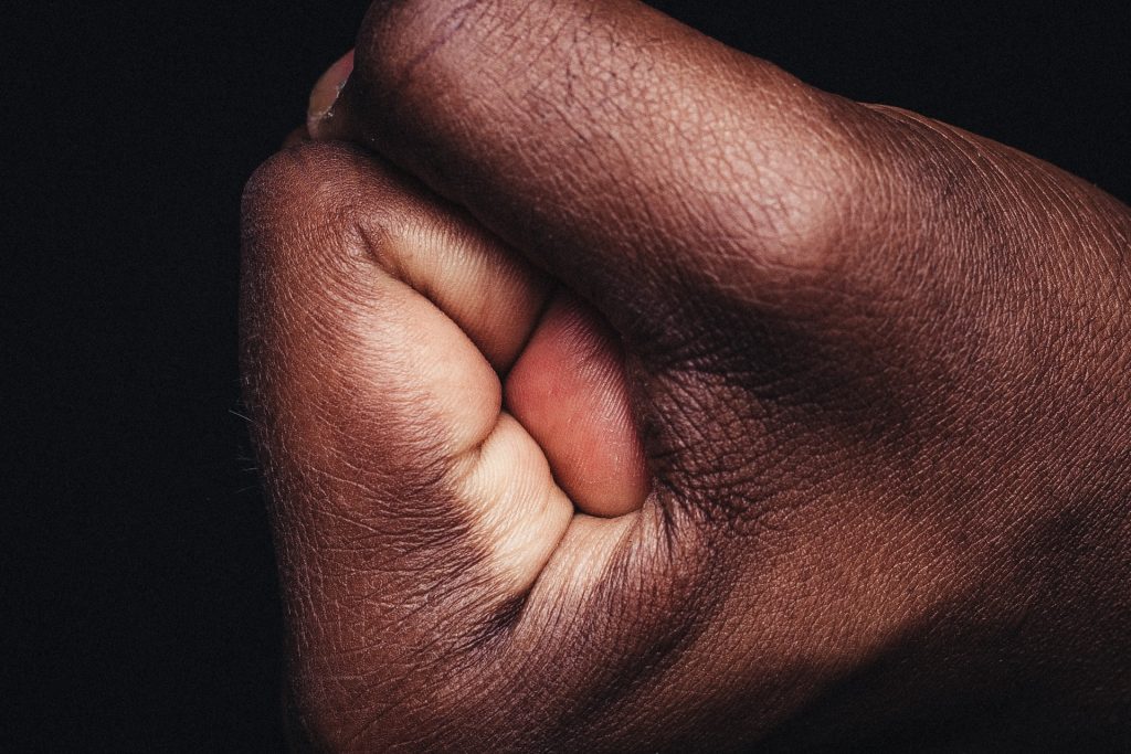
Melanoma is often detected later in people with darker skin complexions – and the consequences can be devastating, according to the results of a Mayo Clinic study published in the Journal of Surgical Oncology.

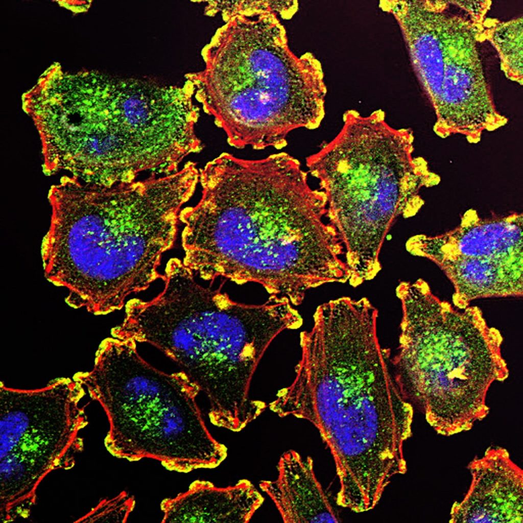
While melanoma may be found less frequently in people with darker complexions than fair ones, this aggressive form of skin cancer, accounting for 75% of all skin-cancer-related deaths, can strike anyone. The study, which consisted of 492 597 patients with melanoma, suggests that added vigilance in early screening is particularly needed for Black men, whose cancers are often found at later stages, leading to worse outcomes compared to white patients or Black women.

“We compared non-Hispanic Black patients to white patients and saw striking differences in how patients presented with the disease,” says surgical oncologist Tina Hieken, MD, senior author of the study and a researcher at Mayo Clinic Comprehensive Cancer Center. “We saw more extremity melanoma, and more later-stage disease.”

Extremity melanoma refers to skin cancer that can develop on the arms, legs, hands and feet. Various factors, including social risk factors and biological components, could be at play, but further research is needed to help determine why these differences exist.

Revealing differences in sex-based immune response

The research found that Black female patients with melanoma fared better than Black male patients. Men tended to be older at diagnosis and more likely to have cancer that had spread to their lymph nodes compared to women. This translated to worse survival rates: the five-year survival for Black men with stage 3 melanoma was only 42% chance, compared to 71% for Black women.

Most research on melanoma hasn’t focused on how race and sex affect outcomes and hasn’t looked at the influence of race and ethnicity across all groups. Dr Hieken says the study highlights the need to understand these differences better, noting that this is the first large study to confirm that sex-based differences in melanoma outcomes exist within the non-Hispanic Black population.

“When we talk about later-stage melanoma patients who are female versus male in that non-Hispanic Black patient cohort who ended up doing worse, some biological things may be going on here that are interesting,” says Dr Hieken.

One theory centres on variations in immune response.

“Several immune signals suggest that women may respond better to some immunotherapies than males,” says Dr Hieken.

Researchers note that more studies focused on melanoma in a broader range of people, including more Black participants in clinical trials, is key to bridging this knowledge gap and potentially identifying more effective treatments.

Healthcare professionals should screen carefully

Dr Hieken notes that this study is a wake-up call for everyone battling to diagnose and cure melanoma, regardless of the patient’s sex or skin tone.

She emphasises that healthcare professionals should carefully examine areas like palms, soles and under fingernails, where melanoma might be more challenging to spot on darker skin.

“We can incorporate screening for skin lesions or lesions under the nails into the visit for patients as part of their regular checkups,” says Dr Hieken. “What we want to do is elevate care for our patients.”

Source: Mayo Clinic

AI-based App can Help Physicians Diagnose Melanomas

3D structure of a melanoma cell derived by ion abrasion scanning electron microscopy. Credit: Sriram Subramaniam/ National Cancer Institute

A mobile app that uses artificial intelligence, AI, to analyse images of suspected skin lesions can diagnose melanoma with very high precision. This is shown in a study led from Linköping University in Sweden where the app has been tested in primary care. The results have been published in the British Journal of Dermatology.

“Our study is the first in the world to test an AI-based mobile app for melanoma in primary care in this way. A great many studies have been done on previously collected images of skin lesions and those studies relatively agree that AI is good at distinguishing dangerous from harmless ones. We were quite surprised by the fact that no one had done a study on primary care patients,” says Magnus Falk, senior associate professor at the Department of Health, Medicine and Caring Sciences at Linköping University, specialist in general practice at Region Östergötland, who led the current study.

Melanoma can be difficult to differentiate from other skin changes, even for experienced physicians. However, it is important to detect melanoma as early as possible, as it is a serious type of skin cancer.

There is currently no established AI-based support for assessing skin lesions in Swedish healthcare.

“Primary care physicians encounter many skin lesions every day and with limited resources need to make decisions about treatment in cases of suspected skin melanoma. This often results in an abundance of referrals to specialists or the removal of skin lesions, which in the majority of cases turn out to be harmless. We wanted to see if the AI support tool in the app could perform better than primary care physicians when it comes to identifying pigmented skin lesions as dangerous or not, in comparison with the final diagnosis,” says Panos Papachristou, researcher affiliated with Karolinska Institutet and specialist in general practice, main author of the study and co-founder of the company that developed the app.

And the results are promising.

“First of all, the app missed no melanoma. This disease is so dangerous that it’s essential not to miss it. But it’s almost equally important that the AI decision support tool could acquit many suspected skin lesions and determine that they were harmless,” says Magnus Falk.

In the study, primary care physicians followed the usual procedure for diagnosing suspected skin tumours. If the physicians suspected melanoma, they either referred the patient to a dermatologist for diagnosis, or the skin lesion was cut away for tissue analysis and diagnosis.

Only after the physician decided how to handle the suspected melanoma did they use the AI-based app. This involves the physician taking a picture of the skin lesion with a mobile phone equipped with an enlargement lens called a dermatoscope. The app analyses the image and provides guidance on whether or not the skin lesion appears to be melanoma.

To find out how well the AI-based app worked as a decision support tool, the researchers compared the app’s response to the diagnoses made by the regular diagnostic procedure.

Of the more than 250 skin lesions examined, physicians found 11 melanomas and 10 precursors of cancer, known as in situ melanoma. The app found all the melanomas, and missed only one precursor. In cases where the app responded that a suspected lesion was not a melanoma, including in situ melanoma, there was a 99.5% probability that this was correct.

“It seems that this method could be useful. But in this study, physicians weren’t allowed to let their decision be influenced by the app’s response, so we don’t know what happens in practice if you use an AI-based decision support tool. So even if this is a very positive result, there is uncertainty and we need to continue to evaluate the usefulness of this tool with scientific studies,” says Magnus Falk.

The researchers now plan to proceed with a large follow-up primary care study in several countries, where use of the app as an active decision support tool will be compared to not using it at all.

Source: Linköping University

Terahertz Biosensor can Accurately Detect Skin Cancer

3D structure of a melanoma cell derived by ion abrasion scanning electron microscopy. Credit: Sriram Subramaniam/ National Cancer Institute

Researchers have developed a revolutionary biosensor using terahertz (THz) waves that can detect skin cancer with exceptional sensitivity, potentially paving the way for earlier and easier diagnoses. Published in the journal IEEE Transactions on Biomedical Engineering, the study presents a significant advancement in early cancer detection, thanks to a multidisciplinary collaboration of teams from Queen Mary University of London and the University of Glasgow.

“Traditional methods for detecting skin cancer often involve expensive, time-consuming, CT, PET scans and invasive higher frequencies technologies,” explains Dr Shohreh Nourinovin, Postdoctoral Research Associate at Queen Mary’s School of Electronic Engineering and Computer Science, and the study’s first author.

“Our biosensor offers a non-invasive and highly efficient solution, leveraging the unique properties of THz waves – a type of radiation with lower energy than X-rays, thus safe for humans – to detect subtle changes in cell characteristics.”

The key innovation lies in the biosensor’s design. Featuring tiny, asymmetric resonators on a flexible substrate, it can detect subtle changes in the properties of cells.

Unlike traditional methods that rely solely on refractive index, this device analyses a combination of parameters, including resonance frequency, transmission magnitude, and a value called “Full Width at Half Maximum” (FWHM). This comprehensive approach provides a richer picture of the tissue, allowing for more accurate differentiation between healthy and cancerous cells and to measure malignancy degree of the tissue.

In tests, the biosensor successfully differentiated between normal skin cells and basal cell carcinoma (BCC) cells, even at different concentrations. This ability to detect early-stage cancer holds immense potential for improving patient outcomes.

“The implications of this study extend far beyond skin cancer detection,” says Dr Nourinovin.

“This technology could be used for early detection of various cancers and other diseases, like Alzheimer’s, with potential applications in resource-limited settings due to its portability and affordability.”

Dr Nourinovin’s research journey wasn’t without its challenges.

Initially focusing on THz spectroscopy for cancer analysis, her project was temporarily halted due to the COVID pandemic. However, this setback led her to explore the potential of THz metasurfaces, a novel approach that sparked a new chapter in her research.

Source: Queen Mary University of London

Scientists Find a Protein That Keeps Melanoma Hidden from the Immune System

3D structure of a melanoma cell derived by ion abrasion scanning electron microscopy. Credit: Sriram Subramaniam/ National Cancer Institute

New research has helped explain how melanoma evades the immune system and may guide the discovery of future therapies for the disease. The study found that a protein known to be active in immune cells is also active inside melanoma cells, helping promote tumour growth. The findings, published in the journal Science Advances, suggest that targeting this protein with new drugs may deliver a powerful double hit to melanoma tumours.

“The immune system’s control of a tumour is influenced by both internal factors within tumour cells, as well as factors from the tumour’s surroundings,” says first author Hyungsoo Kim, PhD, a research assistant professor at Sanford Burnham Prebys in the lab of senior author Ze’ev Ronai, PhD. “We found that the protein we’re studying is involved in both, which makes it an ideal target for new cancer therapies.”

“Immunotherapy is the first-line therapy for several cancers now, but the success of immunotherapy is limited because many cancers either don’t respond to it or become resistant over time,” says Kim. “An important goal remains to improve the effectiveness of immunotherapy.”

To find ways to boost immunotherapy in melanoma, the research team analysed data from patient tumours to identify genes that may coincide with patients’ responsiveness to immunotherapy. This led to the identification of a protein that helps tumours evade the immune system – called NR2F6 – which was found not only in tumour cells, but also in the surrounding noncancerous cells.

“Often we find that a protein has the opposite effect outside of tumours compared to what it does within a tumour, which is less effective for therapy,” says Kim. “In the case of NR2F6, we found that it elicits the same change in the tumour and in its surrounding tissues, pointing to a synergistic effect. This means that treatments that block this protein’s activity could be twice as effective.”

In a mouse model, the researchers then deleted the NR2F6 protein in both melanoma tumours and in the tumours’ environment. This inhibited melanoma growth more strongly, compared to when this effect occurs in either the tumour or its microenvironment alone. The cancer’s response to immunotherapy was also enhanced upon loss of NR2F6 in both tumours and their microenvironment.

“This tells us that NR2F6 helps melanoma evade the immune system, and without it, the immune system can more readily suppress tumour growth,” adds Kim.

To help advance their discovery further, the team is working with the Institute’s Conrad Prebys Center for Chemical Genomics to identify new drugs that can target NR2F6.

“Discovering drugs that can target this protein are expected to offer a new way to treat melanomas, and possibly other tumours, that would otherwise resist immunotherapy,” says Kim.

Source: Sanford Burnham Prebys

Faecal Microbiota Transplants Could Boost Melanoma Immunotherapy

3D structure of a melanoma cell derived by ion abrasion scanning electron microscopy. Credit: Sriram Subramaniam/ National Cancer Institute

In a world-first clinical trial published in the journal Nature Medicine, a multi-centre study has found faecal microbiota transplants (FMT) from healthy donors are safe and show promise in improving response to immunotherapy in patients with advanced melanoma.

While immunotherapy drugs can significantly improve survival outcomes in those with melanoma, they are only effective in 40–50% of patients. Preliminary research has suggested that the human microbiome may play a role in whether or not a patient responds.

“In this study, we aimed to improve melanoma patients’ response to immunotherapy by improving the health of their microbiome through faecal transplants,” says Dr John Lenehan, Medical Oncologist at London Health Sciences Centre’s (LHSC).

A faecal transplant involves collecting stool from a healthy donor, screening and preparing it in a lab, and transplanting it to the patient. The goal is to transplant the donor’s microbiome so that healthy bacteria will prosper in the patient’s gut.

“The connection between the microbiome, the immune system and cancer treatment is a growing field in science,” explains Dr Saman Maleki, senior investigator on the study. “This study aimed to harness microbes to improve outcomes for patients with melanoma.”

The phase I trial included 20 melanoma patients recruited from LHSC, CHUM and Jewish General Hospital. Patients were administered approximately 40 faecal transplant capsules orally during a single session, one week before they started immunotherapy treatment.

The trial found that combining faecal transplants with immunotherapy is safe for patients. The study also found 65% of patients who retained the donors’ microbiome had a clinical response to the combination treatment. Five patients experienced adverse events sometimes associated with immunotherapy and had their treatment discontinued.

“We have reached a plateau in treating melanoma with immunotherapy, but the microbiome has the potential to be a paradigm shift,” says oncologist Dr Bertrand Routy.

The study is unique due to its administration of faecal transplants (from healthy donors) in capsule form to cancer patients – a technique pioneered in London by Dr Michael Silverman.

“Our group has been doing faecal transplants for 20 years, initially finding success treating C. difficile infections. This has enabled us to refine our methods and provide an exceptionally high rate of the donor microbes surviving in the recipient’s gut with just a single dose,” says Dr Silverman. “Our data suggests at least some of the success we are seeing in melanoma patients is related to the efficacy of the capsules.”

The team has already started a larger phase II trial involving centres in Ontario and Quebec.

Source: Lawson Health Research Institute

Researchers Discover an Anti-tumour Regulator on B Cells

Melanoma Cells. Credit: National Cancer Institute

B cells are thought to play a critical role in innate and adaptive immunity, but their exact role in anti-tumour immunity remains unknown. Looking at B cells with a technique called single-cell profiling, which looks at all the genes in the cell, researchers found a protein that – when deleted – reduced tumour growth. The researchers write in Nature that this regulator could be a target for new cancer treatments.

The team, consisting of immunologists at Brigham and Women’s Hospital and dermatologists from Massachusetts General Hospital, identified a subset of B cells that expands specifically in the draining lymph node over time in mice with melanoma tumours.

They found a cell surface receptor called TIM-1 expressed on these B cells during melanoma growth. They also characterised multiple accompanying cell surface proteins that were involved in the B cell’s immune function. Interestingly, they found that deleting a molecule TIM-1, but not any of the other accompanying proteins, dramatically decreased tumour growth. The researchers concluded that TIM-1 controls B cell activation and immune response that combats cancer, including activating another type of the killer tumour-specific T cells for inhibiting tumour growth.

“The collaboration across institutions was extremely fruitful as we combined our immunology expertise at the Brigham with work at David Fisher’s MGH laboratory where seminal discoveries in skin malignancies have been made,” said lead author Lloyd Bod, PhD, of the Department of Neurology at the Brigham, who conducted this work while completing his postdoctoral fellowship at the Brigham. “The collaboration allowed us to test and demonstrate the therapeutic potential of targeting TIM-1 in melanoma models.”

Source: Mass General Brigham

Completion Lymph Node Dissection Shows no Benefit in Advanced Melanoma

Melanoma cells. Source: National Cancer Institute.

A review of data, published in Annals of Surgical Oncology, shows that completion lymph node dissection surgery for patients with stage III melanoma confers no benefit. This is especially true given that immunotherapy has shown success in treating metastases.

For years, surgery for patients with stage III melanoma, where the cancer had metastasised into lymph nodes, involved removing them along with the primary tumour. Known as completion lymph node dissection (CLND), the surgery was meant to ensure that no cancer remained after surgery.

More recently, however, cancer surgeons have discovered that CLND has the potential to cause more problems than it solves. In most cases, patients do better on immunotherapy alone than they do when their surgery involves removal of the lymph nodes, due to potential complications from lymph node surgery.

To address this, researchers reviewed their patient data to determine if immunotherapy alone resulted in better outcomes than CLND.

“In the few years prior to immunotherapy being available, some surgical trials were done asking if regional node dissection by itself improves overall survival for the patients,” said Martin McCarter, MD, a professor of surgical oncology at the University of Colorado (CU). “And the answer came back: no, it did not improve survival. That had been the standard forever, because we didn’t have other effective therapies, but once the definitive trials were done, we learned that CLND wasn’t helping, it wasn’t improving survival. Subsequent trials demonstrated that immunotherapy can improve survival in metastatic melanoma.”

For the study, the researchers looked at data on 90 patients who underwent sentinel lymph node biopsy (a procedure to determine if a skin melanoma has spread microscopically) only for stage III melanoma but did not undergo CLND. Of those patients, 56 received immunotherapy and 34 did not. Those who received immunotherapy had better rates of distant metastasis-free survival, meaning their cancer was less likely to come back.

“As treatments for melanoma have evolved, the standard of care may be evolving as well,” Prof McCarter said. “This study took a look at the patients who had a sentinel lymph node biopsy, so we knew the patient had a positive melanoma metastasis to their regional node. Those folks historically used to go on and get the completion lymph node dissection, but recently, people started to forego doing that lymph node dissection, which did not improve survival, and instead moved directly to immunotherapy, which did improve survival in other clinical trials. We proved that this is acceptable, that we’re not causing more harm to patients by doing it, and that those who do go on to get the immunotherapy seem to benefit from it.”

Forgoing CLND is part of a recent movement in cancer treatment known as de-escalation (or de-implementation) — performing only absolutely needed surgery. It’s especially important when it comes to lymph node surgery, Prof McCarter said, as in addition to the usual surgical risks, CLND has a 20% to 30% risk of permanent lymphoedema.

“If you could avoid that complication and not compromise a patient’s survival, that would be beneficial,” McCarter said. “That’s what we guessed was happening outside of definitive clinical trial evidence, and that’s what we were able to show. We know that we often overtreat patients, and this fits in that paradigm of finding ways to de-escalate unnecessary therapies, which has been done in breast cancer and other cancers as well.”

The researchers hope the findings will sway surgeons for whom CLND is still routine, despite the earlier studies showing that the additional surgery was not improving survival.

“Previous clinical trials with the use of adjuvant immunotherapy for melanoma had required a CLND,” Prof McCarter explained. “This study used real-world data from our stage III melanoma patients who were treated with immunotherapy without having a prior CLND.

“It takes years to change people’s practice patterns. I still have conversations with community surgeons who treat melanoma, asking me, ‘Should I be doing these regional node dissections?’ even though this data has been out for five to 10 years now,” Prof McCarter continued. “They’re afraid to give up what they used to do, and they’re afraid that they are doing a disservice to the patients or not giving them the best chance, when in reality, our understanding of cancer biology has evolved. We now have effective immunotherapy, which is overcoming some of the limitations of surgery while improving outcomes.”

Source:  University of Colorado

A New Understanding of How Moles Turn into Melanoma

Melanoma cells. Source: National Cancer Institute.

Moles and melanomas are both skin tumours that come from melanocytes. Moles are usually harmless, while melanomas are cancerous and often deadly without treatment. A study published in eLife Magazine explains how common moles and melanomas form and why sometimes moles turn into melanoma.

Melanocytes produce melanin that protects the skin against UV radiation. Specific changes to the DNA sequence of melanocytes, called BRAF gene mutations, are found in over 75% of moles. The same change is also found in 50% of melanomas and is common in lung, colon and other cancers. It was thought that when melanocytes only have the BRAFV600E mutation the cell stops dividing, resulting in a mole. When melanocytes have other mutations with BRAFV600E, they divide uncontrollably, turning into melanoma. This model is called ‘oncogene-induced senescence’.

“A number of studies have challenged this model in recent years,” said Robert Judson-Torres, PhD, Huntsman Cancer Institute (HCI) researcher and University of Utah (U of U) assistant professor. “These studies have provided excellent data to suggest that the oncogene-induced senescence model does not explain mole formation but what they have all lacked is an alternative explanation – which has remained elusive.”

The study team took moles and melanomas donated by patients and used transcriptomic profiling and digital holographic cytometry. Transcriptomic profiling lets researchers determine molecular differences between moles and melanomas. Digital holographic cytometry helps researchers track changes in human cells.

“We discovered a new molecular mechanism that explains how moles form, how melanomas form, and why moles sometimes become melanomas,” said Prof Judson-Torres.

The study shows that additional mutations for melanocytes to turn into melanoma are not needed, but environmental signalling can be a trigger for the cells. Melanocytes express genes in different environments, instructing them to either divide uncontrollably or stop dividing altogether.

“Origins of melanoma being dependent on environmental signals gives a new outlook in prevention and treatment,” said Prof Judson-Torres. “It also plays a role in trying to combat melanoma by preventing and targeting genetic mutations. We might also be able to combat melanoma by changing the environment.”

These findings create a foundation for researching potential melanoma biomarkers, allowing doctors to spot cancerous changes in the blood sooner. The researchers are also keen to use these data to better understand potential topical agents to reduce risk melanoma risk, delay development, or stop recurrence, and to detect melanoma early.

Source: Huntsman Cancer Institute

Old Antibiotics as New Weapons against Melanoma

Researchers may have hit upon a new weapon in the fight against melanoma: antibiotics that target a vulnerability in the ‘power plants’ of cancer cells when they try to survive cancer therapy.

“As the cancer evolves, some melanoma cells may escape the treatment and stop proliferating to ‘hide’ from the immune system. These are the cells that have the potential to form a new tumor mass at a later stage,” explains cancer researcher and RNA biologist Eleonora Leucci at KU Leuven, Belgium. “In order to survive the cancer treatment however, those inactive cells need to keep their ‘power plants’—the mitochondria—switched on at all times.” As mitochondria derive from bacteria that, over time, started living inside cells, they are very vulnerable to a specific class of antibiotics. This is what gave us the idea to use these antibiotics as anti-melanoma agents.”

The researchers implanted patient-derived tumors into mice, which were then treated with antibiotics, either as alone or in combined with existing anti-melanoma therapies. Leucci observed: “The antibiotics quickly killed many cancer cells and could thus be used to buy the precious time needed for immunotherapy to kick in. In tumors that were no longer responding to targeted therapies, the antibiotics extended the lifespan of—and in some cases even cured—the mice.”

The researchers made use of nearly antibiotics rendered nearly obsolete because of antibiotic resistance. However, this does not affect the efficacy of the treatment in this study, Leucci explained. “The cancer cells show high sensitivity to these antibiotics, so we can now look to repurpose them to treat cancer instead of bacterial infections.”

However, patients with melanoma should not try to experiment, warned Leucci. “Our findings are based on research in mice, so we don’t know how effective this treatment is in human beings. Our study mentions only one human case where a melanoma patient received antibiotics to treat a bacterial infection, and this re-sensitized a resistant melanoma lesion to standard therapy. This result is cause for optimism, but we need more research and clinical studies to examine the use of antibiotics to treat cancer patients. Together with oncologist Oliver Bechter (KU Leuven/UZ Leuven), who is a co-author of this study, we are currently exploring our options.”

Source: KU Leuven

Journal information: Roberto Vendramin et al, Activation of the integrated stress response confers vulnerability to mitoribosome-targeting antibiotics in melanoma, Journal of Experimental Medicine (2021). DOI: 10.1084/jem.20210571

High Melanoma Rates Reported in Sunny US State

A study reports that melanoma mortality among people in Utah, a sunny, high-altitude state, outpaced that of the rest of the country over 1975 to 2013.

This runs counter to the falling melanoma death rates in recent years in both Utah and the United States, a trend likely due to improved treatments such as immunotherapy. It is still the deadliest skin cancer type, with melanoma diagnosis rate in Utah the highest in the US.

Motivation for the research was recent evidence showing that for most of the 1990s and 2000s, even as melanoma mortality in the United States remained constant, incidence increased six-fold. This increasing incidence without accompanying mortality rise indicates overdiagnosis, ie false cancer diagnoses. 

In the case of melanoma, overdiagnosis may result from increased scrutiny, where increasing numbers of biopsies may find a benign lesion that would have gone undetected.

Since Utah has the highest melanoma incidence, a team from Huntsman Cancer Institute (HCI) at the University of Utah set out to evaluate the state’s melanoma diagnosis and death rate data. Biostatistician Kim Herget analysed data from the Utah Cancer Registry, a National Cancer Institute Survival, Epidemiology, and End Results (SEER) database, and the researchers found that in contrast to the rest of the country, melanoma mortality in Utah rose 0.8% per year from 1975 to 2013. Even though Utah’s melanoma death rates have been falling in recent years, they are still higher than the rest of the country.

“Although we agree that overdiagnosis of melanoma is a growing problem, the sustained increase in melanoma mortality in Utah suggests that at least some fraction of the increasing incidence is real and cannot be attributed solely to overdiagnosis,” said Doug Grossman, MD, PhD, who co-leads the HCI melanoma and skin cancer centre at HCI and serves as professor of dermatology at the University of Utah. “Our research underscores an increased risk for Utahns, and so we must remain vigilant about melanoma. For doctors, this means regular conversations with patients about their skin health and family history. For patients, this means practicing sun-safe behaviors like diligent sunscreen use, wearing sun-protective clothing, and monitoring their skin at home on a monthly basis to reduce risk of skin cancer and optimize early detection.”

Future studies should determine whether this trend in people living in Utah results from increased ultraviolet exposure in a mostly fair-skinned population living in a sunny, high-altitude climate or if it is associated with other factors such as environment or genetics. Melanomas are also found at higher rates in immune compromised patients.

Source: Medical Xpress

Journal information: Doug Grossman et al, The Rapid Rise in Cutaneous Melanoma Diagnoses N Engl J Med 2021; 384:e54 DOI: 10.1056/NEJMc2101980