Tag: B cells

Researchers Discover that Lupus is Triggered by Epstein-Barr Virus

A woman with Systemic Lupus Erythematosus. Source: Wikimedia CC0

Epstein-Barr virus (EBV) one, of humanity’s most ubiquitous infectious pathogens, is to blame for systemic lupus erythematosus Stanford Medicine investigators and their colleagues have found.

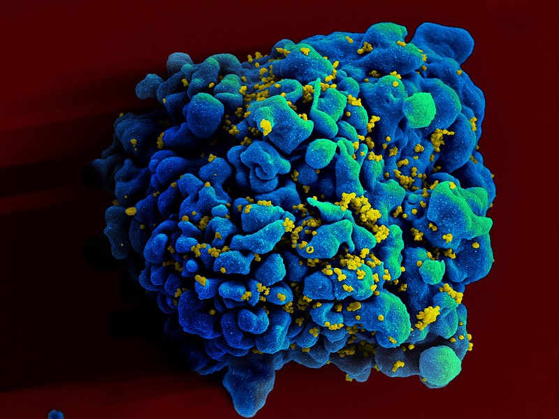
The Epstein-Barr virus (EBV), which resides silently inside 95% of the world’s population, is directly responsible for making a minuscule number of immune cells go rogue and persuade far more of their fellow immune cells to launch a widespread assault on the body’s tissues, the scientists have shown.

The findings were published in Science Translational Medicine.

“This is the single most impactful finding to emerge from my lab in my entire career,” said William Robinson, MD, PhD, a professor of immunology and rheumatology and the study’s senior author. “We think it applies to 100% of lupus cases.”

The study’s lead author is Shady Younis, PhD, professor and instructor in immunology and rheumatology.

About five million worldwide – 90% of them women – have lupus in which the immune system attacks the contents of cell nuclei. This results in damage to organs and tissues throughout the body, with symptoms varying widely among individuals.

Practically the only way to not get EBV is to live in a bubble.”

With appropriate diagnosis and medication, most lupus patients can live reasonably normal lives, but for about 5% of them the disorder can be life-threatening, said Robinson,. Existing treatments slow down disease progression but don’t cure it, he said.

The virus meets the B cell

By the time we’ve reached adulthood, the vast majority of us have been infected by EBV. Transmitted in saliva, EBV infection typically occurs in childhood, from sharing a spoon with or drinking from the same glass as a sibling or a friend, or maybe during our teen years, from exchanging a kiss. EBV can cause mononucleosis, “the kissing disease,” which begins with a fever that subsides but lapses into a profound fatigue that can persist for months.

“Practically the only way to not get EBV is to live in a bubble,” Robinson said. “If you’ve lived a normal life,” the odds are nearly 20 to 1 you’ve got it.

Once you’ve been infected by EBV you can’t get rid of it, Robinson said, even if you remain or become symptom-free. EBV belongs to a large family of viruses, including those responsible for chickenpox and herpes, that can deposit their genetic material into the nuclei of infected cells. There the virus slumbers in a latent form, hiding from the immune system’s surveillance agents. This may last as long as the cell it’s hiding in stays alive. Or, under certain conditions, the virus may reactivate and force the infected cell’s replicative machinery to produce myriad copies of themselves that break out to infect other cells and other people.

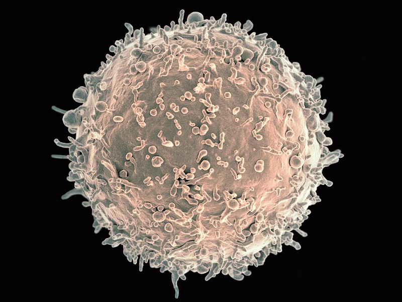
Among the cell types in which EBV takes up permanent residence are B cells, immune cells that do a couple of important things after they ingest bits of microbial pathogens. For one, they can produce antibodies: customised proteins that find and bind immune-system-arousing proteins or other molecules (immunologists call them “antigens”) on microbial pathogens that have infected an individual, or are trying to. For another, B cells are “professional antigen-presenting cells”: They can process antigens and display them on their surfaces to encourage other immune cells to raise the intensity of their hunt for the pathogen in question. That’s a substantial force multiplier for kick-starting an immune response.

Our bodies harbour hundreds of billions of B cells, which, through many cell divisions, develop an enormous diversity of antibodies. In the aggregate, these antibodies can bind an estimated 10 billion to 100 billion different antigenic shapes. This is why we’re able to mount a successful immune response to so many different pathogens.

Oddly, about 20% of the B cells in our bodies are autoreactive. They target antigens belonging to our own tissues – not by design, but due to the random way B-cell diversity comes about: through sloppy replication, apparently engineered by evolution to ensure diversification. Fortunately, these B cells are typically in a dopey state of inertia, and they pretty much leave our tissues alone.

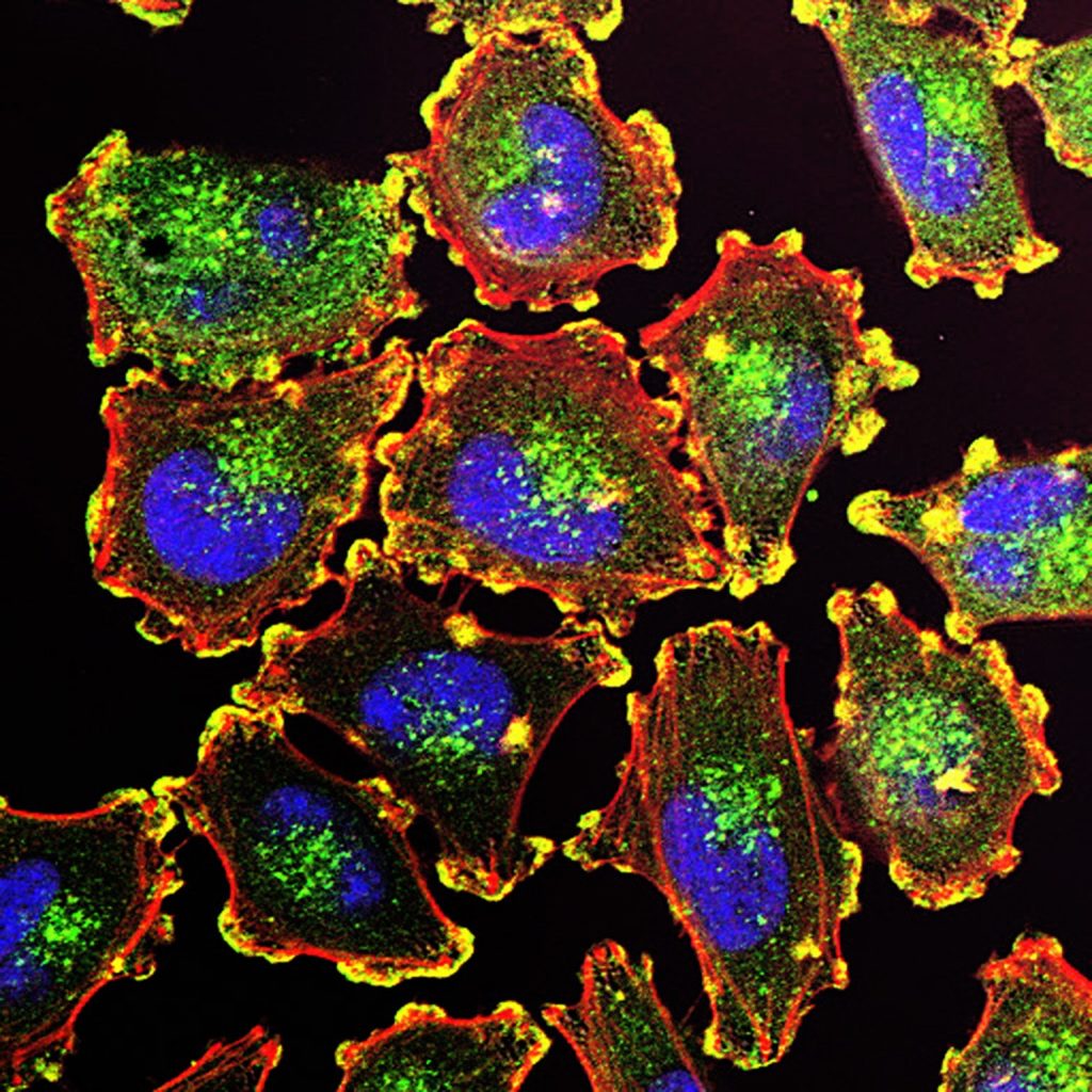
But at times, somnolent autoreactive B cells become activated, take aim at our own tissues and instigate one of the disorders collectively called autoimmunity. Some awakened autoreactive B cells crank out antibodies that bind to proteins and DNA inside the nuclei of our cells. Such activated “antinuclear antibodies” — the hallmark of lupus — trigger damage to tissues randomly distributed throughout the body, because virtually all our body’s cells have nuclei.

The vast majority of EBV-infected people (most of us, that is) have no idea they’re still sheltering a virus and never get lupus. But essentially everyone with lupus is EBV-infected, studies have shown. An EBV-lupus connection has been long suspected but never nailed down until now.

The antinuclear B cell gets ornery

Although latent EBV is ubiquitous in the sense that almost everybody carries it, it resides in only a tiny fraction of any given person’s B cells. As a result, until the new study, it was virtually impossible for existing methods to identify infected B cells and distinguish them from uninfected ones. But Robinson and his colleagues developed an extremely high-precision sequencing system that enabled them to do this. They found that fewer than 1 in 10,000 of a typical EBV-infected but otherwise healthy individual’s B cells are hosting a dormant EBV viral genome.

Employing their new EBV-infected-B-cell-identifying technology along with bioinformatics and cell-culture experimentation, the researchers found out how such small numbers of infected cells can cause a powerful immune attack on one’s own tissues. In lupus patients, the fraction of EBV-infected B cells rises to about 1 in 400 — a 25-fold difference.

It’s known that the latent EBV, despite its near-total inactivity, nonetheless occasionally nudges the B cell it’s been snoozing in to produce a single viral protein, EBNA2. The researchers showed that this protein acts as a molecular switch – a “transcription factor” – activating a battery of genes in the B cell’s genome that had previously been at rest. At least two of the human genes switched on by EBNA2 are recipes for proteins that are, themselves, transcription factors that turn on a variety of other pro-inflammatory human genes.

The net effect of all these genetic fireworks is that the B cell becomes highly inflammatory: It dons its “professional antigen-presenting cell” uniform and starts stimulating other immune cells (called helper T cells) that happen to share a predilection for targeting cell-nuclear components. These helper T cells enlist multitudes of other antinuclear B cells as well as antinuclear killer T cells, vicious attack dogs of the immune system.

When that militia bulks up, it doesn’t matter whether any of the newly recruited antinuclear B cells are EBV-infected or not. (The vast majority of them aren’t.) If there are enough of them, the result is a bout of lupus.

What comes next?

Robinson said he suspects that this cascade of EBV-generated self-targeting B-cell activation might extend beyond lupus to other autoimmune diseases such as multiple sclerosis, rheumatoid arthritis and Crohn’s disease, where hints of EBV-initiated EBNA2 activity have been observed.

The million-dollar question: If about 95% of us are walking around with latent EBV in our B cells, why do some of us, but not all of us, get autoimmunity? Robinson speculates that perhaps only certain EBV strains spur the transformation of infected B cells into antigen-presenting “driver” cells that broadly activate huge numbers of antinuclear B cells.

Many companies are working on an EBV vaccine, and clinical trials of such a vaccine are underway. But that vaccine would have to be given soon after birth, Robinson noted, as such vaccines are unable to rid an already-infected person of the virus.

Stanford University’s Office of Technology Licensing has filed a provisional patent application on intellectual property associated with the study’s findings and technologies used to obtain them. Robinson, Younis and a third study co-author, Mahesh Pandit, PhD, a postdoctoral scholar in immunology and rheumatology, are named inventors on the application. They are co-founders and stockholders of a company, EBVio Inc., a company exploring an experimental lupus treatment, ultradeep B-cell depletion. This procedure involves total annihilation of all circulating B cells, which are replaced over the following few months by new, EBV-free B cells born continually in the bone marrow. Robinson is also a director of EBVio Inc. and a co-founder and shareholder of Flatiron Bio, LLC.

Source: Stanford Medicine

Multiple Sclerosis Drug Ocrelizumab Works by Reshaping the Immune System

Myelin sheath damage. Credit: Scientific Animations CC4.0

When ocrelizumab became the first FDA-approved treatment for early forms of multiple sclerosis (MS) in 2017, it offered patients immense hope. The long-awaited drug is a monoclonal antibody that depletes B cells – the immune cells that drive MS progression. Exactly how ocrelizumab does this, however, remains unclear.

In a new study published in The Journal of Clinical Investigation, Yale scientists begin to answer this question. By using single-cell RNA sequencing, a technique that provides a window into the gene expression in individual cells, the researchers laid out a detailed view of how ocrelizumab achieves its therapeutic effects.

“The surprise was that the drug doesn’t work at all the way we thought it was working,” says David A. Hafler, MD, Professor of Neurology at Yale School of Medicine, who led the study. “We knew what the end result was and that the drug was enormously effective in patients. But what’s driving the drug’s action is a type of white blood cell in the central nervous system. No one would ever hypothesise that.”

The roles of T cells and B cells in multiple sclerosis

B and T cells have closely intertwined roles in the immune system. B cells are critical cells that recognise foreign objects, bind them, and present them to T cells, which then signal other immune cells to take action. But this relationship goes awry in disease.

Scanning electron micrograph of a B cell. Credit: NIH

In MS, abnormally active B cells trigger T cells to attack the myelin sheath, the protective layer of nerve fibres, leading to neurological symptoms, such as loss of vision, muscle weakness, and cognitive impairment. More than two decades ago, Hafler and his team discovered this was due to defects in regulatory T cells, which normally put the brake on immune responses, but when defective, unleash immune cells that mistakenly target the body’s own tissues.

In the early stages of MS, both B and T cells are deemed to be the drivers of the disease. Once the disease progresses to a neurodegenerative stage, other inflammatory processes become more prominent.

“Once you enter the neurodegenerative phase of the disease, it is much more difficult to stop the process,” Hafler says. “What we’ve learned is that the earlier you treat the disease, the better the outcome.”

Ocrelizumab binds to the surface of B cells, leading to their destruction. And especially for people in the early stages of MS, it can be quite effective. “The drug works incredibly well,” Hafler says. But Hafler and his team found that ocrelizumab was doing far more than just controlling B cells.

In the new study, the researchers analysed the blood and cerebrospinal fluid of 18 patients, all of whom had an early-onset form of multiple sclerosis in which patients cycle between periods of disease remission and relapse. The scientists measured the cell type-specific changes in protein expression before and after the patients received six months of ocrelizumab, in an effort to identify immune molecules that might change in response to the drug.

They discovered that the reduction in B cells driven by ocrelizumab led to an increase in the pro-inflammatory molecule TNF-α. This was unexpected because TNF-α has been shown to trigger the immune system and exacerbate inflammation in certain diseases. In fact, medications that block the activity of TNF-α are typically used for treating various autoimmune diseases such as rheumatoid arthritis and inflammatory bowel disease.

As they looked further, the researchers found that by inducing TNF-α, ocrelizumab led to an increase in a specific type of regulatory T cell. This, in turn, curbed the circulation of T cells that attack the myelin.

“This unpredicted increase in TNF-α shows that ocrelizumab works in a paradoxical way,” says Hafler.

Understanding the cause of multiple sclerosis

One of the current working models of MS suggests that the disease originates from the Epstein-Barr virus. “How the Epstein-Barr virus triggers the disease is a point that we don’t yet understand,” Hafler says. However, there is a strong body of evidence to show that the virus infects B cells. Therefore, understanding how a B cell-depleting drug affects T cell activity may lead to further explanations.

The current finding also explains why a fifth of the genes linked to MS risk involve the TNF pathway and why many of those genetic changes are protective in other diseases, such as inflammatory bowel diseases.

“This shows that biology has a richness to it,” Hafler says. “When these molecules are made, where they’re made, and what cell they’re working on have very different effects.”

Hafler suspects that ocrelizumab might be acting through other mechanisms as well, an inkling that motivates his lab to continue their investigation. “For something to work that well, there must be other things going on,” he says.

The team is now beginning to study the pathogenesis of MS in a large cohort of women who have at least one parent with the disease. By following the genetic evolution of the disease, the scientists are hoping to better understand how B cells change the immune landscape in real time.

“This study is only one piece of the puzzle,” Hafler says. “We’ll continue to look for other pieces.”

Source: Yale School of Medicine

Scientists Discover Immune Key for Chronic Viral Infections

Colourised scanning electron micrograph of HIV (yellow) infecting a human T9 cell (blue). Credit: NIH

Australian researchers have discovered a previously unknown rogue immune cell that can cause poor antibody responses in chronic viral infections. The finding, published in the journal, Immunity, may lead to earlier intervention and possibly prevention of some types of viral infections such as HIV or hepatitis.

One of the remaining mysteries of the human immune system is why ‘memory’ B cells often only have a weak capacity to protect us from persistent infections.

In an answer to this, researchers from the Monash University Biomedicine Discovery Institute have now discovered that chronic viral infection induces a previously unknown immune B memory cell that does not produce high levels of antibodies.

Importantly the research team, led by Professor Kim Good-Jacobson and Dr Lucy Cooper, also determined the most effective time during the immune response for therapeutics such as anti-viral and anti-cancer drugs to better boost immune memory cell development.

“What we discovered was a previously unknown cell that is produced by chronic viral infection. We also determined that early intervention with therapeutics was the most effective to stop this type of memory cell being formed, whereas late intervention could not,” Professor Good-Jacobson said.

According to Dr Cooper, chronic viral infections have been known to alter our ability to form effective long-term protective antibody responses, but how that happens is unknown.

“In the future, this research may result in new therapeutic targets, with the aim to reduce the devastating effect of chronic infectious diseases on global health, specifically those that are not currently preventable by vaccines,” she said.

“Revealing this new immune memory cell type, and what genes it expresses, allows us to determine how we can target it therapeutically and whether that will lead to better antibody responses.”

The research team are also looking to see whether this population is a feature of long COVID, which results in some people having a reduced capacity to fight off the symptoms of COVID infection long after the virus has dissipated.

Source: Monash University

‘Junk Cells’ Actually Have a Powerful Role against Malaria

Red blood cell Infected with malaria parasites. Colourised scanning electron micrograph of red blood cell infected with malaria parasites (teal). The small bumps on the infected cell show how the parasite remodels its host cell by forming protrusions called ‘knobs’ on the surface, enabling it to avoid destruction and cause inflammation. Uninfected cells (red) have smoother surfaces. Credit: NIAID

Researchers from The Australian National University (ANU) have discovered a previously unknown ability of a group of immune system cells, known as Atypical B cells (ABCs), to fight infectious diseases such as malaria.

The discovery, published in Science Immunology, provides new insight into how the immune system fights infections and brings scientists a step closer to harnessing the body’s natural defences to combat malaria.

The scientists say ABCs could also be key to developing new treatments for chronic autoimmune conditions such as lupus. According to the researchers, ABCs have long been associated with malaria, as malaria patients have more of these cells in their system compared to the general population.

“In this study, we wanted to understand the mechanisms that drive the creation of ABCs in the immune system, but also find out whether these cells are good or bad for us when it comes to fighting infection,” lead author Dr Xin Gao, from ANU, said.

“Although ABCs are known to contribute to chronic inflammatory diseases and autoimmunity, we’ve discovered a previously unknown ability of these cells to fight disease. In this sense, ABCs are like a double-edged sword.

“Contrary to past belief, ABCs are not junk cells; they are more important than we thought.

“Our research found that ABCs are also instrumental in developing T follicular helper cells. These helper cells generate powerful antibodies that help the body fight malaria parasites.

“Antibodies can block parasites in the blood as they travel from the site of the infectious mosquito bite to the liver, where the infection is first established.”

In 2022, malaria killed more than 600 000 people worldwide. Although the disease is preventable and curable, scientists face an uphill battle to find long-lasting treatments as malaria parasites continue to find new ways to build resistance to current therapies.

Using gene-editing technology on mice, the ANU researchers discovered a gene called Zeb2 is crucial to the production of ABCs.

“We found that manipulating the Zeb2 gene disrupted the creation of ABCs in the immune system,” study co-author Professor Ian Cockburn, from The ANU John Curtin School of Medical Research, said.

“Importantly, we found that mice without the Zeb2 gene were unable to control malaria infection.

“Therefore, the findings show that ABCs play a crucial role in fighting malaria infections.”

The researchers say targeting ABCs could also pave the way for new treatments for certain autoimmune diseases such as lupus.

“ABCs also appear in large numbers in many autoimmune diseases, including lupus, which can be life-threating in severe cases,” Professor Cockburn said.

“By developing a better understanding of the role of ABCs in the immune system and the cells’ role in fighting disease, it could bring us a step closer to one day developing new and more effective therapies.”

Source: Australian National University

B Cells Are Elevated in PCOS – But Are not The Cause

Phot by Sora Shimazaki on Pexels

While previous research have shown that while more B cells are present in women in polycystic ovary syndrome (PCOS), a new study by researchers at Karolinska Institutet has ruled out B cells as the cause of this common syndrome. The study is published in eLife.

Affecting 10–15% of women of fertile age, PCOS is linked to irregular ovulation and menstruation, pregnancy complications, and insulin resistance, all of which are worsened by being overweight. PCOS increases with rising body mass index, and in women with severe obesity, it is around 25%. While the root cause of the condition is unknown, a driver is a surplus of androgens.  

A recent study has shown that women with PCOS have a higher number of B cells in their blood and proposed that these contribute to the development of PCOS through the production of autoimmune antibodies.

“We’ve now examined how B cells affect the development of PCOS with the goal to find new ways of treating the conditions,” says the study’s last author Elisabet Stener-Victorin, research group leader for reproductive endocrinology and metabolism at the Department of Physiology and Pharmacology at Karolinska Institutet.

First, blood from women with PCOS was examined and showed abnormal variations in the frequency of different populations of B cells compared to healthy women. Among these were the so called double negative B cells, a heterogeneous cluster, where some have been described as poised to develop autoimmune functions.

To study whether B cells may cause PCOS, the researchers transferred antibodies from women with PCOS to mice to see if they developed the syndrome. While this proved not to be the case, they did put on weight.

Mice unaffected by B cells

The next step was to transfer B cells from PCOS-like mice (induced by continuous exposure to androgens) to mice lacking B cells to test the hypothesis that B cells drive the development of the disease. However, the recipient mice were unaffected by this transfer.

To see if B cells play an essential role in the development of PCOS, mice lacking B cells were exposed to androgens. These mice were not protected as expected, but developed the same characteristics as normal mice acquire when exposed to androgens.

Finally, androgen exposed PCOS-like mice displayed altered B cell frequencies, as women with PCOS, and simultaneous treatment with a drug that blocks androgen receptors, prevents these alterations of B cells in both blood and tissues, such as ovaries and endometrium.

The researchers conclude that androgens are necessary for the condition to form, but not B cells, the role of which remains unclear.

“B cells are clearly affected in the syndrome, which could contribute to a higher susceptibility to some comorbidities, but they don’t cause PCOS,” says Sara Torstensson, PhD student at Institutet and shared first author.

“We are now studying how other immune cells are affected and how this influences reproductive and metabolic function in PCOS,” says Angelo Ascani, guest PhD student at Graz University, Austria and also first author.

Source: Karolinska Institutet

Researchers Discover an Anti-tumour Regulator on B Cells

Melanoma Cells. Credit: National Cancer Institute

B cells are thought to play a critical role in innate and adaptive immunity, but their exact role in anti-tumour immunity remains unknown. Looking at B cells with a technique called single-cell profiling, which looks at all the genes in the cell, researchers found a protein that – when deleted – reduced tumour growth. The researchers write in Nature that this regulator could be a target for new cancer treatments.

The team, consisting of immunologists at Brigham and Women’s Hospital and dermatologists from Massachusetts General Hospital, identified a subset of B cells that expands specifically in the draining lymph node over time in mice with melanoma tumours.

They found a cell surface receptor called TIM-1 expressed on these B cells during melanoma growth. They also characterised multiple accompanying cell surface proteins that were involved in the B cell’s immune function. Interestingly, they found that deleting a molecule TIM-1, but not any of the other accompanying proteins, dramatically decreased tumour growth. The researchers concluded that TIM-1 controls B cell activation and immune response that combats cancer, including activating another type of the killer tumour-specific T cells for inhibiting tumour growth.

“The collaboration across institutions was extremely fruitful as we combined our immunology expertise at the Brigham with work at David Fisher’s MGH laboratory where seminal discoveries in skin malignancies have been made,” said lead author Lloyd Bod, PhD, of the Department of Neurology at the Brigham, who conducted this work while completing his postdoctoral fellowship at the Brigham. “The collaboration allowed us to test and demonstrate the therapeutic potential of targeting TIM-1 in melanoma models.”

Source: Mass General Brigham

Pseudomonas Aeruginosa Locks out Immune Cells

Pseudomonas
Scanning Electron Micrograph of Pseudomonas aeruginosa. Credit: CDC/Janice Carr

Pseudomonas aeruginosa bacteria are a common menace in hospital wards, causing life-threatening infections, and are often resistant to antibiotics. Researchers have discovered a mechanism that likely contributes to the severity of P. aeruginosa infections, which could also be a target for future treatments. The results were recently appeared in the journal EMBO Reports.

Many bacterial species use sugar-binding molecules called lectins to attach to and invade host cells. Lectins can also influence the immune response to bacterial infections. However, these functions have hardly been researched so far. A research consortium led by Prof Dr Winfried Römer at the University of Freiburg and Prof Dr Christopher G. Mueller at the CNRS/University of Strasbourg has investigated the effect of the lectin LecB from P. aeruginosa on the immune system. It found that isolated LecB can render immune cells ineffective: The cells are then no longer able to migrate through the body and trigger an immune response. The administration of a substance directed against LecB prevented this effect and led to the immune cells being able to move unhindered again.

LecB blockades immune cells

As soon as they perceive an infection, cells of the innate immune system migrate to a nearby lymph node, where they activate T and B cells, triggering a targeted immune response. LecB, according to the current study, prevents this migration. “We assume that LecB not only acts on the immune cells themselves in this process, but also has an unexpected effect on the cells lining the inside of the blood and lymph vessels,” Römer explains. “When LecB binds to these cells, it triggers extensive changes in them.” Indeed, the researchers observed that important structural molecules were relocated to the interior of the cells and degraded. At the same time, the cell skeleton became more rigid. “The cell layer thus becomes an impenetrable barrier for the immune cells,” Römer said.

An effective agent against LecB

Can this effect be prevented? To find out, the researchers tested a specific LecB inhibitor that resembles the sugar building blocks to which LecB otherwise binds. “The inhibitor prevented the changes in the cells, and T-cell activation was possible again,” Mueller said. The inhibitor was developed by Prof Dr Alexander Titz, who conducts research at the Helmholtz Institute for Pharmaceutical Research Saarland and Saarland University.

Further studies are needed to determine how clinically relevant the inhibition of the immune system by LecB is to the spread of P. aeruginosa infection and whether the LecB inhibitor has potential for therapeutic application. “The current results provide further evidence that lectins are a useful target for the development of new therapies, especially for antibiotic-resistant pathogens such as P. aeruginosa,” the authors conclude.

Source: University of Freiburg

Complex Surface Features on B Cells may Hold Immune Secrets

Image credit: Albert Ludwigs University of Freiburg

Using new microscopic methods in combination with machine learning-based image analysis, researchers from Albert Ludwigs University of Freiburg have discovered new structures on the surface of living B cells that affect the distribution and possibly the function of their antigen receptors. The researchers’ study has been published in The EMBO Journal.

B cells recognise pathogens through specialised receptors on their surface. For the first time, scientists were able to observe how these receptors are distributed on the surface of living and moving cells. They found that the B cell surface is shaped into a characteristic landscape of interconnected ridges and protrusions. On this landscape, the IgM-class B cell antigen receptors (IgM-BCR) accumulate in specific areas. The precision of the receptors’ localisation and their clustering into larger units likely constitute a mechanism that controls receptor signalling and facilitates antigen sensing and thereby the activation of B cells.

In most immunological textbooks, lymphocytes are depicted as round, ball-like cells whose smooth surface carries randomly distributed receptors. The notion of a smooth unstructured B cell surface has already been challenged by electron micrographs of fixed and frozen lymphocytes, revealing thin membrane protrusions called microvilli on the cells’ surface. These tentacle-like structures help immune cells to search for molecular markers of pathogens, so-called antigens. B lymphocytes recognise such antigens through different classes of their B cell antigen receptors (BCR). These antigen receptors are complex molecular machines that, when activated, interact with other molecules to initiate a signalling cascade, leading to the differentiation of B cells into plasma cells and the production of protective antibodies.

The research group of Prof. Dr. Michael Reth collaborated with other imaging specialists to analyse how the IgM-BCR is distributed across the 3D surface of living B cells. For this, they used a technique called lattice light sheet microscopy, LLSM for short. “This method can capture volumetric images of living cells at a very high speed,” explains Dr. Deniz Saltukoglu from Freiburg University, the first author of the study. “In other types of high-resolution microscopy, cells need to be attached to a flat surface, which completely alters the B cells’ outer structures. LLSM allowed us to observe the cells in an environment that mimics biological tissues, meaning that the structures and movements that we saw were largely undisturbed,” she says.

The researchers then developed custom image analysis tools to quantify and objectively characterise the microscopic data. “We needed to segment the images and isolate morphological features,” says Saltukoglu. “So far this had only been done with two-dimensional data, so we had to develop new computational tools for volumetric, time course data.” For this, the researchers drew inspiration from algorithms that are used to map geographical data for archaeological surveys. With this approach, they found that the B cell surface carries a network of elevated ridges, with microvilli growing from the intersections of the network. Within this “cellular landscape”, the IgM-BCRs form clusters that concentrate along the ridges, in close proximity to the bases of the microvilli. The position of these clusters was linked with the dynamic movement of the ridges on the cells’ surface.

“We think that the 3-D location of the antigen receptors controls their activity,” says Reth. “Localisation at the microvilli base may prevent their unwanted activation. Once B cells receive a danger signal, they extent their microvilli and we assume that the IgM-BCR clusters then get recruited to the tip where they are localized in an optimal position for antigen sensing.”

This hypothesis is in line with other findings from Reth’s group, which suggest that the IgM-BCRs are regulated via lateral interactions with regulatory coreceptors. This means that the position and distribution of antigen receptors likely represent additional control mechanisms that affect signalling and activation of cells of the immune system.

Source: Albert Ludwigs University of Freiburg

A Step Closer to a Once-off Treatment for HIV

HIV invading a human cell
HIV invading a human cell: Credit NIH

Researchers from Tel Aviv University have demonstrated success of a novel technology that may be developed into a one-time vaccine to treat people with HIV and AIDS. Using CRISPR technology, the researchers engineered B cells that in turn stimulate the immune system to produce HIV-neutralising antibodies.

Published in Nature, the study was led by Dr Adi Barzel and PhD student Alessio Nehmad and conducted in collaboration with additional researchers from Israel and the US.

“Based on this study,” said Dr Barzel, “we can expect that over the coming years we will be able to produce a medication for AIDS, additional infectious diseases and certain types of cancer caused by a virus, such as cervical cancer, head and neck cancer and more.”

He explains that the treatment can become a kind of permanent medication, lingering in the body to fight the virus. “We developed an innovative treatment that may defeat the virus with a one-time injection, with the potential of bringing about tremendous improvement in the patients’ condition. When the engineered B cells encounter the virus, the virus stimulates and encourages them to divide, so we are utilising the very cause of the disease to combat it. Furthermore, if the virus changes, the B cells will also change accordingly in order to combat it, so we have created the first medication ever that can evolve in the body and defeat viruses in the ‘arms race’.”

When they mature, the antibody-generating B cells move into the blood and lymphatic system and from there to the different body parts.

Dr Barzel explained: “Until now, only a few scientists, and we among them, had been able to engineer B cells outside of the body. In this study, we were the first to do this within body and then make those cells generate the desired antibodies. The genetic engineering is conducted with viral carriers derived from viruses that were also engineered. We did this to avoid causing any damage, and solely bring the gene coded for the antibody into the B cells in the body.”

“Additionally, in this case we have been able to accurately introduce the antibodies into a desired site in the B cell genome. All lab models that had been administered the treatment responded, and had high quantities of the desired antibody in their blood. We produced the antibody from the blood and made sure it was actually effective in neutralising the HIV virus in the lab dish.”

Source: Tel Aviv University

Immunotherapy Flop Leads to Cancer Treatment Breakthrough

Human B cell
Scanning Electron Micrograph of a human B Cell. Credit: NIH

When patients in the UK showed adverse side effects during a cancer immunotherapy trial, researchers went back through the data and worked with patient samples to see what went wrong. Published in Nature, their findings provide clues into the dangerous side effects of many immunotherapies – and point to a better strategy for treating solid tumours.

“This work shows the importance of learning from early stage clinical trials,” says La Jolla Institute for Immunology (LJI) Professor Pandurangan Vijayanand, MD, PhD, who co-led the new research with Christian H. Ottensmeier, MD, PhD, FRCP, a professor with the University of Liverpool.

“In the oncology world, immunotherapy has revolutionised the way we think about treatment,” said Prof Ottensmeier. “We can give immunotherapies to patients even with metastatic and spreading disease, and then just three years later wave goodbye and tell them their cancer is cured. This is an astounding change.”

Unfortunately, only 20–30% of solid cancer patients given immunotherapies go into long-term remission. Some people see no change after immunotherapy, but others develop serious side effects, which can be debilitating, even fatal, and these patients are forced to discontinue the immunotherapy.

The researchers worked with samples from a recent UK clinical trial for head and neck cancers. The patients were given an oral cancer immunotherapy – a PI3Kδ inhibitor. At the time, PI3Kδ inhibitors had proven effective for B cell lymphomas but had not yet been tested in solid tumours.

PI3Kδ inhibitors are a new to cancer immunotherapy, but they hold promise for their ability to inhibit ‘regulatory’ T cells (Tregs). Tregs normally try to stop other T cells, called effector T cells, from targeting the body’s own tissues. Oncologists inhibit Tregs inside tumours so effector T cells can let loose and generate cancer-killing CD8+ T cells.

“Having an oral tablet that can take off the brakes – the Tregs– can be a great asset for oncologists,” said Prof Vijayanand.

Unfortunately, 12 of the 21 patients in the trial had to discontinue treatment early because they developed inflammation in the colon, a condition called colitis. “We thought this drug wouldn’t be toxic, so why was this happening?” said Prof Vijayanand.

Simon Eschweiler, PhD, an instructor at La Jolla Insitute, led the review into exactly how PI3Kδ inhibitor treatment affected immune cells in these patients. Gene sequencing showed that in the process of increasing tumour-fighting T cells in tumours, the PI3Kδ inhibitor also blocked a specific Treg cell subset from protecting the colon. Without Tregs patrolling there, pathogenic T cells, called Th17 and Tc17 cells, moved in and caused inflammation and colitis.

It was clear that a larger than needed PI3Kδ inhibitor dose had been given, and had disrupted the immune cell balance in the gut.

The pathway that leads to the toxicity seen in the new study may be broadly applicable to other organs harbouring similar Treg cells, and to other Treg cell-targeting immunotherapies like anti-CTLA-4, Eschweiler says.

The team found that intermittent dosing could be a valid treatment strategy that combines sustained anti-tumour immunity with reduced toxicity. The researchers are now designing a human clinical trial to test the intermittent dosing strategy in humans.

Why the lack of toxicity in trials for B cell lymphomas? Dr Eschweiler noted that in previous studies, lymphoma patients had been given several prior therapies leading to an overall immunocompromised state. This means the lymphoma patients didn’t have the same type or magnitude of immune response upon PI3Kδ inhibition. Meanwhile, the head and neck cancer patients were treatment-naive. Since their immune system was uncompromised, the immune-related adverse events were more rapid and pronounced.

Overall, the new study shows the importance of studying not just personalised therapies but personalised therapy doses and schedules.

Source: La Jolla Institute