Tag: 30/6/26

Experimental Drug Offers Hope for Huntington’s Disease

The image shows two halves of mouse brains: on the left, brain of an untreated mouse; on the right, brain of a mouse treated with the drug anle138b. It is clearly visible that the treated mouse has significantly fewer aggregates of mutant huntingtin. (Image: Miguel da Silva Padilha)

An experimental drug has been shown to alleviate symptoms of Huntington’s disease and extend lifespan in mouse models. Further studies are required to determine whether these results may also apply to humans.

The hereditary disorder Huntington’s disease has so far been considered incurable. Its clinical manifestations include impaired motor control and psychiatric symptoms. A new study offers promising insights. It shows that a specific drug candidate called anle138b can significantly reduce the toxic protein clumps in the brain that are characteristic of the disease.

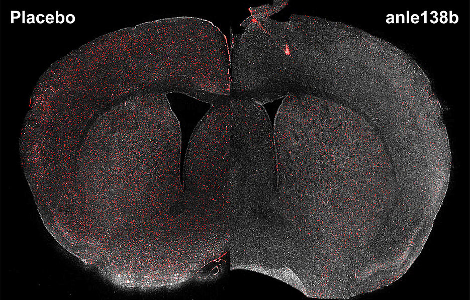
Affected mice that were administered this compound retained their mobility for a longer time, their brains shrank less, and their lifespan was extended compared to untreated mice. Importantly, the compound not only alleviates symptoms but also addresses the underlying cause of the disease by preventing disease-specific harmful protein clumps from destroying nerve cells and their connections. These results were also confirmed in experiments with human stem cells from Huntington’s patients.

Promising Therapeutic Candidate

These are the key findings of a study that has now been published in the journal EMBO Molecular Medicine. The study was led by Professor Irina Dudanova, who holds the Chair of Anatomy and Cell Biology I at the University of Würzburg since April 2026, and her doctoral student Miguel da Silva Padilha. The substance was developed by the teams of Christian Griesinger, Director at the Max Planck Institute of Multidisciplinary Sciences in Göttingen, and Armin Giese from the Ludwig-Maximilians University in Munich, now at MODAG GmbH. Other participants of the study come from the Max Planck Institute of Biological Intelligence in Martinsried and the University of Cologne.

“Our data show that specifically targeting toxic protein aggregates with the compound anle138b is a promising approach for stabilizing neuronal health in the long term,” says Irina Dudanova, commenting on the study’s findings.

Cellular waste destroys nerve cells

Background: Huntington’s disease is an inherited movement disorder caused by a defect in a specific section of DNA, the gene that encodes the protein huntingtin. According to the health insurance organization AOK, approximately 10,000 people in Germany are affected by the disease. Several hundred new cases are diagnosed each year. A faulty repetition of the genetic code (known as CAG repeats) causes the huntingtin protein to take on an abnormal shape and form clumps.

The resulting protein aggregates can be thought of as a form of cellular waste that accumulates inside neurons. The protein aggregates disrupt vital cellular communication and lead to cell death, particularly in brain regions involved in for movement and cognition. An effective therapy that targets the underlying causes is not available. This is where the compound investigated anle138b comes into play, as it prevents the formation of the harmful structures.

The researchers investigated the efficacy of anle138b in two different mouse models: While one suffered from a severe, early-onset form of the disease, the other model mirrored the genetic situation in adult patients. The compound showed beneficial effects in both models.

A characteristic feature of Huntington’s disease is the loss of the protein PDE10A, which is found almost exclusively in the nerve cells that die in this disease. The amount of PDE10A decreases dramatically long before patients show the first severe symptoms. “If PDE10A levels drop, that is a clear signal that the disease is progressing. The protein is therefore well-suited as a biomarker for Huntington’s disease,” explains Miguel da Silva Padilha. If less nerve cells die, then the PDE10A levels stay high. This is exactly what the scientists observed: as a result of anle138b treatment, the concentration of PDE10A remained high in both mouse models.

Efficacy Demonstrated in Human Stem Cells

A key milestone of the study is the confirmation of these effects in human cells. “In our experiments with induced pluripotent stem cells – that is, precursor cells derived from Huntington patients’ cells – we also observed that the addition of anle138b reduced the amount of huntingtin aggregates,” says Irina Dudanova.

Since the compound targets a fundamental mechanism of protein aggregation, it is also of interest for research on other neurodegenerative diseases. Corresponding studies in mouse models have been so promising that two years ago a large clinical trial was started for the treatment of multiple system atrophy – a Parkinson’s-like disorder characterised by severe impairments of motor function, balance, and the autonomic nervous system.


Original publication

Anle138b ameliorates pathological phenotypes in mouse and cellular models of Huntington’s disease. Miguel da Silva Padilha, Seda Koyuncu, Evangeline Chabanis, Sergey Ryazanov, Andrei Leonov, David Vilchez, Rüdiger Klein, Armin Giese, Christian Griesinger and Irina Dudanova. EMBO Molecular Medicine, DOI: 10.1038/s44321-026-00459-9

Source: Universität Würzburg

Irregular Heartbeat Risk Much Lower in Super‑fit People than Previously Thought


Our study shows that atrial fibrillation risk in very fit people is smaller than previously thought. Pexels Photo by Freestocksorg

Axel Carl Carlsson, Karolinska Institutet; Marcel Ballin, Uppsala University, and Peter Nordström, Uppsala University

Exercise is important for your heart and cardiovascular health. But for years, research has suggested that very fit people – particularly young men – have a higher risk of developing atrial fibrillation (an irregular heartbeat) later in life than less athletic people do.

But our new large-scale study of more than one million young Swedish men challenges this long-held concern about fitness and heart health. The study shows that atrial fibrillation risk in very fit people is smaller than previously thought. And, importantly, the benefits of being fit – such as having lower rates of other cardiovascular diseases, such as stroke and heart attack – clearly outweigh the potential downsides.

To conduct our study, we analysed data from over one million Swedish men. Each of these men had completed a military conscription test between 1972 and 1995 when they were around 18 years old. During conscription, fitness was estimated using a maximal ergometer bicycle test.

We followed the participants using national health registries until 2023. Information was collected on diagnoses in specialised outpatient and inpatient care, as well as information on cause of death where relevant.

Nearly half a million of these men were siblings, allowing the team to compare brothers with different fitness levels. This method also helped control for shared genetics and upbringing to better understand risk.

In the population-wide analysis, where all men were compared based on their fitness levels, the fittest men did show a higher risk of atrial fibrillation until age 40. After 45, however, the benefits – fewer other heart problems – started to outweigh the risk.

But the sibling analysis told a different story. When comparing brothers, the link between high fitness and atrial fibrillation weakened significantly. The net positive effect of fitness on overall heart and cardiovascular health, although also weakening in the sibling analysis, was still evident from the start.

These benefits grew larger as the men aged. Across five decades of follow-up – from the age of 18 until the age of 65 – the benefits of being highly fit outweighed the overall risk of developing atrial fibrillation at all ages.

An advantage of our study is that we were able to compare siblings. Photo by Andrea Piacquadio on Pexels

While the higher risk of atrial fibrillation in the fittest men weakened in the sibling analysis, an elevated risk did remain. However, this risk was small overall. The potential risk of developing atrial fibrillation was also far outweighed by the positive benefits that being fit had on reducing risk of other cardiovascular diseases.

There was also some evidence that the increased risk of atrial fibrillation seemed to be driven by typically milder forms of atrial fibrillation.

Cardiovascular benefits of fitness

These findings suggest that earlier studies may have overestimated the risk of developing atrial fibrillation if you’re very fit because they couldn’t fully account for shared familial factors – such as genes and shared environmental factors – that may influence both fitness and heart health.

Because our study was able to account for these shared factors, we were able to show that we should potentially tone down the message that being very fit or engaging in endurance exercise poses a big risk to heart health. The risk of atrial fibrillation isn’t zero, but the benefits to heart health more generally are far greater.

Our results have also been confirmed in previous studies. However, our research builds upon this prior evidence by incorporating the sibling comparisons in the analyses, showing that the atrial fibrillation risk becomes weaker.

Our study also performed a more detailed examination of risk over time by following up with participants over a longer period of time, while also considering the benefits of high fitness. As such, our research has performed a more direct comparison of benefits and harms throughout life.

Overall, our research illustrates just how beneficial physical fitness is for overall cardiovascular health. Although very fit people do have a slightly higher risk of developing atrial fibrillation compared to people who are less fit, this risk of far lower than previously thought.

Plus, we show that the overall net benefits of fitness on cardiovascular health more broadly far outweigh any potential risk.

Axel Carl Carlsson, Researcher, Department of Neurobiology, Karolinska Institutet; Marcel Ballin, Associated Researcher, Department of Public Health and Caring Sciences, Uppsala University, and Peter Nordström, Professor, Department of Public Health and Caring Sciences, Uppsala University

This article is republished from The Conversation under a Creative Commons license. Read the original article.

Young UP Researchers Develop Citrus-based Supplement to Fight Diabetes and Obesity

Dr Kadima Tshiyoyo, Marni Oberholzer and Ryan Bosch

A team of young researchers at the University of Pretoria (UP) is developing an innovative health supplement that transforms citrus peel waste into a potentially powerful tool against lifestyle diseases such as diabetes, obesity and high cholesterol, while also improving the affordability and accessibility of preventative health products for low-income communities.

The NutraPectin project is led by postdoctoral research fellow Dr Kadima Tshiyoyo, alongside recent Master of Science in Biochemistry graduates Ryan Bosch and Marni Oberholzer. Together, the team from UP’s Faculty of Natural and Agricultural Sciences (NAS) is converting citrus processing waste into a pectin-rich nutraceutical using green extraction technology. When consumed with food, pectin can help slow sugar absorption, reduce cholesterol levels and support gut health.

“South Africa’s growing burden of lifestyle diseases and abundance of agricultural waste inspired our work,” Dr Tshiyoyo said. “NutraPectin uses sustainable green methods to extract bioactive compounds from waste, which are valuable and essential in the management of lifestyle diseases such as diabetes and obesity.”

Turning science into solutions

The development of NutraPectin addresses two growing challenges: the rising prevalence of lifestyle-related diseases and the environmental cost of agricultural and food processing waste. Citrus peel, which is typically discarded in large volumes by the citrus industry, is rich in pectin and other bioactive compounds that can be recovered and repurposed.

Bosch says the NutraPectin research was his honours-degree project under Prof Samkelo Malgas of UP’s Department of Biochemistry, Genetics and Microbiology. “I was drawn to the project as I am passionate about maintaining the environment, and I was excited to investigate how food waste could be converted into useful healing products.”

For Bosch and Oberholzer, the project represents more than laboratory research. It is an opportunity to demonstrate how student-led science can be translated into solutions with tangible social and economic impact, particularly in a country where both healthcare access and food system sustainability remain pressing concerns.

“Sustainability should both underlie and overarch most research if we want to see true economic and social growth,” Oberholzer said. “South Africa has so much potential to sustainably improve and empower our agricultural industry by targeting circularity through waste valorisation. UP was foundational in this development, most markedly through our excellent supervisor’s guidance. We were allowed to explore beyond the strict boundaries of our degrees.”

The project is rooted in the Biocatalysis and Processing Research Group in the Department of Biochemistry, Genetics and Microbiology within the NAS Faculty, under the supervision of Prof Malgas. He says the work highlights the importance of university-based research in advancing both innovation and entrepreneurship, while also addressing societal challenges through science-driven solutions.

“This pectin extraction from citrus waste represents a remarkable advancement in enzyme biotechnology. The student-led innovation model effectively merges hands-on research with mentorship, equipping students with essential skills in biotechnological methods,” Prof Malgas said. “It demonstrates that active student involvement is crucial in driving innovation and developing talent. The technology promises significant environmental benefits by promoting sustainable waste use and reinforcing the circular economy. Additionally, it has the potential to drive economic growth for South Africa’s citrus industry through the production of high-value pectin.”

Catalyst competition winner

A major milestone for the project came in 2024 when NutraPectin was named the winner of the Catalyst competition, an early-stage biotech start-up pitch event organised by Immobazyme in partnership with UVU Bio. The competition brought together seven finalist teams developing biotech solutions aimed at real-world challenges.

NutraPectin stood out to judges for its dual impact: addressing lifestyle diseases while simultaneously offering a scalable approach to waste valorisation in the agricultural sector. The win signalled growing confidence in the commercial viability of the technology, moving it beyond academic research into the early stages of commercialisation.

Indeed, the Catalyst win also unlocked a structured support package designed to accelerate start-up development. This includes access to laboratory space, a reagent and consumables credit facility, mentorship from industry executives, business development training, and specialised technical support to help refine and scale production processes.

The team has also secured a Technology Innovation Agency Seed Grant, which will further support the optimisation of production methods and the transition from prototype to scalable manufacturing.

However, NutraPectin remains in the early stages of development. The current focus is on developing a production pathway that can support larger-scale manufacturing, enabling the product to be brought to market.

Looking ahead, the team envisions NutraPectin as a locally produced, widely accessible nutraceutical that supports preventative health while creating value from South Africa’s agricultural resources.

To move from a laboratory success to a market-ready product, the team is now seeking additional funding and industry partnerships to support pilot-scale production and regulatory development.

“It is envisioned that NutraPectin can move from optimisation and scale-up to a market-ready prototype supported by partnerships and collaboration to expand within South Africa,” Dr Tshiyoyo said. “Our long-term goal is to make the product affordable and accessible while creating sustainable health and commercial impact. We see NutraPectin growing into a versatile innovation with potential in the supplement, health additive, cosmetic, and pharmaceutical industries.”

As South Africa’s Youth Month draws to a close, the NutraPectin team’s work reflects the growing role of young researchers in developing solutions that integrate health, sustainability and innovation. Dr Tshiyoyo advises other young researchers to stay focused and work steadily towards their goals. “My message to the youth is think long term but act today; many consistent steps over the years can lead to your breakthrough,” he said.

Provided by University of Pretoria

Researchers Develop a New Approach for Delivering Healing Treatment Deep into Wounds

A bandage-like device delivers a naturally occurring gas directly to wounds, with the goal of improving blood flow and helping patients heal more effectively. Journal of Biological Engineering, 2026.

When wounds fail to heal, a common problem in patients with diabetes, the result can be infection, tissue damage and long-term complications. Now, researchers at the Texas A&M College of Veterinary Medicine and Biomedical Sciences (VMBS) are developing an approach that could improve healing by increasing blood flow directly at the wound site.

The research, which is published in the Journal of Biological Engineering, focuses on delivering hydrogen sulfide, a gas naturally produced in the body, through a device similar to a bandage that applies the gas directly to injured tissue. The device is being developed in collaboration with Exhalix, a company focused on developing diagnostic and therapeutic technologies for vascular health and wound healing. By targeting the wound itself rather than the entire body, the approach aims to improve circulation where it is needed most – a key factor in whether a wound is able to heal.

“If we can improve healing in these hard-to-treat wounds, it could make a real difference,” said Dr Cristine Heaps, interim head of VMBS’ Department of Physiology and Pharmacology. “People are losing limbs to wounds most of us would never think twice about.”

Improving blood flow to support healing

Hydrogen sulfide plays an important role in the body’s ability to heal by improving circulation by helping vasodilation and promoting angiogenesis. Together, these processes increase blood flow, allowing oxygen and nutrients to reach damaged tissue and support recovery.

This is especially important in patients with limited circulation, such as those with diabetes or ischemia, a condition in which tissues do not receive enough oxygen due to reduced blood flow. Chronic wounds are a significant challenge in these patients, often requiring ongoing treatment and increasing the risk of serious complications. Without adequate circulation, even minor wounds can struggle to heal.

“In many of these patients, the body just isn’t getting enough blood to the wound to support healing,” Heaps said.

Current treatments often involve cleaning the wound, managing infection and using specialised dressings or therapies to encourage healing, but they do not directly address the underlying lack of blood flow. The bandage-like device the research team is developing would, instead, apply hydrogen sulfide directly to the wound, using a specialised coating that generates controlled amounts of the gas at the wound site, a localised approach Heaps says is critical for safety.

For example, hydrogen sulfide distributed throughout the body could cause blood vessels to widen too much, reducing blood flow to vital organs like the brain.

“If that effect happened throughout the entire body, your blood pressure would drop too much because your vessels would all widen at once,” Heaps said. “By keeping it localised, we can target the wound without affecting the rest of the body.”

Early findings suggest that their device keeps the gas concentrated at the wound site, allowing it to act directly on damaged tissue without circulating through the rest of the body.

Building on existing treatments

In addition to developing this new approach, researchers are also exploring how it could work alongside current standards of care. One of the most widely used treatments for chronic wounds is negative pressure wound therapy, which uses controlled suction to remove fluids and promote healing.

“We’re using our device in combination with negative pressure wound therapy to see if we can improve outcomes beyond what either approach can do on its own,” Heaps said.

In this approach, hydrogen sulfide is delivered for a set period before suction is reintroduced, allowing the gas to act on the wound without being immediately removed. The team is studying whether this combined strategy can further improve blood flow and healing.

“This could apply to a wide range of patients – not just those with diabetes, but anyone with wounds that are slow to heal,” Heaps said. “It could also help surgical patients and those recovering from injuries that limit blood flow.”

The research remains in the early, preclinical stage and has yet to be evaluated in human patients.

“We’re still working to understand the best dosing and how often it should be delivered to get the greatest benefit,” Heaps said.

While more research is needed, early findings suggest that delivering hydrogen sulfide directly to wounds could offer a new way to improve healing – especially for patients who face the greatest challenges in recovery.

Source: Texas A&M University

Why Antibiotics Fail Against a Common Dental Implant Disease

By Alexmit artOwn work, CC BY-SA 4.0, Link

Dental implants have given tens of millions of people something dentures never could: a full set of fixed and fully functioning teeth. Unfortunately, 10% to 20% of implant patients eventually experience an aggressive jawbone infection called peri-implantitis. 

Antibiotics usually fail to stop the infection for reasons that researchers have never understood – until now.

A new study in PNAS Nexus by researchers with the Rutgers School of Dental Medicine found that bacteria corrode implants, causing them to shed microscopic titanium particles into the surrounding tissue. Those particles hijack the immune cells sent to clear the infection and lock them into a state of inflammation that destroys the jawbone they are supposed to protect.

Working with human tissue samples, cultured human immune cells and a genetically engineered mouse model, the team pinpointed a specific calcium channel in the body’s bacteria-eating macrophages that the titanium particles activate. Switching that channel off in mice prevented the disease. The result is the first credible drug target for a condition that affects up to one in five implant recipients and costs the global health system more than a billion dollars a year.

“For the first time, we show why all the antibiotic treatments that work around teeth do not work around implants,” said Georgios Kotsakis, the study’s senior author and the assistant dean for clinical research at the dental school. “Now that we know the cause, we can start developing therapeutics.”

Peri-implantitis has long been a puzzle because it initially looks like its counterpart in natural teeth, which is called periodontitis and begins with the same oral bacteria. In patients with natural teeth, antibiotics and routine cleaning resolve the infection. In patients with implants, the same drugs against the same bacteria succeed less than half the time, while the bone underneath continues to disappear.

Most research over the past 20 years has focused on the bacteria. Members of Kotsakis’ lab took a different approach and began looking at the implants. Bacteria living on the implant surface produce acidic biofilms that slowly corrode the titanium, releasing billions of particles smaller than a red blood cell. The same shedding can occur during routine cleaning, especially with instruments that dentists typically use on natural teeth.

Inside the gum, those particles get coated with a bacterial toxin called lipopolysaccharide. To the immune system, they suddenly look like enormous, indigestible bacteria – but macrophages cannot digest metal. The cells become trapped in a hyperinflammatory state, pumping out signalling molecules including interleukin-1 beta, an inflammatory protein also implicated in rheumatoid arthritis and Alzheimer’s disease. 

That inflammation eats away at bone. Worse, the immune cells lose their ability to deal with the original infection. In the lab, macrophages exposed to titanium particles took up less than half as many bacteria as unexposed cells. 

“These particles are little magnets that attract the bacterial toxin, and they hijack the immune system, preventing it from clearing bacteria,” said Kotsakis. “You have a perfect storm that defies antibiotics.”

Team members traced the cascade to a calcium channel (a specialised, pore-forming protein structure within cell membranes) called TRPC1. In mice engineered without it, the immune cells handled the same titanium-plus-bacteria challenge normally: abscesses were dramatically smaller, inflammatory cytokines dropped, and bacterial clearance was restored. 

Funded by the National Institutes of Health, members of Kotsakis’ group are testing drug candidates that target the same pathway in human cells.

For people who already have implants, the most useful finding may be a quieter one. The strongest known protective factor is regular professional cleaning, but the kind of cleaning matters. Until roughly a decade ago, many dentists scraped implants with the metal scalers used on teeth, a method the Rutgers lab and others have shown can itself corrode the implant and accelerate the disease. Nonabrasive techniques are now standard. 

By Andrew Smith

Source: Rutgers University

How the Brain’s Physical Shape Guides its Internal Wiring

Brain showing areas related to ADHD. Credit: Scientific Animations CC4.0

A breakthrough study led by Monash University researchers has shed light on the factors shaping the intricate wiring of our brains. The research, published in the world-leading journal Cell, reveals that the brain’s complex wiring diagram, known as the cortical connectome, does not form at random. Instead, a new mathematical model shows that connections preferentially form between locations that support natural, shape-driven “resonant patterns”.

Lead author Francis Normand, from the Turner Institute for Brain and Mental Health at Monash University, likens the brain to a musical instrument, such as a bell or a drum.

“Just as the physical shape of a bell or a drum determines its vibrations and the music that it produces, the physical geometry of the brain constrains the patterns of neural activity it can support,” he said.

Mr Normand conducted the research, alongside Professor Alex Fornito and Dr James Pang, both from the Turner Institute for Brain and Mental Health at Monash University. By testing their mathematical formula against publicly available datasets, the research team showed that this geometric rule holds true across various species, from mice through to humans. This demonstrates that the physical shape of the brain has served as a blueprint in guiding its internal wiring for at least 90 million years of mammalian evolution.

Significantly, the researchers showed that the formula successfully predicts both how the brain is wired: its “topology”; and where the wires physically go: its “topography”, which are important properties that previous theories have failed to predict.

Mr Normand said that while the general idea of physical space constraining the brain has long been recognised, this study is the first to formalise and mathematically quantify the rule using a framework called neural field theory.

“Traditional models treat the brain as a collection of distinct regions sending signals through their connections. Our model suggests that the cortex can be treated like a continuous physical medium through which waves of activity propagate,” Mr Normand said.

“The model assumes that connections are strengthened between locations that show coordinated activity fluctuations when the brain expresses certain resonant patterns that it prefers due to its shape, much like the ripples formed by a raindrop will be influenced by the shape of a pond.

“Crucially, our model suggests the brain wires itself in an energy-efficient way to support these resonant patterns, strongly favouring low-frequency patterns, resembling a deep, low hum rather than a high-pitched chirp. These broad, brain-wide patterns require far less energy to sustain,” he said.

The research opens new doors for brain modelling in the future and could help understand how structural changes or malformations alter the brain’s wiring in psychiatric or neurological disorders.

“The fact that a single mathematical formula can accurately predict brain networks in both a tiny mouse and a human reveals just how powerful physical geometry is in shaping brain connectivity,” Mr Normand said.

Read the research paper: http://doi.org/10.1016/j.cell.2026.05.048

Source: Monash University