

Tag: 21/9/26

Human Brain is Two Separate Organs, New Research Finds

A new study led by Stanford Medicine found the brain is two separate organs adjacent to one another. The finding could aid research into devastating neurological diseases.
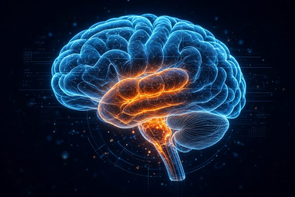
AI image generated with Grok

For centuries, scientists have thought of the brain as a single, unified organ. But new research led by Stanford Medicine reveals that what we call the brain is two distinct organs that evolved independently over hundreds of millions of years.

The discovery overturns a prevailing model of brain development. For decades researchers have subscribed to the theory that there is a single progenitor cell early in development that gives rise to the entire brain. This model suggested all parts of the brain shared a common developmental origin.

The new research finding shows that the human brain consists of two ancient nervous systems cleverly packaged together — a more primitive part that regulates our hearts’ beating, our breathing and other functions, and another that makes us distinctly human, capable of poetry, mathematics and wondering about our own origins.

The discovery could help explain why scientists have struggled for decades to grow certain types of brain cells in the laboratory — and it opens new avenues for studying devastating diseases that affect the brain stem, such as spinal muscular atrophy (also known as SMA) and amyotrophic lateral sclerosis (also known as ALS or Lou Gehrig’s disease).

“We’ve shown for the first time that the front of the brain arises from a totally different progenitor cell than the back of the brain,” said Kyle Loh, PhD, associate professor of developmental biology. “Our discovery means that we can now grow neurons from the back of the brain, the hindbrain, in a petri dish and study their functions.”

The findings were published in Nature Neuroscience Sept. 18. Loh is the senior author. Graduate students Carolyn Dundes and Rayyan Jokhai are co-first authors of the research.

Two brains

The adult brain has three main regions: the forebrain, midbrain and hindbrain. The forebrain handles higher-level thinking — language, consciousness and abstract reasoning. In contrast, the hindbrain, located at the back of the skull and often called the brain stem, controls essential, automatic functions that keep us alive: breathing, sleeping, and regulating our heartbeat and hunger urges. The hindbrain neurons also control the muscles of the face, tongue and throat, which affect speech and swallowing.

Despite the critical importance of the hindbrain, scientists have struggled for decades to generate human hindbrain neurons in the laboratory. This gap has hampered research into devastating diseases affecting the brain stem, including spinal muscular atrophy and amyotrophic lateral sclerosis.

SMA is a leading genetic cause of death in children under 1 year of age. ALS, which is often diagnosed between the ages of 40 and 70, affects both the forebrain and the hindbrain. In both disorders, certain hindbrain neurons gradually cease to function, and the patient loses the ability to swallow, which can cause pneumonia when food or liquid is inhaled into the lungs; eventually, patients lose the ability to breathe.

The researchers’ breakthrough came from studying the earliest moments of embryonic development, during a stage called gastrulation when the body first takes shape. Jokhai and Dundes discovered that the hindbrain follows a separate developmental path, running in parallel to — rather than branching off from — the pathway that creates the forebrain and midbrain.

The researchers learned this from examining developing mouse embryos. They identified two different brain progenitor cells. One, which expresses a gene called Otx2, is destined to become the forebrain and midbrain. The other, which expresses a gene called Gbx2, is committed to forming the hindbrain. They showed that these two cell populations never overlap; they are mutually exclusive from the earliest stages of development.

The team then examined the DNA packaging, or chromatin, in these cells. Chromatin is a way cells determine which genes can be easily accessed and which are bundled away out of reach. What they found was striking: The anterior neural ectoderm (future forebrain and midbrain) and posterior neural ectoderm (future hindbrain) have fundamentally different chromatin configurations. These differences essentially locked each progenitor cell into its respective fate, like travelers on parallel tracks that never cross.

“Previous attempts to make hindbrain neurons likely tried to coax forebrain and midbrain progenitors into hindbrain cells, which our study shows is not possible,” Jokhai said.

This revelation explained decades of frustration in the field — scientists had been trying to turn one type of progenitor cell into another that it is fundamentally incapable of becoming.

“In stem cell biology, people are always fixated with creating the end cell type, like the neuron,” Jokhai said. “But it’s important to begin at the earliest stages of embryonic development. Our careful attention to that early time point allowed us to find this fundamental split in brain development.”

Growing hindbrain neurons

Armed with this knowledge, the researchers for the first time successfully coaxed human pluripotent stem cells (a kind of cell that can create any cell in the human body) to become functional hindbrain motor neurons in the laboratory. These lab-grown neurons displayed all the hallmarks of authentic hindbrain cells: They exhibited waves of electrical activity called action potentials and made proteins that identify the segments of the hindbrain that control facial and swallowing muscles.

Finally, the researchers looked back over 550 million years of evolutionary time. They found the same two-origin brain pattern in chickens; zebrafish; and, remarkably, in acorn worms, tiny creatures living on the ocean floor that share a distant common ancestor with humans. Jellyfish, which diverged from humans about 600 to 700 million years ago, have two nervous systems at different ends of their body.

“Our research suggests that evolution took two existing neural systems and pushed them together spatially,” Loh said. “Having the brain as one organ would probably be more efficient, but we rely on this primordial way to make the brain as two separate pieces.”

“I was surprised at our findings because the word ‘brain’ implies a contiguous organ that likely has a singular origin,” Jokhai said. “But even 500 million years ago, there were these separate neural systems, which now almost operate as one, which is very cool.”

The research also has implications for investigating treatments for SMA, ALS and other conditions affecting the brain stem. Until now, studying these diseases has been nearly impossible because scientists cannot obtain brain stem tissue from living patients. The ability to grow these neurons in a dish opens new possibilities for understanding what goes wrong. There’s even an unexpected connection to obesity treatment: The hindbrain contains circuits that regulate hunger — which is precisely how weight-loss drugs like semaglutide work.

The researchers would like to extend their studies to determine the developmental origins of the spinal cord and to learn exactly how SMA and ALS compromise the function of hindbrain neurons.

“Now we have a model to better understand these devastating diseases, and work toward regenerative therapies for them,” Jokhai said. “This is a very exciting new frontier in brain research.”

Original written by Krista Conger

Source: Stanford Medicine

Can Vitamin C Improve Outcomes in People with Pre-cancer Blood Disorders?

Phase 2 trial results warrant further study in a phase 3 trial.

Photo by Diana Polekhina on Unsplash

Vitamin C boosts the activity of cellular proteins called TET enzymes that help control which genes are turned on or off. Because decreased function of TET enzymes is a common driver of certain forms of blood cancer, researchers tested the effects of vitamin C supplements in patients at risk of developing such malignancies. Results of the investigators’ phase 2 clinical trial are published by Wiley online in CANCER, a peer-reviewed journal of the American Cancer Society.

The randomised, double-blind, placebo-controlled EVITA trial enrolled 109 patients in Denmark and the United States who had either a blood condition that can turn cancerous or a low-risk form of blood cancer. At the start of the trial, 55 patients were randomly assigned to receive oral vitamin C (1000mg/day) and 54 were assigned to receive placebo, for a total of 12 months.

Although the primary endpoint (growth rate of precancerous or cancerous cells) was similar between groups, participants who received vitamin C experienced changes in inflammatory signaling that aligns with better outcomes. Also, anaemia, pneumonia, acute aseptic arthritis, and internal bleeding were less frequent (although gastrointestinal problems were more frequent) in patients taking vitamin C compared with those taking placebo.

At a median follow-up of 33.6 months (nearly 3 years) in the intention-to-treat population, 35 deaths were recorded, including 24 in the placebo group and 11 in the vitamin C group. An exploratory analysis of these data found that participants in the vitamin C group were more likely to survive during follow-up than those in the placebo group. This finding requires confirmation in a larger phase 3 clinical trial.

“The EVITA trial gives us a strong rationale to continue exploring if and how vitamin C might benefit people with certain pre-cancer or early-stage blood cancers. More work is needed but we are cautiously optimistic that these findings could inform future strategies to intercept leukemia development,” said co–senior author Peter A. Jones, PhD, DSc (hon), of Van Andel Institute, in Grand Rapids, Michigan. Jones is co-leader of the Van Andel Institute–Stand Up To Cancer (VAI–SU2C) Epigenetics Dream Team, which led the EVITA trial.

“We are encouraged by our findings and what they ultimately could mean for people with these early-stage blood disorders. Although it is too soon to make recommendations based on our results, we are hopeful that a larger study will give us more definitive answers,” added co–senior author Kirsten Grønbæk, MD, PhD, of Rigshospitalet, Copenhagen University Hospital in Denmark. Grønbæk is a longtime member of the VAI–SU2C Epigenetics Dream Team.

Source: Wiley

Cardiac Rehabilitation Cuts Hospital Admissions and Heart Attack Risk

A new Cochrane review of 107 trials shows that exercise-based cardiac rehabilitation is associated with numerous benefits and helps people return to healthier, more active lives

Photo by Barbara Olsen on Pexels

Exercise-based cardiac rehabilitation reduces hospital admissions by a third and heart attack risk by more than a quarter, as well as remaining beneficial in the long term, a new Cochrane review finds. 

The evidence also suggests that newer home-based and digitally supported cardiac rehabilitation programmes can be just as effective as traditional centre-based services. 

Coronary heart disease is the single most common cause of death globally. It can lead to chest pain, heart attacks, or stroke. Cardiac rehabilitation programmes are widely recommended following a cardiac event, with exercise recognised as a core component.  

Effective and inexpensive intervention 

The researchers, led by the University of Glasgow, analysed 107 randomised controlled trials involving 26 886 people with coronary heart disease. Most participants had experienced a heart attack, undergone heart procedures, or experienced some sort of chest pain. Women were underrepresented, making up only 17% of participants despite being included in most studies.  

The overall results show that, compared with no programme, exercise-based cardiac rehabilitation reduces heart attacks by 28%, reduces all-cause hospital admissions by 35%, and probably reduces deaths. 

Participants also reported better physical functioning, general health, vitality, social functioning, and mental wellbeing. These benefits were seen across several validated quality-of-life measures.  

“Our review shows that exercise-based cardiac rehabilitation can reduce the risk of heart attack and hospital admission while helping people return to healthier, more active lives,” says Dr Grace Dibben, lead author from the University of Glasgow.  

“Many people lose confidence after a heart attack or cardiac procedure and worry that physical activity could be dangerous,” Dr Dibben adds. “Appropriately prescribed exercise is not only safe for most patients, but can play a vital role in recovery and long-term heart health.” 

Growing evidence for modern rehabilitation models

This new update included 22 new trials featuring just under 3500 participants, incorporating more recent data into the final results. The more recent studies included participants based in low- and middle-income countries (LMICs) and tested home-based and digital cardiac rehabilitation programmes.  

“This review confirms that exercise is a great tool for improving the overall health of people with coronary heart disease,” says Professor Rod Taylor, senior author from the University of Glasgow. “By adding newer studies to this update, we’re able to see that digital programmes work as well as in-person programmes, which really changes the game here. These approaches may help us reach people who cannot attend traditional centre-based services.” 

Several studies in this update were conducted in LMICs, examining different types of exercise in different countries; for example, yoga in India and tai chi in China. The authors suggest this not only improves the relevance of the evidence base, but that some populations may find rehabilitation built around traditional and cultural forms of exercise more appealing than conventional, machine-based rehab. 

Low uptake despite evidence-backed benefits 

Despite strong evidence supporting cardiac rehabilitation, uptake remains a challenge in many healthcare systems. Various guidelines, including NICE in the UK, recognise cardiac rehabilitation as a key component following a cardiac event. However, cardiac rehabilitation continues to be widely underused with generally low participation rates. According to the 2025 National Audit of Cardiac Rehabilitation (NACR) report, the uptake of cardiac rehabilitation for patients with acute coronary syndrome was 44.5% in England and 71.3% in Wales.

The authors explain that many patients face barriers to attending traditional hospital-based programmes, including work commitments, transport challenges, and caring responsibilities. This can disproportionately impact groups like women, older adults, and deprived communities the hardest.  
 

Exercise-based cardiac rehabilitation is an inexpensive and cost-effective intervention, but right now it’s underfunded and unprioritised.

We want this option to be available to everyone. This new research shows you don’t even have to go into a rehab centre to benefit – you can participate from home and still see the benefits.

– Dr Grace Dibben, University of Glasgow.  

Sex Differences in Attention are Present at Birth, Study Finds

Photo by Tim Bish on Unsplash

Cambridge scientists have shown that girls and boys show differences in what they pay attention to, even at birth. Since these differences are present so early, it is possible they emerge due to prenatal factors.

It’s nature and nurture, not nature or nurture

Simon Baron-Cohen

The findings suggest that some psychological differences between males and females (such the development of social skills) may be influenced by both prenatal biology as well as later experience.

The study, published today in Biology of Sex Differences, involved testing 130 newborn infants only hours after birth. It builds on previous research by the same lab and uses robust methods to demonstrate the differences.

For years, research has shown that girls, on average, learn to talk earlier and develop social skills faster than boys. Scientists have long debated whether these differences are due to culture or biology. One way to answer this question is to test if sex differences are present soon after birth, before society and culture have influenced a child’s development. 

A study from the University of Cambridge published in 2000 found girls at birth on average looked longer at faces, and boys on average looked longer at objects. Since then, scientists in the field called for a replication of this study. However, no research team has attempted this until now, since it is very challenging to carry out research on newborn infants.   

In this new study, infants were presented with side-by-side videos containing a social stimulus, namely a human face with natural movement, and a non-social object, namely a set of metallic balls swinging due to gravity and their mechanical properties (a Newton’s cradle). 

The team found that, regardless of sex, newborn infants generally preferred to look at the social stimulus. However, on average, female infants spent a greater proportion of the time looking at the social stimulus compared to male infants, while male infants spent a greater proportion of time looking at the non-social stimulus compared to female infants.

Since this is within hours of birth, before much postnatal experience (such as social expectations based on gender) has occurred, it is likely that prenatal biological factors that differ between the sexes during pregnancy (such as hormone levels and genetics) contribute to the observed on-average sex differences. 

Yumnah Khan, a PhD student at the Autism Research Centre, University of Cambridge and the lead researcher on the study, said: “Our recent research has also shown that on-average sex differences are present in brain structure at birth and emerge prenatally. This new study tells us that there are not only on-average sex differences in the brain but also in attention and behaviour.” 

Professor Sir Simon Baron Cohen, Director of the Autism Research Centre, University of Cambridge, who supervised the study, said: “This study addresses the age-old question of whether nature plays any role at all in shaping sex differences in the human mind and brain. The fact that we observe these differences within hours of birth, before extensive postnatal experience has occurred, suggests that prenatal factors play a role, and that these prenatal factors – sex hormones and/or genes – go on to interact with postnatal experience. In other words, it’s nature and nurture, not nature or nurture.”

The new study used more robust methods compared to the original 2000 study. In particular, it included more controlled experimental testing conditions as well as a more reliable method to monitor infant gaze. Crucially, the researchers who analysed the videos of the baby’s eyes were blind to the baby’s sex and whether the baby was looking at the social or the non-social stimulus, which ensured that they could not bias the results. 

Dr Alex Tsompanidis, Assistant Research Professor at the Autism Research Centre and a member of the research team, said: “Sex differences at birth are important as starting points, but we need to remember that they don’t fully determine how children learn and develop in later life. This is because biological and social factors always interact, contributing to the rich diversity of human societies.”

Professor Carrie Allison, Deputy Director of the Autism Research Centre and another member of the research team, said: “The differences we see don’t apply to all male or all female infants, and are only seen when you compare groups of male and female infants, on average. There’s a lot a variation within, and overlap between, each sex. Our future research needs to better understand that variation.” 

The team is now interested to find out if these early sex differences contribute to later differences in children’s minds, brains and behaviour. To test this requires a longitudinal (follow-up) study design, which is underway. The researchers hope that by understanding the origins of the earliest sex differences, scientists can better understand neurodiversity and conditions such as autism, which is diagnosed about twice as often in boys than in girls.

This research was supported by the Wellcome Trust, the Simon Foundation Autism Research Initiative (SFARI), and a PhD studentship from Trinity College, Cambridge and the Cambridge Trust. A follow-up study is funded by the K. Lisa Yang Centre for Autism Research at Cambridge, which is part of the Yang Tan Collective.

Reference

Khan, YT, Tsompanidis, A, Hymanson, E, Wickramanayake, S, APEX Consortium, Austin, T, Allison, C, & Baron-Cohen, S. Sex differences in social attention at birth. Biology of Sex Differences; 21 Sept 2026; DOI: 10.1186/s13293-026-00976-9

Republished from the University of Cambridge under a Creative Commons licence. Read the original article,