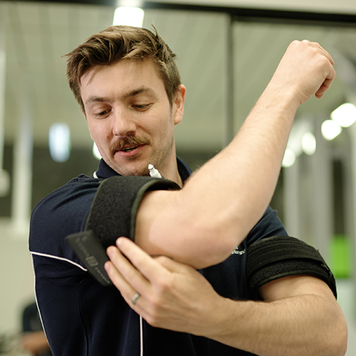
The novel exercise involves applying a pneumatic cuff to restrict the flow of blood. Credit: University of South Australia
New research from the University of South Australia is offering fresh hope to people living with rheumatoid arthritis (RA).
Evaluating the effectiveness of a novel form of exercise – blood flow restricted resistance training – among people with RA, researchers found that this alternative workout method not only improved their strength and physical performance, but also reduced their pain.
Blood flow restricted resistance training involves placing a pneumatic cuff – much like a blood pressure cuff – around the top of the working limb. The cuff is then inflated so that it restricts blood flow out of the limb, creating a highly metabolic environment which forces the muscles to work harder, even when using lighter weights or less effort.
The Arthritis Australia-funded study is the first to trial blood flow restricted resistance training on both the upper and lower limbs in people with RA, using five exercises – leg press, machine hamstring curl, machine knee extension, cable tricep extension, and cable bicep curl – with gradually increasing weights.
All participants in the study reported that they “liked” the programme, and the group showed clear improvements in strength, movement and pain levels.
Lead researcher UniSA’s Dr Hunter Bennett says the training offers a practical and achievable option for people with RA.
“RA can cause a loss of muscle mass and strength, which affects day-to-day activities, independence, and increases the risk of falls and fractures,” he says.
“Resistance training is one of the best ways to rebuild that strength, but for people with RA, using heavy weights can be difficult or harmful due to pain, fatigue or injury risk. This is where blood flow restricted resistance training can help.”
Dr Bennett says this approach is ideal for people who need to do resistance exercises but find it hard to lift weights.
“Many people with health conditions are understandably deterred by exercise, yet it is often one of the best things they can do to improve their condition,” he says.
And while this exercise might look unusual, the research shows that it works.
“This kind of training could be a game-changer for people with rheumatoid arthritis.
“It offers a way to build strength and reduce pain without pushing through discomfort – and that’s incredibly empowering for people who’ve often been limited by their condition.”
While this was a small-scale trial, researchers say the results are promising and lay the foundations for a larger trial comparing blood flow restricted resistance exercise to more traditional exercise approaches.
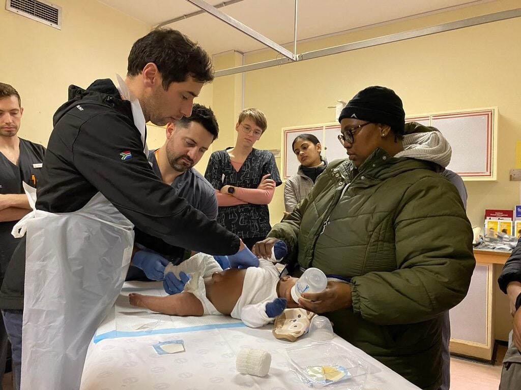
Dr Saul Kaplan (left) stands next to Dr Kenny Beck, with mother Zukiswa Panyaza as her baby receives a full leg cast at the clubfoot clinic in Tygerberg Hospital’s Division of Orthopedic Surgery, while medical students observe. (Photo: Sue Segar/Spotlight)
By Sue Segar
When Karen Mara Moss’s son was diagnosed with clubfoot, she travelled to the US in search of a life-changing treatment. She made a promise to bring it home and two decades on, her non-profit is at the heart of a remarkable success story.
“I looked at those tiny feet. They were turned over and rigidly pointing inwards,” recalls Karen Mara Moss about the day her son Alex was born in 2003.
For her, the memory is as vivid today as it was then. Within moments of his birth, a concerned obstetrician commented on Alex’s feet. Then the paediatrician diagnosed Alex with bilateral clubfoot, a condition in which a baby is born with one or both feet twisted inward and downward.
“I remember thinking: Will he walk with a limp? Will people mock him?” Moss tells Spotlight. “It was a traumatic time.”
She says the paediatrician told her not to worry. “He said they’d have to cut his feet and straighten them, and it would all be perfect,” says Moss.
Despite having several prenatal scans and tests, the condition had not been picked up before birth.
The most common form of clubfoot present at birth is idiopathic clubfoot, medically known as talipes equinovarus. It is when a baby’s foot is pointed in and down because the tissues connecting the muscles to the bone are shorter than usual, leading to pain and reduced mobility if left untreated, according to a review study published in The Lancet medical journal. In most cases, the cause of this congenital anomaly which ranges from mild to severe, is unknown, baby boys are twice as likely to be born with clubfoot as baby girls, and about half of children with clubfoot have it in both feet. Globally, an estimated 176 000 babies are born with the condition every year.
Eight days after Alex was born, Moss says she met with a paediatric orthopaedic surgeon. She says he told her he’d fixed many clubfeet using the Kite method and even had one patient playing first-team rugby. The Kite method was developed in the 1930s and uses manipulations and castings to achieve a sequential and gradual full correction of the forefoot, then the hindfoot, and finally, the ankle. After the casting is done, the baby wears a special splint to keep the feet pointing slightly outward and upward, but, critically, many would also require further surgery.
Back then, the standard of care for clubfoot was surgical management, says Dr Pieter Maré, an orthopaedic surgeon who heads up the clubfoot clinic at Greys Hospital in Pietermaritzburg, Kwazulu-Natal. “The reality was that a large number of children required extensive surgery before the Ponseti method,” he says.
Moss followed the doctor’s advice, and during that first appointment, she says he began applying casts up to Alex’s knees. “He started wrenching Alex’s foot, holding the back, whilst pushing the front of the foot, and plastering the foot. Alex was blue in the face from screaming. I was crying while holding him down,” she says.
Another way
But after two months and seven casts, she says there was little improvement in Alex’s feet. That is when Moss began searching for answers herself. Doing research on the internet, she discovered the University of Iowa Children’s Hospital website, where she read about a technique developed by Dr Ignacio Ponseti, which he claimed could help children have pain-free, functional feet without surgery.
The Ponseti method was developed in the 1950s but only became more widely used in the United States in the 1990s, and later in much of the rest of the world. The technique uses gentle manipulations and plaster casts to correct the midfoot, hindfoot, and forefoot simultaneously, while the ankle is treated afterwards. In some cases, before the last cast is applied, it may require a percutaneous tenotomy which is a minimally invasive procedure to cut the heel cord that is resistant to stretching. A brace is then fitted the same day as the last cast is taken off.
“The Kite method was developed to correct clubfoot but over time it was realised that this method was using the wrong anatomical methods,” explains Professor Anria Horn, a consultant orthopaedic surgeon at the Red Cross Children’s Hospital in Cape Town.
“There are multiple joints in the foot and the Kite method was, effectively, manipulating the wrong joint in an attempt to bring about the correction in the foot. Ponseti discovered that the manipulation should occur at a different joint,” she says.
Back in 2003, Moss emailed Ponseti, and a few days later called his office. “I was put through to a man with a Spanish accent. He said he’d read my email, and that he’d seen the photos I sent of my son’s feet; that what we’d done was not the way his method worked. He suggested I go to Iowa because nobody in South Africa was practising his method,” she says.
Not long after this, Moss and her husband travelled with ten-week-old Alex for 10 000 miles from sunny South Africa to an unseasonable snowstorm in Iowa.
The idea of travelling to a foreign country to see a “special” doctor that one read about on the internet, with a treatment carrying his name, may raise red flags for some. There are after all no shortage of quacks out there exploiting vulnerable people with just this type of story of an underutilised treatment. Ponseti, however, was a serious scientist and, even by 2003, his method had performed well in several studies and had been quite widely adopted by doctors in the United States.
Moss says in that first visit, Ponseti eventually did a cast all the way up Alex’s leg. “He looked like a little turtle with his legs sticking out. By the time he’d done the second cast, Alex was asleep,” Moss recalls.
“Dr Ponseti’s normal protocol was to remove the cast every week, then re-manipulate the foot into a different position, and reapply the cast. For out-of-town patients, he accelerated the treatment and changed the cast every five days,” she adds.
After just one cast, Alex’s foot looked different, says Moss. “They did another cast, and five days later, it was time for the third cast. Dr Ponseti took the second cast off and then did the percutaneous tenotomy, as well as the third and final cast.”
After this procedure and with Alex now in his final casts, they were told they could return to South Africa and take the casts off three weeks later. Moss said an orthotist measured Alex’s feet before the tenotomy and gave her instructions on how to fit the clubfoot brace he would wear for four years while sleeping.
Three weeks later, back home, Moss soaked the casts off and started to put the brace on at night. She says Alex’s feet were straight.
‘A parting gift’
On her final day in Iowa, Moss recalls Ponseti telling her: “You’re the first South African that’s ever been here. Please go back home and tell the doctors not to operate on clubfoot”. He gave her his book, copies of his research papers, and CDs demonstrating his casting method – a parting gift that would shape the course of her life.
Determined to share her what she had learnt, Moss created a website to provide information on clubfoot. The website gained traction and soon she started getting requests from parents across southern Africa for help to access the Ponseti method.
At the time, Moss says she knew of only one doctor using the method, whom she recommended parents consult. “I’d met him soon after my return to South Africa in 2003 and had lent him Dr Ponseti’s book and papers. He’d then gone to the US to attend a Ponseti training workshop and started using the method. I was sending everyone to him.”
The founding of STEPS
Moss realised the best solution was to bring the training directly to South Africa. In 2005, despite having no experience in running a non-profit organisation, she founded STEPS, driven by her commitment to introduce the Ponseti method across the country.
Moss says STEPS held its first two-day Ponseti training course in 2006, with about 60 paediatric orthopaedic surgeons attending. “Three Ponseti experts came from Canada, Brazil and the UK to give the training. They taught a lot of theory and used bone models to demonstrate the method,” she says.
The second STEPS Ponseti workshop in 2007 focused on public health facilities. Moss says the training took place at the Charlotte Maxeke Johannesburg Academic Hospital.
Partly due to the workshops, partly due to the strength of the accumulating scientific evidence, the method caught on in the country. In 2012, the South African Paediatric Orthopaedic Society officially endorsed the method. A Cochrane Review published in the same year found that, while the available evidence was far from complete, it did indicate that the method works well. Cochrane Reviews are a highly regarded type of study that attempts to assess evidence from all randomised clinical trials relating to a specific medical question.
“The Ponseti method has become the gold standard for the treatment of idiopathic clubfoot,” stated an article published in the World Journal of Orthopedics in 2014. And according to the Lancet study cited earlier, “the Ponseti method is widely recognised as an effective conservative treatment approach for clubfoot that avoids corrective surgery in over 90% of cases”.
Today, Horn says the Kite method isn’t used in South Africa any more, having been replaced by the Ponseti method. “STEPS has played a big part in promoting the Ponseti method in South Africa, as well as providing training, workshops and conferences and supporting clubfoot clinics across the country. Our job would have been much harder without the support that STEPS provides,” she adds.
Ponseti in the public sector
Given the equipment and know-how involved, making the Ponseti method available in South Africa’s public sector was a challenge. In 2013, Moss launched a support programme to help government clinics offer the treatment. STEPS began by partnering with just six clinics. With support from donors, they recruited staff to visit each clinic weekly to guide families or trained someone on-site to do so. They also provided educational materials to help raise awareness. Over time, this led to STEPS helping develop standard systems and processes for running the clinics, making care more consistent and accessible. When some clinics couldn’t provide braces, STEPS arranged for it to be donated.
Today, STEPS has 48 partner clinics across South Africa, ranging from a tiny rural clinic in Lusikisiki in the Eastern Cape to bigger clinics in Gauteng and the Western Cape. “Lusikisiki might see three patients a week, and Chris Hani Baragwanath Academic Hospital could see 80. They all open once a week, except for some small, rural clinics,” Moss says.
Based on stats that STEPS collected, Moss estimates that at least 2 000 children are born every year with clubfoot in South Africa. Through the help of her organisation, she says: “More than 20 500 children have accessed effective treatment. We’ve … distributed 22 628 clubfoot braces. In 2024, we supported 4 592 children at partner clinics in different stages of the four-year treatment protocol.”
Moss adds that STEPS has conducted over 20 training sessions across South Africa, Namibia, Botswana, and the Seychelles, with more than 2 000 healthcare professionals. “Parents were bringing their children over the border as they couldn’t access treatment back home. We worked with the ministries of health in those countries to teach the Ponseti method there,” she says.
Though separated by an ocean, Moss says she stayed in close contact with Ponseti. She says the last time they saw each other was at a clubfoot symposium in Iowa in 2007. Two years later, he passed away at the age of 95.
“I felt as if I’d lost a member of my family,” Moss says. “He was the master, and he inspired me in my work to improve the lives of children born with clubfoot.” She said she would always carry the ache of missing him, but bringing his method to South Africa, just as she had promised, was something that gave her a deep sense of purpose and peace.
That promise, purpose and peace started with Alex who is today in his final year of a Bachelor of Commerce degree and who, in his own words, “enjoys being active outdoors with my friends”, likes playing padel, and going on hikes.
*This article is part of Spotlight’s 2025 Women in Health series, featuring the remarkable contributions of women to healthcare and science.
A world-first Swinburne-led study into young adults’ brain activity has found that TV and gaming are associated with increased focus, while social media is associated with decreased focus.
In this study, published in Nature, 18-25 year olds exposed to phone screens for only three minutes experienced changes in mood, energy, tension, focus and happiness, explains one of the lead researchers Swinburne’s Dr Alexandra Gaillard.
“Our study was the first to record brain activity during different forms of screen use on young adults using functional near-infrared spectroscopy (fNIRS). We found that different forms of screen use, including social media, are associated with distinct patterns in activity and mood states.”
“Almost everyone owns a smart phone which they use for at least three hours a day for entertainment. Mood disorders are increasing in prevalence worldwide and we shouldn’t rule out the possibility that phones are a contributor.”
The study found that oxygenated haemoglobin (HbO) levels increased more following social media use and gaming compared to TV viewing, while deoxygenated haemoglobin (HbR) levels increased more following gaming.
“These findings suggest that interactive types of entertainment really do get the brain more engaged,” says Dr Gaillard.
“Interestingly, though, when it came to social media, people reported feeling less focused—and those who felt less focused also showed lower levels of brain activity. On the flip side, gaming actually helped boost focus and showed a rise in deoxygenated haemoglobin, which means the brain was actively using more of the oxygen it was getting. In other words, gaming seemed to get the brain working harder in a good way.”
With six months to go until Australia’s impending teen social media ban, there are still no clear pathways for age-checking tools and the positive impacts of the policy on different types of technology and platforms.
Dr Gaillard says that while this study looked at young adults, these findings suggest a similar outcome to teenagers which should be considered by experts when implementing the ban.
“If this is the effect on a fully developed brain, we urgently need to consider the impacts on teenagers and children who are increasingly using these technologies.”
The Swinburne research team is calling for further research to understand the complex and nuanced relationship between screen activities and how they engage they brain.
“Excessive screen time can negatively impact cognitive abilities, attention and executive functioning, but we also know how invaluable they can be in forming connections and a sense of belonging as well as improving educational outcomes.”
“This isn’t a call for blanket reductions; screens certainly serve a purpose for unwinding and leisure. We ask that young people are conscious of how their activity impacts them and that they make choices that are right for them.”
But women doctors in this attire are often misidentified as nurses or medical assistants. And preferences for doctors’ dress strongly influenced by clinical context/medical specialty
Patients are still more likely to trust doctors and consider them more professional when they wear white coats, although women doctors in this attire are often misidentified as nurses or medical assistants, finds a review of the available research on the topic, published in the open-access journal BMJ Open.
But patient preferences for doctors’ attire seem to be strongly influenced by clinical context and medical specialty, with a growing acceptance of scrubs, especially in emergency or high-risk settings, the findings indicate.
In the 19th century, doctors primarily wore black, because medical encounters were viewed as serious and formal occasions. But with the growing emphasis on hygiene and scientific advancements in medicine, white coats represented cleanliness and professionalism and became standard attire for doctors worldwide in the 20th century, note the researchers.
Since the publication of a previous comprehensive review of published research on the impact of doctors’ attire at work, which highlighted the significant role this has in patient satisfaction, trust, and adherence to medical advice, health care practices and societal expectations have evolved, they explain.
The researchers therefore wanted to update those findings, broadening the scope to include diverse clinical contexts, in a bid to gain a deeper understanding of how doctors’ attire might influence interactions with patients and treatment outcomes.
They scoured research databases for relevant studies published between January 2015 and August 2024. From an initial haul of 724 studies, 32 were eligible for inclusion in the review.
Most of the studies were carried out in the U.S. (17); two each came from Japan, China, and Pakistan; and the rest were carried out in Indonesia, India, Ethiopia, Korea, Germany, Malaysia, Saudi Arabia, Singapore and Switzerland.
Clinical setting had some bearing on patient preferences. For example, palliative care doctors’ attire didn’t affect patients’ trust, or assessments of the practitioner’s capabilities, but patients preferred emergency care doctors to wear white coats or scrubs. And they were happy for primary care doctors to be dressed casually and to wear white coats.
Specialty also seemed to influence perceptions, with patient preferences for doctors to wear white coats observed in orthopedics, surgery, dermatology, ophthalmology, and obstetrics and gynecology, for example.
The included studies indicated a distinct preference among patients for male doctors to wear suits. One study indicated that accessories, such as watches and glasses worn by male doctors, significantly enhanced perceived professionalism and trustworthiness, which aligns with the findings of previously published studies, note the researchers.
The UK, however, has implemented a ‘bare below the elbows’ policy, which bans doctors from wearing white coats, watches, ties and long sleeves to minimize infection risks.
And even when male and female doctors wore identical attire, female doctors were still more likely to be misidentified as nurses or medical assistants—a misidentification that was consistently observed across different cultural settings.
But patients preferred white coats over business or casual attire for both male and female doctors, and irrespective of gender, they favored doctors wearing white coats, perceiving these practitioners to be trustworthy, respectful, skilled, communicative and empathetic.
Notably, male surgeons wearing a white coat over scrubs were looked on less favorably than those wearing a suit with a white coat, scrubs, or just a suit. On the other hand, female surgeons in white coats over scrubs were preferred over those in suits or casual attire.
“This preference aligns with recent evidence indicating that female physicians are often judged more on appearance than their male counterparts. The way female physicians dress significantly influences perceptions of competence and professionalism, highlighting the gendered expectations that patients hold,” write the researchers.
“These gender-specific preferences for surgical attire were each supported by a single study and warrant further validation… Nonetheless, gender-related perceptions of physician attire were consistently reported across studies with a low risk of bias, supporting the robustness of this theme,” they add.
A few studies explored doctors’ attire during the COVID-19 pandemic, when numerous doctors reported a preference for wearing scrubs to prioritize hygiene and ease of movement.
These studies consistently reported a shift in patient preference towards practical and hygienic attire, such as scrubs and face masks, reflecting heightened sensitivity to infection control, say the researchers.
The researchers acknowledge various limitations to their findings, in particular the fact that most of the included studies were carried out in the U.S., there were none from South America, and only a few from European, Asian, and African countries. Many of the studies also relied on self-reported data, and none included children or patients with mental health issues.
“While the traditional white coat is seen as a symbol of professionalism and trust, patients have increasingly accepted scrubs, especially in emergency or high-risk settings,” they write.
“The expectations regarding attire are often gendered, particularly affecting the recognition and respect given to female physicians, which highlights the importance of institutional initiatives aimed at reducing bias and fostering equitable perceptions among patients,” they add.
Medical institutions should consider adopting flexible dress codes that align with patient preferences for different clinical environments and medical specialties, they suggest.
Creatine is popularly known as a muscle-building supplement, but its influence on human muscle function can be a matter of life or death. But getting it to one particular organ that needs it – the brain – is challenging.
“Creatine is very crucial for energy-consuming cells in skeletal muscle throughout the body, but also in the brain and in the heart,” said Chin-Yi Chen, a research scientist at Virginia Tech’s Fralin Biomedical Research Institute at VTC.
Chen is part of a research team working to develop a technique that uses focused ultrasound to deliver creatine directly to the brain. The work, being conducted in the lab of Fralin Biomedical Research Institute Assistant Professor Cheng-Chia “Fred” Wu, will be supported by a $30 000 grant from the Association for Creatine Deficiencies.
Creatine plays a vital role in the brain, where it interacts with phosphoric acid to help create the key energy molecule adenosine triphosphate (ATP). In addition to its role in energy production, creatine also influences neurotransmitter systems.
For example, creatine influences the brain’s major inhibitory pathways that use the neurotransmitter gamma-aminobutyric acid (GABA), which limits neuronal excitability in the central nervous system. It may play a role in a variety of functions, including seizure control, learning, memory, and brain development.
A growing body of research suggests that creatine may itself function as a neurotransmitter, as it is delivered to neurons from glial cells in the brain and can influence signalling processes between other neurons. While creatine deficiency disorders can weaken the skeletal muscle and the heart, they can also severely affect the brain. Many patients see increased muscle mass and body weight with creatine supplements, but they often continue to face neurodevelopmental challenges that can hinder their ability to speak, read, or write.
This is largely caused by the brain’s protective blood-brain barrier preventing creatine entry.
Wu studies therapeutic focused ultrasound, which precisely directs sound waves to temporarily accessed areas of the brain. The process allows drugs to reach diseased tissue without harming surrounding healthy cells. While Wu is investigating this method as a potential treatment for paediatric brain cancer, he also sees potential in applying it to creatine deficiency.
“Through the partnership between Virginia Tech and Children’s National Hospital, I was able to present our work in focused ultrasound at the Children’s National Research & Innovation Campus,” Wu said. “There, I met Dr Seth Berger, a medical geneticist, who introduced me to creatine transporter deficiency. Together, we saw the promise that focused ultrasound had to offer.”
The Focused Ultrasound Foundation has recognised Virginia Tech and Children’s National as Centers of Excellence. Wu said the two organisations bring together clinical specialists, trial experts, and research scientists who can design experiments that could inform future clinical trials.
“It was a moment that made me really excited – that I had found a lab where I could move from basic research to something that could help patients,” Chen said. “When Fred asked me, ‘Are you interested in this project?’ I said, ‘Yes, of course.’”
Because creatine deficiencies can impair brain development, the early stages of Chen’s project will concentrate on using focused ultrasound to deliver creatine across the blood-brain barrier. Chen hopes the technique will restore normal brain mass in models of creatine deficiency.
Crohn’s disease, a debilitating inflammatory bowel disease, has many known contributing factors, including bacterial changes in the microbiome that foster an inflammatory environment. Now, for the first time, Crohn’s disease has been tied to a virus – specifically, Epstein-Barr virus (EBV), best known for causing infectious mononucleosis (mono).
Researchers had already observed increased levels of EBV in the intestine of patients with Crohn’s disease and had also found associations between EBV and autoimmune diseases other than Crohn’s, including lupus, multiple sclerosis, and rheumatoid arthritis.
Nandy and colleagues analyzed data from a cohort of initially healthy military recruits, aged 20 to 24, who provided periodic serum samples throughout their service. They tested the samples with VirScan, a high-throughput assay developed by study coauthor Stephen Elledge, PhD, at Harvard Medical School. VirScan enables detection of antibodies against a wide range of viruses, providing insights into viral exposures.
Compared with healthy controls, military personnel whose samples were positive for anti-EBV antibodies were three times more likely to eventually develop Crohn’s disease. Interestingly, evidence of EBV exposure preceded their Crohn’s diagnosis by five to seven years.
“We went into this study not looking for EBV; we were looking for any virus that might elicit inflammatory bowel disease,” says Scott Snapper, MD, PhD, the study’s senior investigator and director of the Inflammatory Bowel Disease Center at Boston Children’s. “Then, when EBV was a hit, we looked very specifically at immune responses to EBV with more detailed tests.”
These tests bore out and strengthened the VirScan findings.
The team next looked for anti-EBV antibodies in a second cohort – more than 5000 children (median age; 11 years) who were first-degree relatives of people with Crohn’s disease. In this cohort, EBV was not a statistically significant predictor of a subsequent Crohn’s disease diagnosis. Snapper speculates that having first-degree relatives with Crohn’s could already have put them at increased risk due to shared genetic or environmental factors, muddying the association with EBV.
EBV and the immune system
Another possibility is that EBV affects children’s immune systems differently, perhaps because children are less likely to develop infectious mono when exposed. “Responding to certain organisms early in life may boost the immune system in a way that prevents immune-mediated disease,” Snapper says.
Nandy and Snapper now want to figure out what EBV does to people at a molecular level to make them more susceptible to Crohn’s disease.
One possibility is that the virus has certain genes or molecules that interact with human genes involved in susceptibility to autoimmune conditions. Another may relate to an anti-inflammatory protein produced by EBV, remarkably similar to mammalian IL-10. People exposed to that protein may make antibodies against it, preventing their own IL-10 from working and leaving them susceptible to inflammatory disease.
“Mechanistically, we need to understand exactly how EBV alters the immune system leading to Crohn’s disease,” Snapper says. “If you could figure out the mechanisms, you could come up with new therapies.”
Squamous cancer cell being attacked by cytotoxic T cells. Image by National Cancer Institute on Unsplash
By measuring interactions between cells, the method offers insights into how the human body fights viral infections, how malfunctions can lead to autoimmune diseases and why immunotherapies work for some people but not others.
A healthy immune system is trained to detect and destroy infections and cancer cells. This defence is based on a complex communication system at cellular level, in which different immune cells each perform a specialised task: recognising infectious agents, alerting other immune cells, and eliminating harmful cells or pathogens. Problems arise when communication between different cell types is disrupted, potentially leading to a variety of diseases.
For example, cancer cells often develop strategies to specifically disrupt or circumvent the exchange of information in the immune system – this allows them to evade immune surveillance and grow unhindered. “Modern immunotherapies have fundamentally changed the treatment of certain types of cancer by restoring or specifically strengthening communication between immune cells,” explains Prof Simon Haas, one of the leaders of the study.
Dr Daniel Hübschmann, also head of the study, adds: “However, not all patients respond equally well to these therapies and reliable methods for predicting which patients will benefit most are still lacking.”
Decoding immune cell communication for personalised cancer therapies
Scientists have now developed a technology that overcomes many of these hurdles through a better understanding of immune cell communication. With this method, made possible by interdisciplinary collaboration, millions of cell-cell interactions can be measured quickly and cost-effectively, both in research laboratories and in the clinic.
The scientists are using the newly developed technology to investigate the behaviour and kinetics of immunotherapies and to gain insights into how these therapies work at the level of cell-cell interactions. They were able to show that the approach enables the prediction of individual therapy responses and can thus create a central basis for personalised immunotherapies and targeted therapy decisions.
In addition, the researchers were able to use their new technology to visualise, in high resolution, how cells of the immune system interact with each other during viral infections and autoimmune diseases. The results allow them to develop dynamic maps of immune cell networks, illustrating for the first time how the immune defence is coordinated in different tissues.
Together with clinical partners, the team is now working on translating these findings from research into practice, for example to better predict treatment success and utilise immunotherapies in a more personalised manner.
Sanofi has reaffirmed its commitment to South African healthcare with the appointment of two senior leaders and a renewed focus on supporting healthcare professionals, expanding local partnerships, and improving access to essential treatments in key areas such as vaccines, immunology, and rare diseases.
“This is a new chapter for Sanofi in South Africa,” says Jean-Baptiste Bregeon, Sanofi South Africa’s Country Lead and Head of Vaccines. “We are focused on making a real impact through practical support for healthcare professionals, expanding access to important medicines and vaccines, and strengthening our ties with partners across the system.”
Focused leadership
Bregeon brings over 20 years of international experience, with senior roles in commercial and clinical operations across the Middle East, North Africa, and Eurasia. As former General Manager for North Africa at Sanofi Vaccines, he led efforts to improve vaccine access and build local capacity. He has also advised Sanofi’s global leadership on operational strategy and commercial planning.
Bregeon will oversee Sanofi’s overall strategy in the country and lead its vaccine division, which remains a core part of the company’s healthcare offering.
Lenisha Maharaj, newly appointed Head of Pharma for South Africa, Namibia and Botswana, is a seasoned Pharmaceutical Executive with over 20 years’ experience in the pharmaceutical and healthcare sectors. At Sanofi, she will oversee growth across the general medicines and specialty care portfolios, supporting both primary and specialist care.
“Sanofi understands the realities that healthcare professionals face,” says Maharaj. “Our goal is to support them with the right tools, training, and access to innovative treatments so they can keep delivering high-quality care.”
Strengthening partnerships and access
A key part of Sanofi’s long-term strategy is to build strong, lasting partnerships. Its collaboration with Biovac, a local vaccine manufacturer, is a core example. This will help to boost domestic vaccine production capacity and reduce reliance on global supply chains. Such public-private partnerships support healthcare resilience as well as local economic and scientific development.
Beyond vaccines, Sanofi is investing in other areas that have a direct impact on healthcare professionals and the patients they serve:
Clinical trials in South Africa: Expanding its local clinical research footprint to increase representation of African patients in global trials and ensure that treatment decisions are informed by local data.
Access to innovative medicines: Working to bring advanced treatments in rare diseases and immunology to South Africa, along with patient support programmes and diagnostic assistance, to help doctors and patients navigate access challenges.
Medical education and digital engagement: Offering CPD-accredited education, clinical webinars, and digital tools to help healthcare professionals stay updated on the latest treatment protocols and deliver more streamlined care.
Policy engagement and system support: Actively involved in discussions with policymakers and funders to help shape health policies that improve medicine access and promote fair, sustainable healthcare.
Supporting healthcare professionals
Sanofi says it recognises the ongoing pressures facing South African healthcare workers, from dealing with supply shortages and reimbursement hurdles to managing the growing burden of chronic and complex diseases.
“We see our role as being part of the solution,” says Bregeon. “We are here to support healthcare professionals, not only by ensuring a reliable supply of medicines, but by providing the training, tools, and partnerships they need to do their jobs effectively.”
The company is also prioritising engagement with healthcare professionals through regular updates, field team support, and opportunities for joint problem-solving around system-level challenges.
Sanofi says it is committed to staying in close conversation with the healthcare community and building a more resilient, inclusive, and patient-focused system.
“We want to work with doctors, nurses, pharmacists, policymakers and funders to build something long-term,” Bregeon adds. “We believe that by working together, we can improve outcomes for patients and strengthen the foundation of healthcare in this country.”
A two-drug combination for treating advanced kidney cancer had sustained and durable clinical benefit in more than five years of follow-up, according to a study published August 1 in Nature Medicine.
The study reports final clinical data and biomarker analyses from the Phase 3 KEYNOTE-426 trial, which compared the drug combination pembrolizumab plus axitinib versus the single drug sunitinib for patients with previously untreated advanced clear cell renal cell carcinoma, the most common type of kidney cancer.
“KEYNOTE-426 was the first trial to combine a PD-1 inhibitor immunotherapy (pembrolizumab) with a VEGF receptor inhibitor antiangiogenic drug (axitinib) in the first-line setting for advanced renal cell carcinoma. It therefore has the longest follow-up duration among the various trials comparing these types of drug combinations,” said Brian Rini, MD, a medical oncologist at Vanderbilt-Ingram Cancer Center, Professor of Medicine and the study’s lead and corresponding author.
Immunotherapy drugs like pembrolizumab stimulate the immune system to kill tumour cells. VEGF receptor inhibitors like axitinib and sunitinib block angiogenesis — the development of blood vessels that tumours need to grow and spread. Pembrolizumab plus axitinib and other immunotherapy-antiangiogenic drug combinations are now standard first-line treatments for advanced kidney cancer.
“Before the development of antiangiogenic drugs and immunotherapies, advanced renal cell carcinoma had a very poor prognosis. These drug combinations have dramatically improved treatment options and outcomes for patients,” said Rini.
The first interim analysis of outcomes from KEYNOTE-426, published Feb. 16, 2019, in the New England Journal of Medicine, demonstrated that trial participants treated with pembrolizumab plus axitinib had longer overall and progression-free survival, and higher objective response rates compared to those taking sunitinib. The median follow-up was 12.8 months.
Now, with a median follow-up of 67.2 months, the current analysis confirms and extends the interim analysis and provides valuable information about biomarkers that could help guide treatment decisions.
The study in Nature Medicine reports that pembrolizumab plus axitinib had longer overall survival (47.2 months versus 40.8 months for sunitinib) and longer progression-free survival (15.7 months versus 11.1 months for sunitinib). The objective response rate was 60.6% for pembrolizumab plus axitinib and 39.6% for sunitinib.
The researchers reported a variety of associations between the expression of biomarkers and outcomes (overall survival, progression-free survival, objective response rate). The biomarkers they evaluated included an 18-gene T-cell-inflamed expression profile, angiogenesis signature, and PD-1 ligand expression.
“There is an unmet need for biomarkers that are predictive of patient outcomes following treatment with available first-line therapies for advanced renal cell carcinoma,” Rini said. “Although our analysis showed potential clinical utility of some RNA signatures in identifying patients who are likely to benefit the most from each treatment, further prospective clinical studies are needed.”
Pembrolizumab plus axitinib is a first-line treatment option for patients with advanced renal cell carcinoma regardless of biomarker subtypes, he noted.
Scanning electron micrograph of Yersinia pestis, which causes bubonic plague, on proventricular spines of a Xenopsylla cheopis flea. Credit: NIAID
Researchers from the UK and Madagascar, in collaboration with Madagascar’s health services and national plague programme, have conducted the world’s first rigorous clinical trial of treatments for bubonic plague.
The IMASOY trial provides the first robust evidence of the efficacy and safety of two treatment regimens. They also found that a ten-day course of an oral antibiotic (ciprofloxacin) is a highly effective and safe alternative to existing treatment with injectable antibiotic requiring hospitalisation.
Plague has scourged humanity for millennia. Though cases have been declining, it remains a high-threat pathogen of pandemic potential, due to its widespread animal reservoir and potential for being weaponised via deliberate release. Most continents see sporadic cases of bubonic plague every year, with Madagascar reporting approximately 80% of global cases. Bubonic plague is a life-threatening disease with mortality ranging from 15-25%.
The results from the trial, published in the New England Journal of Medicine, are the outcome of a hugely ambitious clinical trial conducted over the last five years in rural Madagascar.
With many cases occurring in remote villages and with outbreaks being unpredictable, the team deployed dozens of research assistants, and trained over 230 local doctors and nurses and 1300 village health workers. The trial was embedded within Madagascar’s national health service and was conducted with the support of the Ministry of Public Health.
Several treatment options are included in the current international and national guidelines, but none of them have been rigorously tested in humans. Regulatory approval has been based solely on data from animal studies and human safety data.
Researchers designed the IMASOY trial to test two treatments for plague. Patients were randomly allocated to receive either a ten-day oral regimen of ciprofloxacin (an antibiotic tablet which can be taken at home), or a regimen requiring three days’ injectable gentamicin followed by seven days’ ciprofloxacin, (with the intravenous (IV) therapy requiring patients to be hospitalised).
450 patients with clinically suspected bubonic plague were enrolled between 2020-2024 at 47 sites in 11 districts in Madagascar and treated with either regimen. Of these, 222 plague infections were confirmed by laboratory testing.
Key findings: • Both regimens were found to be highly effective and safe, with overall treatment success rates of about 90% and an overall mortality rate of about 4%. • A ten-day ciprofloxacin regimen has many advantages over a regimen requiring three days of injectable antibiotics. It alleviates bed space (particularly in health centres with minimal bed availability for the surrounding villages), reduces healthcare worker workload, and is much cheaper – costing about one-tenth of the current treatment regimen, depending on the route of administration.
With funding from Wellcome and the UK Foreign, Commonwealth, and Development Office, researchers from the University of Oxford, Institut Pasteur de Madagascar, Centre Hospitalier Universitaire Joseph Raseta Befelatanana, Centre d’Infectiologie Charles Mérieux, London School of Hygiene and Tropical Medicine, along with Madagascar’s health services and national plague programme, collaborated through the International Severe Acute Respiratory and emerging Infection Consortium (ISARIC) to fill this gap in knowledge.
Piero Olliaro, Professor at the Pandemic Science Institute, University of Oxford and senior author of the study said: ‘Despite its deadly history, we’ve had little clinical evidence on treating bubonic plague – until now. Thanks to the patients and healthcare workers in the trial, we now have real-world proof of effective, safe treatment. Ongoing data analysis will deepen our understanding of the disease, including risk factors, symptoms, and diagnostics.’
Mihaja Raberahona, physician at CHU Joseph Raseta Befelatanana and researcher at the Centre d’Infectiologie Charles Mérieux Madagascar said: ‘In Madagascar, where plague cases occur in remote rural locations with limited healthcare infrastructure, taking a straightforward oral antibiotic is vastly preferable to a treatment requiring injections. It alleviates healthcare worker workload and is also much cheaper – costing about one-tenth of the current treatment regimen.’
Rindra Vatosoa Randremanana, Medical epidemiologist at Institut Pasteur de Madagascar said: ‘The trial was a massive operational undertaking. Many patients were in remote villages and the unpredictable nature of the outbreaks made the research very challenging. Recruiting the 450 patients, of whom about half were confirmed with bubonic plague, took five plague transmission seasons and an army of research assistants, doctors and community health workers. We are hugely grateful to everyone who took part.’
The impact of this study is far-reaching. As a result of the trial, the researchers will be working with national and international bodies like the World Health Organization (WHO) to provide the evidence that may be required to update clinical guidelines, thereby translating the results into practice and saving lives.