MRI-guided Radiation Therapy Reduces Side Effects from Prostate Cancer Radiotherapy

A technique that uses MRI as a guide can make radiotherapy safer for prostate cancer patients by better aiming beams at the prostate while sparing nearby tissue in the bladder, urethra, and rectum. That is the finding of a thorough analysis of all published clinical trials of the technique, called magnetic resonance–guided daily adaptive stereotactic body radiotherapy (MRg-A-SBRT). The analysis is published in CANCER.

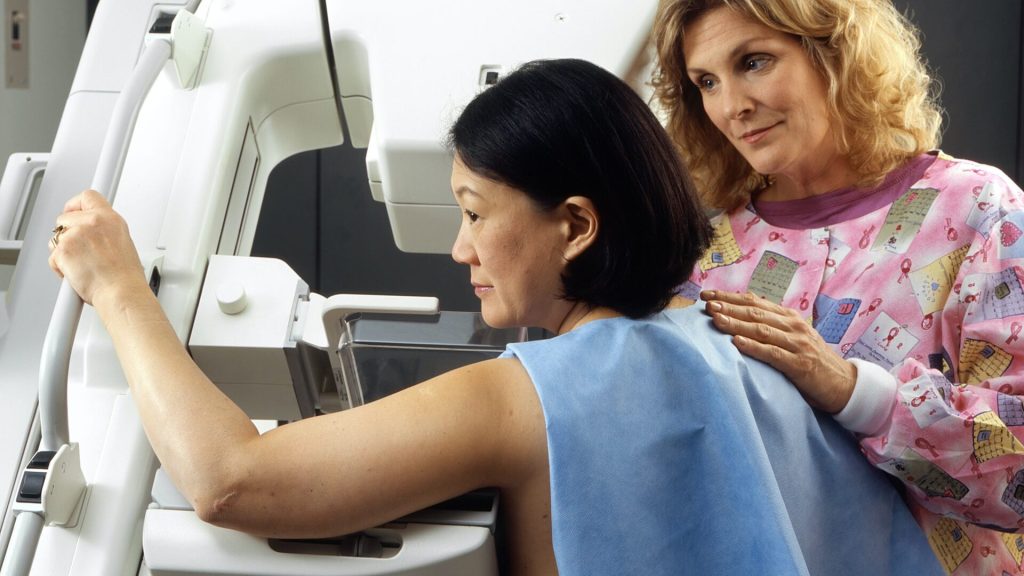
By providing detailed images, MRg-A-SBRT can be used to adjust a patient’s radiation plan every day to account for anatomical changes and to monitor the position of the prostate in real time while the radiation beam is on to ensure that treatment is being directed accurately to the prostate. Although MRg-A-SBRT is becoming more popular and multiple clinical trials have tested it, it is unclear whether the technique, which requires more time and resources than standard procedures, has an impact on clinical outcomes and side effects compared with other ways of delivering radiation.

To investigate, Jonathan E. Leeman, MD, of the Dana-Farber Cancer Institute and Brigham and Women’s Hospital, and his colleagues combined data from 29 clinical trials testing MRg-A-SBRT versus conventional CT-guided treatment, with a total of 2457 patients.

MRg-A-SBRT was associated with significantly fewer urinary and bowel side effects in the short term following radiation. Specifically, there was a 44% reduction in urinary side effects and a 60% reduction in bowel side effects.

“The study is the first to directly evaluate the benefits of MR-guided adaptive prostate radiation in comparison to another more standard and conventional form of radiation, and it provides support for use of this treatment in the management of prostate cancer,” said Dr Leeman.

Dr Leeman noted that the study also raises further questions regarding this type of treatment. For example, will the short-term benefits lead to long-term benefits, which are more impactful for patients? Longer follow-up will help answer this question because MRg-A-SBRT is a relatively new treatment. Also, which aspect of the technology is responsible for the improved outcomes seen in clinical trials? “It could potentially be the capability for imaging-based monitoring during the treatment or it could be related to the adaptive planning component. Further studies will be needed to disentangle this,” said Dr Leeman.

An accompanying editorial discusses the analysis’ findings, weighs the potential benefits and shortcomings of adopting this treatment strategy for patients, and questions the value of broad adoption.

Source: Wiley

Contrary to Prior Belief, T Cells Even Protect the Cornea

Photo by Stormseeker on Unsplash

Researchers have discovered that the immune cells guarding the healthy human cornea from pathogens and inflammation are T cells, and dendritic cells, as previously thought. The discovery, published in PNAS, redefines current understanding of the immune cell landscape in the cornea of a healthy human eye. It builds on the team’s previous research in Cell Reports that showed that T cells protect the eye against virus infection in mice.

The collaborative research team jointly developed a new imaging technique as part of their investigation.

Research leader Professor Scott Mueller, from the Department of Microbiology and Immunology at the Doherty Institute explained that our knowledge of the various immune cell types in the human cornea is important for establishing the eye’s protective mechanisms against pathogens and disease.

“By combining our newly developed imaging technique with other advanced analytical approaches, we were able to discover that a significant number of cells at the surface of the healthy cornea are actually T cells,” said Professor Mueller.

“Until now, these cells were mistakenly classified as dendritic cells based on static imaging. This completely changes the current dogma in the field that only dendritic cells are present in the healthy cornea.”

The study’s first author, University of Melbourne’s Associate Professor Laura Downie said that being able to dynamically capture the cells’ normal behaviour, and in response to inflammation, provides unique understanding into the immune response in the eye.

“Using our non-invasive imaging approach, which we term Functional In Vivo Confocal Microscopy (Fun-IVCM), we have been able to see that these T cells move around quickly and interact with other cells and nerves in the outermost layer of the cornea. We also captured different cell dynamics in response to contact lens wear and in allergic eye disease, and quantified how these behaviours are modulated by drug treatments,” said Associate Professor Downie.

“These findings reshape our understanding of the distinct immune cell subsets in the human cornea, and how they respond to different stimuli. Using Fun-IVCM, we can achieve rapid, real-time insight into the cellular immune responses in living humans, in this accessible peripheral sensory tissue.”

Senior author Dr Holly Chinnery, also of the University of Melbourne, added that the new research will have major implications for the medical and immunology fields, including for patients and practitioners.

“Because this new technique involves non-invasive, time-lapse imaging of the human cornea, Fun-IVCM could be used in clinics directly to assess immune responses and ocular health. It could even be used for general immune system health,” said Dr Chinnery.

“Changes in T cells and behaviour could be used as a clinical biomarker of disease and assist with treatments.”

Source: The Peter Doherty Institute for Infection and Immunity

Daily Statin Reduces Risk of Major Cardiovascular Events in People Living with HIV

Photo by Towfiqu Barbhuiya on Unsplash

A new study published in the New England Journal of Medicine found that statins, a class of cholesterol-lowering medications, may offset the high risk of cardiovascular disease in people living with HIV by more than a third, potentially preventing one in five major cardiovascular events or premature deaths in this population. People living with HIV can have a 50–100% increased risk for cardiovascular disease.

“This research suggests that statins may provide an accessible, cost-effective measure to improve the cardiovascular health and quality of life for people living with HIV,” said Gary H. Gibbons, MD, director of the National Heart, Lung, and Blood Institute (NHLBI), a study funder. “Additional research can further expand on this effect, while providing a roadmap to rapidly translate research findings into clinical practice.”

For the double-blinded phase 3 trial, known as Randomised Trial to Prevent Vascular Events in HIV (REPRIEVE) study, researchers randomised participants into either a treatment group, where they received pitavastatin calcium daily or a control group receiving placebo. The researchers followed participants for about five years, but ended the trial early when they discovered the treatment benefits outweighed potential risks.

To understand the benefits, the researchers compared how often participants in each group experienced major cardiovascular events, including heart attacksstrokes, or surgery to open a blocked artery. They found participants who took daily pitavastatin had 35% fewer major cardiovascular events than those who took a placebo. The researchers also measured the number of deaths in combination with major cardiovascular events during the study period and found participants in the treatment group were 21% less likely than those in the placebo group to experience these events. Additionally, those taking pitavastatin had a 30% reduction in their low-density lipoprotein (LDL) cholesterol levels.

“Lowering LDL cholesterol levels reduces risks for cardiovascular events, like having a heart attack and stroke, but these findings suggest there may be additional effects of statin therapy that explain these reduced risks among people living with HIV,” said Steven K. Grinspoon, MD, the study chair. “Ongoing research about how statin therapy may affect inflammation and increased immune activation among people with HIV may help us better understand the additional benefits we’re seeing with this treatment approach.” 

To support optimal health outcomes among the study participants, normal liver and kidney function were an enrolment criteria. They were also required to take antiretroviral therapy, which itself is critical to reducing the risk of HIV complications and related comorbidities, including cardiovascular disease.

Beginning in 2015, REPRIEVE enrolled 7769 adults, ages 40–75, from 145 sites in 12 countries. Adults in the study were an average age of 50 and had low-to-moderate risks for cardiovascular disease, which meant they normally would not have been prescribed statins. Women accounted for 31% of participants. Approximately 41% of study participants identified as Black, 35% as white, 15% as Asian, and 9% as another race.

Source: National Insitutes of Health

Cadmium Detected in Urine of Women with Endometriosis

Phot by Sora Shimazaki on Pexels

Women with a history of endometriosis had higher concentrations of cadmium in their urine compared to those without that diagnosis, according to a study with a large representative study that suggests the toxic metal, which is also an endocrine disruptor, could be linked to the development of endometriosis. The researchers, from Michigan State University, published these findings in the journal Human Reproduction.

Affecting one in 10 reproductive-age women, endometriosis is when tissue that looks like the lining of the uterus, appears outside of it. Those with endometriosis can experience chronic, painful and debilitating symptoms, which can interfere with all aspects of life, including daily activity, work productivity, school performance and personal relationships.

“Despite the adverse impact of endometriosis on quality of life, it remains an understudied condition,” said Kristen Upson, assistant professor in the Department of Epidemiology and Biostatistics at the MSU College of Human Medicine and senior author of the study.

“By looking at environmental risk factors such as metal cadmium, we are moving the needle closer to understanding risk factors for this condition,” added the study’s first author, Mandy Hall, a data analyst in the MSU Department of Epidemiology and Biostatistics.

Cadmium is a toxic metal and a “metalloestrogen,” meaning it can act like oestrogen. In the US, people are commonly exposed to cadmium by breathing in cigarette smoke and eating contaminated food like spinach and lettuce.

While this is not the first study exploring a potential link between cadmium and endometriosis, the researchers said it’s the largest study to look at cadmium measured in urine, which reflects long-term exposure between 10 and 30 years.

For their study, researchers used data from NHANES, a national US population study between 1999 and 2006. Out of the survey’s more than 41 000 participants, the researchers limited their study population to those 20 to 54 years of age with information on endometriosis diagnosis.

The researchers then analysed the data, dividing the cadmium levels into four classes, or quartiles, with the first quartile being the lowest exposure and the fourth being the largest exposure.

They found that participants in the second and third quartiles were twice as likely to have been diagnosed with endometriosis than those in the first quartile. The data also suggests a 60% increased prevalence of endometriosis based on urinary cadmium concentrations in the fourth quartile.

“The findings are interesting given that cadmium can act like the hormone estrogen, and this hormone is central to the development of endometriosis,” Hall said.

The researchers say further studies are needed to confirm their findings. Upson said this work is part of her larger research looking at everyday factors that may increase toxic metal exposure in women as well as the impact of toxic metals on gynaecologic health. Hall plans to incorporate environmental factors in their ongoing research on endometriosis and other gynaecologic conditions.

Source: Michigan State University

Specialised Omega-3 Lipid Could be a New Treatment for Acute Kidney Injury

Photo by Robina Weermeijer on Unsplash

Researchers from Singapore have identified a potential dietary supplement of omega-3 that may improve recovery following acute kidney injury (AKI). The finding, published in the Journal of Lipid Research, may offer a new way to treat this serious condition, which currently has few therapies.

The study was part of a long-running research programme at Duke-NUS Medical School investigating how cells take up a specialised omega-3 lipid called LPC-DHA.

AKI affects some 13.3 million people globally each year, with a mortality rate of 20 to 50% depending on the economic status of the country and stage of the disease. One of the main causes of AKI is ischaemic reperfusion injury, which occurs when the kidney’s blood supply is restored after a period of restricted blood flow and poor oxygen delivery due to illness, injury or surgical intervention. In particular, it damages a crucial part of the kidney called the S3 proximal tubules that regulate the levels of absorption of water and soluble substances, including salts.

“AKI is a serious health problem with limited treatment options,” said Dr Randy Loke, first author of the study. “We sought to understand how these tubules repair themselves and found that the activity of the protein Mfsd2a, which transports LPC-DHA into cells, is a key factor influencing the rate of recovery of kidney function after ischaemic reperfusion injury.”

In their study, the researchers discovered that preclinical models with reduced levels of Mfsd2a showed delayed recovery, increased damage and inflammation after kidney injury. However, when these models were treated with LPC-DHA, their kidney function improved and the damage was reduced. LPC-DHA also restored the structure of the S3 proximal tubules, helping them function properly again.

“While more research is needed, the potential of LPC-DHA as a dietary supplement is exciting for future recipients who have suffered from AKI,” said senior study author Professor David Silver. “As our results suggest that LPC-DHA could become a safe and effective treatment that offers lifelong protection, its potential can help protect the kidneys and aid in recovery for these individuals.”

In the next phase, the research team plans to continue investigating the beneficial functions of LPC in the kidney and are aiming to initiate clinical testing of LPC supplements to determine their effectiveness in improving renal function and recovery following AKI in patients.

They also plan to continue their investigations of the protein Mfsd2a to learn more about its role in LPC transport and its involvement in diseases affecting other tissues and organs. Previous research by Prof Silver’s group, with collaborators from other institutions, have already highlighted the significance of the protein’s LPC-transporting activities in diseases of other organs, including the liver, lungs and brain.

Source: Duke–NUS Medical School

Quality and Timing of Snacks Determines Their Health Impacts

Credit: Pixabay CC0

Snacking is becoming increasingly popular, with more than 70% of people reporting they snack at least twice a day. In a new study presented at NUTRITION 2023, researchers examined whether snacking affects health and if the quality of snack foods matters.

“Our study showed that the quality of snacking is more important than the quantity or frequency of snacking, thus choosing high quality snacks over highly processed snacks is likely beneficial,” said presenter Kate Bermingham, PhD, a postdoctoral fellow at King’s College London. “Timing is also important, with late night snacking being unfavourable for health.”

The work is part of the ZOE PREDICT project, a group of large in-depth nutritional research studies designed to uncover how and why people respond differently to the same foods.

“Surprisingly little has been published on snacking, despite the fact that it accounts for 20–25% of energy intake,” said Bermingham. “PREDICT followed a large number of people and captured detailed information on their snacking behaviours, allowing this in-depth exploration of snacking on health.”

Using data from just over 1000 participants in the ZOE PREDICT 1 study, the researchers examined the relationship between snacking quantity, quality, and timing with blood fats and insulin levels, which are both indicators of cardiometabolic health.

The analysis showed that snacking on higher quality foods, which contain significant amounts of nutrients relative to the calories they provide, was associated with better blood fat and insulin responses. The researchers also observed that late-evening snacking, which lengthens eating windows and shortens the overnight fasting period, was associated with unfavourable blood glucose and lipid levels. There was no association between snacking frequency, calories consumed, and food quantity with any of the health measures analysed.

“We observed only weak relationships between snack quality and the remainder of the diet, which highlights snacking as an independent modifiable dietary feature that could be targeted to improve health,” said Bermingham.

Source: EurekAlert!

Childhood TV Watching Linked to Metabolic Syndrome in Adulthood

Photo by Victoria Akvarel on Pexels

A new study has added to the evidence that excessive TV watching as a child can lead to poor health in adulthood. The research, published this week in the journal Pediatrics, found that children who watched more television were more likely to develop metabolic syndrome as an adult.

Metabolic syndrome is a cluster of conditions including hypertension, hyperglycaemia, excess body fat, and abnormal cholesterol levels that lead to an increased risk of heart disease, diabetes and stroke.

Using data from 879 participants of the Dunedin study, researchers found those who watched more television between the ages of 5 and 15 were more likely to have these conditions at age 45.

Television viewing times were asked at ages 5, 7, 9, 11, 13 and 15. On average, they watched just over two hours per weekday.

“Those who watched the most had a higher risk of metabolic syndrome in adulthood,” says Professor Bob Hancox, who led the study.

“More childhood television viewing time was also associated with a higher risk of overweight and obesity and lower physical fitness.”

Boys watched slightly more television than girls and metabolic syndrome was more common in men, than women (34% and 20% respectively). The link between childhood television viewing time and adult metabolic syndrome was seen in both sexes however, and may even be stronger in women.

There was little evidence that watching less television as an adult reduced the association between childhood television viewing and adult health.

“While, like any observational study, researchers cannot prove that the association between television viewing at a young age directly causes adult metabolic syndrome, there are several plausible mechanisms by which longer television viewing times could lead to poorer long-term health.

“Television viewing has low energy expenditure and could displace physical activity and reduce sleep quality,” he says.

“Screentime may also promote higher energy intake, with children consuming more sugar-sweetened beverages and high-fat dietary products with fewer fruit and vegetables. These habits may persist into adulthood.”

The results are important because screen times have increased in recent years with new technologies.

“Children today have far more access to screen-based entertainment and spend much more time being sedentary. It is likely that this will have even more detrimental effects for adult health.

“These findings lend support to the World Health Organization recommendation that children and young teenagers should limit their recreational screen time.”

Source: University of Oregon

Elevated Body Temperature Helps Gut Microbiota to Fight Viruses

Researchers from The University of Tokyo have helped unravel the connection between high body temperature and increased viral resistance. Older adults are at a higher risk of contracting viral infections, research shows. Quite notably, they also have lower mean body temperatures – yet the effects of increased body temperature on fighting viral infections remain largely unexplored. The researchers found that higher temperature increased bile acids along with the infection-fighting capability of the gut microbiota. Their study was published in Nature Communications.

To conduct their experiments, the team used mice which were heat- or cold-exposed at 4°C, 22°C, or 36°C a week before influenza virus infection. After the viral infection was induced, the cold-exposed mice mostly died due to severe hypothermia, whereas the heat-exposed mice were highly resistant to the infection even at increasing doses of the virus. “High-heat-exposed mice raise their basal body temperature above 38°C, allowing them to produce more bile acids in a gut microbiota-dependent manner,” remarks Dr Takeshi Ichinohe from the Division of Viral Infection, The University of Tokyo, Japan.

The authors speculated that signalling of deoxycholic acid (DCA) from the gut microbiota and its plasma membrane-bound receptor “Takeda G-protein-coupled receptor 5” (TGR5) increased host resistance to influenza virus infection by suppressing virus replication and neutrophil-dependent tissue damage.

While working on these experiments, the team noticed that mice infected with the influenza virus showed decreased body temperatures nearly four days after the onset of the infection, and they snuggled together to stay warm!

The team noticed similar results after switching the influenza virus with SARS-CoV-2 and the study results were also validated using a Syrian hamster model. Their experiments revealed that body temperature over 38°C could increase host resistance to influenza virus and SARS-CoV-2 infections. Moreover, they also found that such increase in body temperature catalyzed key gut microbial reactions, which in turn, led to the production of secondary bile acids. These acids can modulate immune responses and safeguard the host against viral infections.

Dr. Ichinohe explains, “The DCA and its nuclear farnesoid X receptor (FXR) agonist protect Syrian hamsters from lethal SARS-CoV-2 infection. Moreover, certain bile acids are reduced in the plasma of COVID-19 patients who develop moderate I/II disease compared with the minor severity of illness group.”

The team then performed extensive analysis to gain insight into the precise mechanisms underlying the gut-metabolite-mediated host resistance to viral infections in heat-exposed rodents. Besides, they also established the role of secondary bile acids and bile acid receptors in mitigating viral infections.

“Our finding that reduction of certain bile acids in the plasma of patients with moderate I/II COVID-19 may provide insight into the variability in clinical disease manifestation in humans and enable approaches for mitigating COVID-19 outcomes,” concludes Dr. Ichinohe.

To briefly summarize, the published study reveals that the high-body-temperature-dependent activation of gut microbiota boosts the serum and intestinal levels of bile acids. This suppresses virus replication and inflammatory responses that follow influenza and SARS-CoV-2 infections.

A heartfelt appreciation to the Japanese researchers for placing their trust in their intuition and gut instincts!

Source: University of Tokyo

Cognitive Impairment Persists Two Years after COVID Infection

Image by Fusion Medical on Unsplash

Researchers from King’s College London have found that people with longer-term COVID symptoms including brain fog showed reduced performance in tasks testing different mental processes – up to two years after infection with the virus, according to results published in the journal eClinicalMedicine.

Researchers examined whether COVID infection affected performance in two rounds of online cognitive testing that took place in 2021 and 2022. Data was collected for over 3000 participants of the COVID Symptom Study Biobank study, across 12 tasks that tested memory, attention, reasoning, processing speed and motor control.

The participants whose test scores were most affected by COVID were those who had experienced symptoms related to the virus for 12 weeks or more. In these people, the effect of COVID on test accuracy was comparable in size to the effect of a 10-year increase in age.

There was no significant improvement in these test scores between the two rounds of testing, which took place nine months apart. By the second round of testing, the average time since participants’ initial COVID infection was almost two years.

The researchers then separated participants by whether they felt fully recovered following COVID infection. People who felt fully recovered after COVID infection performed similarly to those who had not had the virus at all. In contrast, participants who did not feel fully recovered after infection had lower task accuracy scores on average.

Lead author Dr Nathan Cheetham, a Senior Postdoctoral Data Scientist at King’s College London said:

“Our findings suggest that, for people who were living with long-term symptoms after having COVID, the effects of the coronavirus on mental processes such as the ability to recall words and shapes are still detectable at an average of almost two years since their initial infection.

“However, the result that COVID had no effect on performance in our tests for people who felt fully recovered, even if they’d had symptoms for several months and could be considered as experiencing ‘long COVID’, was good news. This study shows the need to monitor those people whose brain function is most affected by COVID-19, to see how their cognitive symptoms continue to develop and provide support towards recovery.”

Professor Claire Steves, a Professor of Ageing and Health at King’s College London, added:

“We used sensitive tests to measure speed and accuracy across a range of brain challenges. This study shows that some individuals have measurable changes in these tests after COVID-19 going on for nearly two years. The fact remains that two years on from their first infection, some people don’t feel fully recovered and their lives continue to be impacted by the long-term effects of the coronavirus. We need more work to understand why this is the case and what can be done to help.”

Source: King’s College London

Scientists Create Protein that Blocks Breast Cancer Metastasis

In a paper published in the journal Biomolecules, UK and Chinese researchers report their creation of a biomedical compound that has the potential to stop breast cancer metastasis.

The scientists from the Chemistry and Biochemistry Departments at the University of Liverpool and Nanjing Medical School in China have discovered a possible way to block proteins produced by cancer cells that promote metastasis – the chief impediment to successful cancer treatment.

Prof Philip Rudland from the University of Liverpool explained: “As a general rule, cancer that has spread is treated with chemotherapy, but this treatment can rarely be given without severely harming or becoming toxic to the patient. The importance of our work was to identify a specific and important target to attack, without toxic side effects.”

The University’s research team have in the past discovered that specific proteins are involved in the metastatic process; these proteins are different from those involved in the production of the primary tumour. One such example is a protein called ‘S100A4’, and is the protein chosen by the research team to target for the identification of chemical inhibitors of metastasis, using model systems of cells from the highly metastatic and incurable hormone receptor-free breast cancer.

Using these model systems, researchers at the University’s Department of Biochemistry discovered a novel compound that can specifically block the interaction of this metastasis-inducing protein S100A4 with its target inside the cell. Researchers in the Department of Chemistry then synthesised a simpler chemical and connected it to a warhead which stimulates cells’ normal protein-degrading machinery. This compound now works at very low doses to inhibit properties associated with metastasis, an improvement of over 20 000-fold on the original unarmed inhibitor, with virtually no toxic side effects. Moreover, in collaboration with Chinese researchers at Nanjing Medical School, they have shown that this compound inhibits metastasis in similar metastatic tumours in mice, suggesting a potential therapeutic role.

Dr Gemma Nixon, Senior Lecturer in Medicinal Chemistry at the University of Liverpool said: “This is an exciting breakthrough in our research. We now hope to take the next steps, and repeat this study in a large group of animals with similar metastatic cancers so that the efficacy and stability of the compounds can be thoroughly investigated and if necessary improved by further design and syntheses, prior to any clinical trials.”

“Significantly, this particular protein we’re investigating occurs in many different cancers, which could mean this approach may be valid for many other commonly occurring human cancers.”

Source: University of Liverpool