Category: Surgeries & Procedures

New Imaging Tool in Development Could Revolutionise Neurosurgery
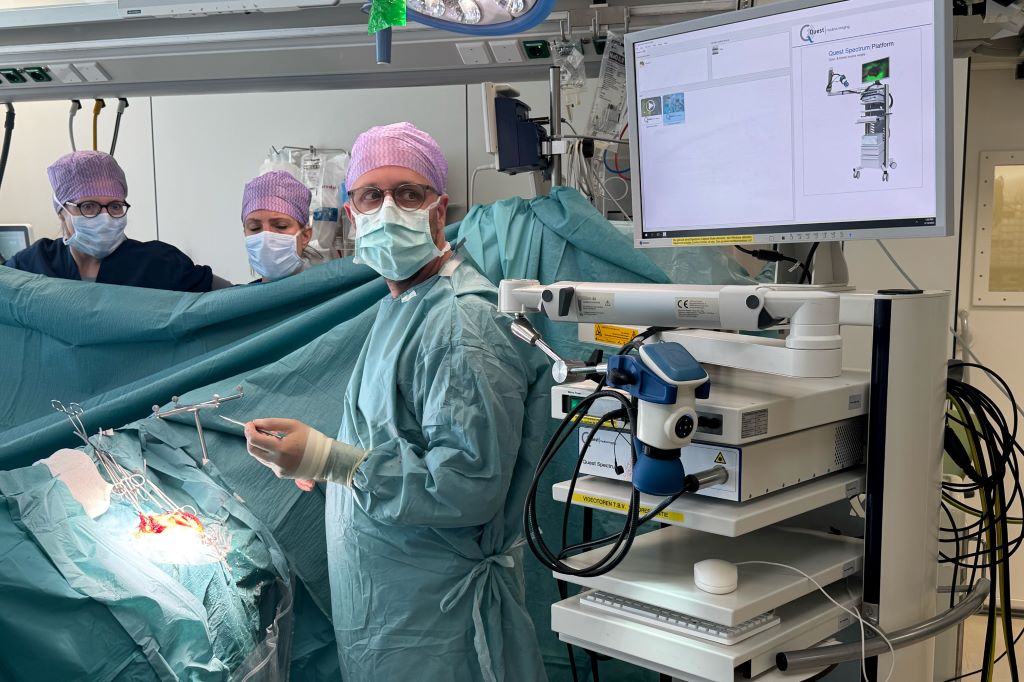
A medical team at Erasmus University Medical Center in the Netherlands uses the new imaging probe with a Quest camera to get a better view of cancerous tumors during non-brain cancer surgery. Photo courtesy of Erasmus University Medical Center

In a significant leap forward for successful cancer surgery, researchers at the University of Missouri and collaborators have developed a new imaging probe to help surgeons more accurately identify and remove aggressive tumours during operations.

The tool is expected to be a critical advancement in the fight against glioblastoma, one of the most difficult-to-treat brain cancers. In the future, it is intended to be expanded for image-guided surgery of various other solid tumours.

Described in a new study in Nature Publishing Group Imaging, the innovation works by pairing a fluorescent dye with a fatty acid molecule that cancer cells readily absorb. When introduced into the body, the compound is taken up by tumour cells, causing them to glow under near-infrared light, revealing cancer that might otherwise remain hidden.

Glioblastoma is considered surgically incurable because the tumour doesn’t stay in one place – it spreads and invades healthy brain tissue in a diffuse, microscopic way. This makes it impossible to remove completely without risking serious damage to brain function.

“Surgery remains one of the primary treatments for many cancers,” Elena Goun, associate professor of chemistry in the College of Arts and Science and one of the lead authors of the study, said. “In breast or prostate cancer, surgeons can often remove the tumour along with surrounding tissue. In brain cancer, that’s simply not possible. You must preserve healthy brain tissue. But if even a few cancer cells are left behind, the disease will return.”

That dilemma is especially acute with glioblastoma, which doesn’t form a neatly contained mass. Instead, it sends out microscopic extensions — finger-like projections that blend into healthy brain tissue and are invisible to the naked eye.

Because of this, surgeons must walk a fine line: removing as much tumour as possible while avoiding harm to vital brain areas. The more thoroughly the tumour is removed, the more effective follow-up treatments like radiation and chemotherapy tend to be.

The new small-molecule probe, known as FA-ICG, is engineered to solve that problem. It links a natural long-chain fatty acid (FA) to indocyanine green (ICG), an FDA-approved near-infrared dye widely used in surgical imaging. This fatty acid-based approach means the probe is highly selective: glioblastoma cells, which thrive on fatty acids, absorb it more than normal brain cells. That makes the cancer stand out more clearly.

The result is a tool that takes advantage of cancer’s altered metabolism to highlight tumour cells from within.

“Surgeons would view a monitor during surgery showing where the probe is lighting up,” Goun explained. “If they still see fluorescent signals, it means cancer is still present and more tissue needs to be removed. When the light disappears, they would know they’ve cleared the area.”

In the operating room, surgeons already use a variety of tools to guide tumour removal – including microscopes, ultrasound and fluorescent dyes. Of those, fluorescent dyes are particularly useful because they make otherwise invisible tumour cells light up under special lighting.

Right now, the only approved imaging dye for glioblastoma surgery is 5-ALA, which fluoresces under blue light. But 5-ALA comes with major limitations: The operating room must be darkened in order to see it, tissue penetration is shallow and the fluorescent signal is often weak and non-specific.

It also comes with side effects, including photosensitivity, meaning patients must avoid bright light exposure after surgery due to the risk of skin and eye damage.

That’s where the FA-ICG probe shines – both literally and functionally.

Compared to 5-ALA, FA-ICG is brighter, works under normal surgical lighting, and offers real-time visualisation under the microscope – no need to turn the lights off mid-surgery. This saves time and makes procedures more efficient. The signal-to-background ratio is also higher, meaning it’s easier to distinguish tumour tissue from healthy brain.

The FA-ICG probe is not only easier to see, it’s also easier to use. Its longer half-life allows more flexibility in scheduling surgeries, and the logistics of administration are simpler than with current probes.

“The upside of fluorescence-guided surgery is that you can make little remnants much more visible using the light emitting properties of these tumour cells when you give them a dye,” said Rutger Balvers, a neurosurgeon at Erasmus University Medical Center in the Netherlands, who is expected to lead human clinical trials of the probe. “And we think that the upside of FA-ICG compared to what we have now is that it’s more select in targeting tumour cells. The visual properties of the probe are better than what we’ve used before.”

Michael Chicoine is a neurosurgeon at MU Health Care and chair of Mizzou’s School of Medicine’s Department of Neurosurgery. While he’s not directly involved in the research, Chicoine understands the potential benefits firsthand.

Currently, he said, MRIs are the gold standard for imaging tumours; however, they’re expensive and time-consuming, especially when required during an operation.

“This fluorescent metabolically linked tool gives you real-time imaging,” he said. “We could merge techniques, using the probe during surgery and saving the MRI for a sort of final exam. It’s definitely an exciting advancement.”

Researchers are also excited about other uses for the probe, including for other types of cancers and for use during follow-up treatments.

“After radiation or chemotherapy, it becomes very difficult to distinguish between scar tissue and active tumor,” Chicoine said. “This probe could give us a definitive answer – helping doctors know whether to continue treatment or adjust it, or consider another surgery. Eliminating the current uncertainty would be really helpful.”

Another promising use of the probe could be in photodynamic therapy either during or after surgery. Since the dye also has light-activated properties that can kill cancer cells, researchers are exploring whether it could double as a treatment tool, not just a diagnostic one.

Clinical trials for use in glioblastoma cases are expected to start in Europe, with strong interest already growing among neurosurgical teams.

The upcoming Phase 1 trial will focus on how patients tolerate the probe, whether there are any side effects at an effective dose and how its performance compares to existing tools. Ultimately, the goal is to make brain tumour surgery safer, helping surgeons remove all cancerous tissues while preserving as much healthy brain tissue as possible.

If results are positive, future studies could expand the use of FA-ICG beyond brain tumours to other cancers with high fatty acid metabolism, such as pancreatic cancer, according to fellow corresponding author Laura Mezzanotte from the Erasmus’ Department of Radiology and Nuclear Medicine.

Source: University of Missouri

Self-esteem Increases by 131% Within One Year of Bariatric Surgery

Photo by I Yunmai on Unsplash

Self-esteem scores more than doubled within one year of weight-loss surgery, according to a new study* presented at the American Society for Metabolic and Bariatric Surgery (ASMBS) 2025 Annual Scientific Meeting.

Researchers from Geisinger Medical Center found that after bariatric surgery self-esteem scores rose to 77.5 from 33.6 – a more than 40-point increase. The higher the score on a scale from 0 to 100, the higher the level of self-esteem and quality of life. The amount of weight loss appears to fuel the increase in self-esteem — scores were highest among those who lost the most weight despite demographics differences including gender, age, and race or type of bariatric procedure.

Researchers used a prospectively maintained database to identify 5,749 patients aged 18 and older with body mass index (BMI) of 35 or more who had metabolic and bariatric surgery between 2006 and 2019. Patients completed the Impact of Weight Quality of Life (iwQOL) survey pre-operatively and 12 months after the operation to assess weight stigma and their quality of life.

“Understanding weight stigma and psychosocial factors associated with obesity is essential to offering holistic care. While these factors should not dictate the decision to have bariatric surgery, they should be an important part of the conversation,” said study co-author Justin Dhyani, MD, Geisinger Medical Center in Danville, PA.

Weight stigma is associated with adverse health outcomes including depression, anxiety, disordered eating, and low self-esteem. Among adults with obesity, the prevalence of weight discrimination is 19% to 42%, with higher rates reported among those with higher BMIs and women.

“Weight stigma is a serious issue that places an extra psychological burden on patients struggling with obesity and there is no excuse for it,” said Ann M. Rogers, MD, MD, FACS, FASMBS, President, ASMBS, who was not involved in the study. “This study shows we need to understand what patients are going through and be supportive and empowering of them as they navigate their health and make decisions about treatment.”

Source: American Society for Metabolic and Bariatric Surgery

Blood Conservation Technique Does Not Reduce Need for Transfusions, New Study Finds

Photo by Charliehelen Robinson on Pexels

An international study published in the New England Journal of Medicine sheds light on acute normovolaemic haemodilution, also known as ANH, a blood conservation technique. ANH involves removing a patient’s blood before the patient goes on heart-lung bypass, and then reinfusing it toward the end of heart surgery. Long debated for its effectiveness, ANH does not appear to reduce the need for red blood cell transfusion, according to results of the global clinical trial.

Nevertheless, study co-author Kenichi Tanaka, MD, professor and chair of the Department of Anesthesiology at the University of Oklahoma College of Medicine, said the outcomes necessitate further scrutiny and may not be applicable to US patients.

More than 2 million patients worldwide undergo heart surgery annually, and about 35% require at least one unit of red blood cells. Blood transfusions can lead to complications, are costly, and are sometimes affected by shortages. Previous retrospective studies and meta-analyses have shown mixed results about whether ANH reduces the need for blood transfusions in heart surgery.

Although the study did not show that ANH decreased infusions, several factors should be taken into consideration, said Kenichi Tanaka, MD, who co-led the United States’ participation in the trial along with researchers from the University of Virginia. Altogether, the study recruited 2010 patients from 11 countries in Europe, Asia, South America and North America.

“This trial may reduce the interest in doing another large, randomised study, but I think it does not exclude the possibility that ANH could help some patients,” he said.

Tanaka pointed to several aspects that may make the study less applicable to the U.S., where about 20% of medical centers use ANH:

  • US centres did not begin enrolling patients in the study until at least half of all participants had been enrolled. He estimates that less than 5% of participants are from the United States.
  • US patients are typically larger than patients from other countries and, paradoxically, larger body size is protective against surgical bleeding and the need for transfusions.
  • Smaller patients have less blood to give during ANH. This means a smaller volume is available to treat anaemia and bleeding that can occur while the patient is on a heart-lung bypass machine.
  • There was no standardised protocol for ANH or transfusion in the trial. Countries like the United States are more likely to use special clotting factor concentrates in combination with ANH, which has been shown to reduce the need for transfusions.
  • In supplemental information not included in the study, patients receiving ANH showed a slight trend toward better overall survival rates compared to those who received transfusions only.

Tanaka is preparing to publish a retrospective study showing that ANH reduced the need for transfusions in a US population.

“I plan to continue practising ANH,” he said. “It is also an option for Jehovah’s Witness patients whose beliefs stipulate they cannot receive blood transfusions (from donor blood). At the least, the study proved that there is no downside for ANH. I believe ANH can be part of a multi-pronged blood conservation strategy in the US, where blood product costs are very high.”

Source: University of Oklohoma

Surprising Shifts in Who’s Getting What in South African Cosmetic Surgery

Professor Chrysis Sofianos

Twenty years ago, cosmetic surgery was still a subject of taboo, whispered about behind closed doors. Today, that silence has been replaced by honest conversations about beauty, confidence, ageing, empowerment, and personal choice, as South Africans openly embrace a wide range of aesthetic enhancements. And what South Africans are choosing might surprise you.

Leading plastic and reconstructive surgeon Professor Chrysis Sofianos shares insights from his own practice, revealing how cosmetic surgery trends are evolving across age and race – and what that means for the future of beauty.

“Cosmetic surgery is no longer about fitting a single mould, but about choosing how we, as individuals, want to age, restore, or refine. Today’s patients are more informed, intentional, and unapologetic about their tastes, and wanting to feel good in their own skin,” he says.

Aesthetic choices by age

Gone are the days where cosmetic procedures were the exclusive domain of patients of a certain age. According to Professor Sofianos, the modern approach views aesthetics as a journey, although age still plays a major role in shaping the types of treatments individuals may seek:

  • 20s: Patients in their twenties are often navigating early self-image issues, driven by social media influences. The most requested procedures include breast augmentations, lip fillers, and occasional rhinoplasties to address long-held insecurities.
  • 30s: Often post-pregnancy or in the midst of career and family life, this group leans toward tummy tucks, mommy makeovers, and the start of anti-ageing injections, cosmetic fillers, and preventative treatments. Restoration without exaggeration is usually the goal.
  • 40s: This is the decade of refinement. Liposuction, eyelid lifts, and more assertive facial rejuvenation procedures become common, as patients seek to stay ahead of midlife volume loss and skin laxity.
  • 50s and beyond: For those who want long-term, natural results, the deep plane facelift, neck lifts, and biostimulators become top of mind. “Our approach is structural, not superficial,” he notes. “It’s about restoring facial harmony without compromising identity.”

“What fascinates me is how differently my patients may define beauty. A 22-year-old influencer and a 55-year-old executive might sit in the same waiting room, but their goals couldn’t be more different. We’re not selling a standard look – we’re facilitating personal choices, and sometimes, those choices may even challenge conventional aesthetic norms.”

Understanding patient preferences: A cultural lens

Perhaps the most significant change is how different communities and racial groups are approaching enhancement in South Africa, reflecting new trends in beauty ideals. For example, African women are increasingly choosing breast reductions, prioritising comfort and opting for a different aesthetic from more traditional beauty standards which often favour a fuller figure or more pronounced curves.

Indian patients are leading the way in tummy tucks and body contouring, while Caucasian patients tend to focus on breast augmentations and high-definition liposuction, particularly around the waistline and abdomen, to refine their silhouette and definition.

“What’s beautiful about this shift is how it reflects our diversity. We’re not seeing patients trying to look like someone else – they’re choosing procedures that enhance their natural features and fit their lifestyles,” says Sofianos. “Critically, there is no one-size-fits-all in aesthetic medicine. The more we understand the nuances of each patient’s background and goals, the better we can serve them.”

Why winter is the most strategic time for surgery

No matter your personal objectives, an insider tip could make a huge difference in your surgical recovery: while people mistakenly tend to plan procedures for December – unaware that most surgeons do not operate after November due to holiday closures and the necessity of post-operative care – smart patients are booking for winter.

Cooler temperatures help to reduce swelling, promote easier healing, and generally mean less downtime. Patients also benefit from increased coverage beneath bulkier clothing, allowing them to recover more discreetly while still going about their daily lives. With fewer social commitments and sunnier distractions, winter becomes the ideal window for surgical recovery.

“If you want to look and feel your best by summer, now is the time to plan,” he advises. “Winter is a strategic opportunity for recovery.”

As aesthetic surgery continues to evolve in South Africa, Professor Sofianos believes that education, access, and authenticity are key pillars moving forward.

“My role is to guide patients through their options, help them make informed choices, and deliver results that are both technically excellent and emotionally empowering. The most rewarding part of my practice isn’t about creating one particular look, but about helping people become more confident versions of themselves. Whether that means a subtle enhancement or a more significant change, the choice is truly personal.”

Surgeons Perform World-first Bladder Transplant

UCLA Health surgical team has performed the first-in-human bladder transplant. 

The surgery was successfully completed at Ronald Reagan UCLA Medical Center on May 4, 2025. The team was led by Dr Nima Nassiri, a urologic transplant surgeon and director of the UCLA Vascularized Composite Bladder Allograft Transplant Program, with assistance from Dr Inderbir Gill, founding executive director of USC Urology.

“Bladder transplantation has been Dr Nassiri’s principal academic focus since we recruited him to the UCLA faculty several years ago,” said Dr Mark Litwin, UCLA Urology Chair, “It is incredibly gratifying to see him take this work from the laboratory to human patients at UCLA, which operates the busiest and most successful solid-organ transplant program in the western United States.”

“This first attempt at bladder transplantation has been over four years in the making,” Nassiri said. “For the appropriately selected patient, it is exciting to be able to offer a new potential option.” 

The patient had lost most of his bladder during a tumour removal, leaving the remainder too small and compromised to work. Both of his kidneys were also subsequently removed due to renal cancer in the setting of pre-existing end-stage kidney disease. As a result, he was on dialysis for seven years. 

The biggest risks of organ transplantation are the body’s potential rejection of the organ and side-effects caused by the mandatory immune suppressing drugs given to prevent organ rejection.

“Because of the need for long-term immunosuppression, the best current candidates are those who are already either on immunosuppression or have an imminent need for it,” Nassiri said. 

Nassiri and Gill collaborated for several years to develop the surgical technique. Numerous pre-clinical procedures were performed at USC and OneLegacy, Southern California’s organ procurement organisation, to prepare for the first human bladder transplant. 

During the complex procedure, the surgeons transplanted the donated kidney, following that with the bladder. The new kidney was then connected to the new bladder using the technique that Nassiri and Gill pioneered. The entire procedure lasted approximately eight hours. 

“The kidney immediately made a large volume of urine, and the patient’s kidney function improved immediately,” Nassiri said. “There was no need for any dialysis after surgery, and the urine drained properly into the new bladder.”

Current treatment for severe terminal cases of bladder dysfunction or a bladder that has been removed due to various conditions includes replacement or augmentation of the urinary reservoir. These surgeries use a portion of a patient’s intestine to create a new bladder or a pathway for the urine to exit the body. 

While these surgeries can be effective, they come with many short-and long-term risks that compromise a patient’s health such as internal bleeding, bacterial infection and digestive issues.

“A bladder transplant, on the other hand, results in a more normal urinary reservoir, and may circumvent some short- and long-term issues associated with using the intestine,” Nassiri said.

As a first-in-human attempt, there are naturally many unknowns associated with the procedure, such as how well the transplanted bladder will function immediately and over time, and how much immunosuppression will ultimately be needed. 

Bladder transplants have not been done previously, in part because of the complicated vascular structure of the pelvic area and the technical complexity of the procedure. As part of the research and development stage, Nassiri and Gill successfully completed numerous practice transplantation surgeries at Keck Medical Center of USC, including the first-ever robotic bladder retrievals and successful robotic transplantations in five recently deceased donors with cardiac function maintained on ventilator support.

The two surgeons also undertook several non-robotic trial runs of bladder recovery at OneLegacy, allowing them to perfect the technique while working closely with multi-disciplinary surgical teams. 

The bladder is strictly within the domain of urologists. At UCLA, kidney transplantation is also housed within the department of urology. This is why the combined kidney and bladder transplant was ultimately performed at UCLA, which has the necessary infrastructure, clinical expertise, and multidisciplinary support to carry out the procedure and manage the patient from pre-transplant evaluation through post-transplant care, all within the one department.

The procedure was performed as part of a UCLA clinical trial. Nassiri hopes to perform more bladder transplants in the near future. 

UCLA Urology has long been at the frontier of urologic transplantation, with pioneering research in kidney transplantation and now, bladder transplantation. 

Source: UCLA Health

Tested and Safe? Court Battle over Circumcision Device

Photo by cottonbro studio

By Tania Broughton

A tender for a circumcision device, set to be used in all provincial health care centres and the military, is under legal scrutiny amid claims that the device is untested and unsafe.

Unicirc Pty Ltd has filed papers in the Pretoria High Court seeking to review and set aside the award of the tender to CircumQ RF Pty Ltd amid claims that the CircumQ device is “vastly inferior” compared to its own and others.

In his founding affidavit, Dr Cyril Norman Parker said that the application was “in the public interest” to ensure only safe and proven surgical devices are used in circumcision procedures. 

“There is no publicly available information even to suggest that CircumQ’s device is such a device,” he said.

Parker – who has extensive circumcision experience – and his wife, Elizabeth Pillgrab-Parker, co-founded and continue to work in two primary health care centres they established in Mitchells Plain and Sea Point in the Western Cape under the auspices of Simunye Health Care.

Parker says he has worked in the area of male circumcision for 30 years, in particular as an HIV prevention strategy.

They are also the co-directors of Unicirc, which has the licence to distribute and sell a single-use circumcision device for safe and cost effective circumcision. The device “has significant capacity for scaling up circumcision procedures”, Parker said.

Unicirc bid for the tender for the supply of a surgical aid to be used at the nine departments of health and the Department of Defence.

Parker said he and his wife set up the Simunye health care centres when HIV prevalence was high.

“The conclusive results of three landmark clinical trials gave cause for optimism that circumcision could reduce female to male transmission of HIV by between 50 and 60%,” he said.

In terms of a policy decision taken by the National Department of Health, circumcision services were offered to all males aged ten and above.

“Our role, as service providers, is to ensure that we provide that service safety. In so far as ten to 14-year-olds, this means the strict use of device-based methods that avoid the need for sutures which brings complication rates down to less than 1.5%.”

He said he had performed over 3000 circumcisions across all age groups using multiple techniques.

“What is absolutely clear is that in order to provide services to everyone in need, a surgical-only approach has to be abandoned in favour of a device approach. But not all devices are the same.

“We conceptualised the development of a new circumcision device to improve safety, efficiency and accessibility.”

He said the Unicirc device, which is manufactured overseas, allowed for a complete circumcision in one visit, performed by a single health care provider, using a local anaesthetic and without any sutures.

The whole procedure is completed in about ten to 12 minutes and with proper training, it can also be performed by nurses.

“It has now been used for more than ten years by a range of different health care providers. More than 7,500 procedures have been performed in all ages in both public and private health care sectors. No severe adverse events have been reported and excellent cosmetic results have been achieved.

“It has resulted in the doubling of the number of circumcisions that can be performed safely in a day, a significant reduction in complications and increased client satisfaction,” Parker said.

When the World Health Organisation (WHO) published its (device) guidelines in 2020, only the Unicirc device came close to meeting the requirements. The manufacturer had now started the process of securing WHO pre-qualification. (WHO pre-qualified devices have to meet strict standards of quality, safety and efficacy.)

The device had also been tested in medical trials.

In contrast, Parker said, very little was known publicly about the CircumQ device.

“I am not aware of any peer-reviewed publications that consider its use. It has not been reviewed in any of the WHO literature I have perused or in any systematic review of circumcision devices that I have read. This is in contrast to the Unicirc device as well as other products.

“While I have come across two studies, I have not been able to find out anything about this research and I have not seen any evidence to suggest that it is close to being prequalified or even evaluated by WHO.”

Parker said however, he had studied its design, read training material and spoken to various experts and researchers and health care workers who had used that device and later attended training on the Unicirc device.

“It is vastly inferior. Sutures are required, increasing healing time and requiring a follow up visit. It increases patient discomfort and the risk of infection, the procedure takes longer and it’s more difficult to scale up because it requires two operators,” he said.

“But it’s not just a question of which device is better. There is simply no scientific data supporting the use of it.

“In the absence of that, it would be highly irresponsible to recommend its use, especially in the vulnerable ten to 14 age group. It places young boys at unnecessary risk of potentially irreparable harm and undermines the circumcision programme as a whole.”

He said while the tender was awarded in August 2023, “the public health system was far from ready to implement it”.

This was apparent from a letter from National Treasury, dated March 2025, in which it was stated that because of delays in training, service providers were allowed to continue using the conventional dorsal slit surgical method.

Parker submitted that the tender should be reviewed and set aside, and a new bidding process should start afresh.

Unicirc has called on the Treasury to provide a record of its decision-making process after which it may file a further affidavit. This will be provided by 30 May.

So far only the Treasury respondents have filed notices of opposition and they have yet to file affidavits.

Republished from GroundUp under a Creative Commons Attribution-NoDerivatives 4.0 International License.

Read the original article.

SA Surgeon Breaks Down the Deep Plane Facelift Surge Amid Global Aesthetic Trends

Professor Chrysis Sofianos

Kris Jenner’s “new face” has everyone talking – and it’s widely speculated to be the result of a deep plane facelift, a procedure now dubbed the gold standard in facial rejuvenation. Let’s not even get into how Khanyi Mbau’s face broke the internet. The demand for natural-looking, sophisticated facial transformations has reached an all-time high, both globally and here in South Africa.

A global weight loss boom, fuelled by miracle diabetes drugs, has led to an unexpected side effect: the ‘O weight loss face’, marked by hollowed cheeks, sagging jowls, and prematurely aged skin. In turn, this phenomenon has sparked a significant rise in demand for advanced facial rejuvenation, and particularly the deep plane facelift – what experts term ‘the facelift that lifts where it matters most’.

“One of the most common complaints I hear from patients is, ‘I finally have the body I wanted, but my face looks 10 years older,’” says Professor Chrysis Sofianos, one of South Africa’s leading plastic surgeons and experts in deep plane facelifts.

The Gauteng-based specialist notes that he has seen a meteoric increase in consultations from patients seeking to reverse the facial deflation and laxity caused by rapid weight loss.

“This is just one instance where the deep plane facelift truly shines, as it restores natural facial harmony by lifting and repositioning deep tissues, not just tightening the skin. By addressing the deeper structural changes caused by significant weight loss, this approach delivers far more natural outcomes and a timeless look.

The Facelift No One Can See

What sets the deep plane facelift apart? Unlike traditional facelifts that only address the skin’s surface, the deep plane technique lifts beneath the superficial musculoaponeurotic system (SMAS) layer of tissue to reposition and support the foundational structures of the face. This approach not only rejuvenates the face more effectively than other techniques, but also avoids the tell-tale ‘pulled’ look, offering a more subtle, organic-looking transformation.

Professor Sofianos further enhances his results with the Vertical Restore method, lifting facial tissues vertically – in harmony with the way gravity naturally impacts the face over time. This technique provides holistic rejuvenation of the midface, jawline, neck, and brow, with results that restore youthful contours and expressions.

“Only a handful of surgeons currently offer this breakthrough procedure, and I can confidently state that the deep plane facelift is the gold standard for natural, long-lasting revitalisation.”

According to Professor Sofianos, deep plane facelifts offer several major advantages compared to conventional facelifts:

  • Superior, long-lasting results: By repositioning deeper facial structures rather than merely tightening the skin, the deep plane facelift offers longer-lasting, more authentic outcomes.
  • Natural look and movement: The technique avoids excessive skin tension, ensuring the face remains expressive and vibrant – even in motion.
  • Comprehensive rejuvenation: From sagging jowls and hollow cheeks to neck laxity, the deep plane facelift addresses multiple problem areas in one single, unified procedure.

The Ultimate One-Stop Shop for Aesthetic Excellence and Care

Professor Sofianos’s practice is not only a leader in surgical innovation but also a complete one-stop destination for all aesthetic needs. His clinic offers a full suite of surgical and non-surgical procedures – from advanced facial surgeries to injectables, laser treatments, skin rejuvenation, and body contouring. Each treatment is bespoke and delivered with meticulous care to ensure natural, balanced, and long-lasting results.

What truly sets Professor Sofianos apart, however, is his absolute commitment to patient care. Every facelift is supported by a holistic pre- and post-operative care programme, designed to optimise recovery and results.

A signature component of this is the integration of hyperbaric oxygen therapy (HBOT), which is included as standard in his facelift packages. HBOT begins around 7–10 days after surgery, dramatically improving oxygen delivery to tissues, speeding up wound healing, reducing bruising and swelling, and significantly shortening overall healing time.

As a result of these additional post-surgical interventions, the typical recovery period is two to three weeks, with most patients able to resume social activities within a month. With comprehensive aftercare – including HBOT and nutrient therapies – patients benefit from accelerated healing and refined results that continue to improve for several months post-surgery.

“The success of a procedure isn’t just about the surgery itself, but about guiding patients through a complete journey from start to finish,” he explains. “My patients receive comprehensive, full-spectrum care – including pre-operative preparation, expert surgical techniques, post-operative hyperbaric therapy, scar management, and continuous follow-ups. It’s this level of commitment that ensures optimal outcomes, supporting patients through every step of their transformation and helping them feel comfortable and secure throughout.”

A Lasting Solution for Facial Rejuvenation in the Weight Loss Era

As weight loss medications continue to help patients address issues with weight management and obesity, experts predict that the demand for facial rejuvenation will only rise.

“This trend has created both challenges and opportunities for plastic surgeons, and in many ways is reshaping our field in real time. The good news for patients is that we’re at the beginning of a new chapter in aesthetic medicine – where a combination of sophisticated surgical techniques and aftercare procedures are helping people achieve what was once thought impossible: natural-looking transformations that truly turn back the clock.”

For those seeking to restore their youthful appearance with confidence and discretion, the deep plane facelift offers an unmatched, cutting-edge solution – delivered with precision, artistry, and unparalleled care by Professor Chrysis Sofianos and his dedicated team.

How Might ACL Surgery Increase the Risk of Knee Osteoarthritis?

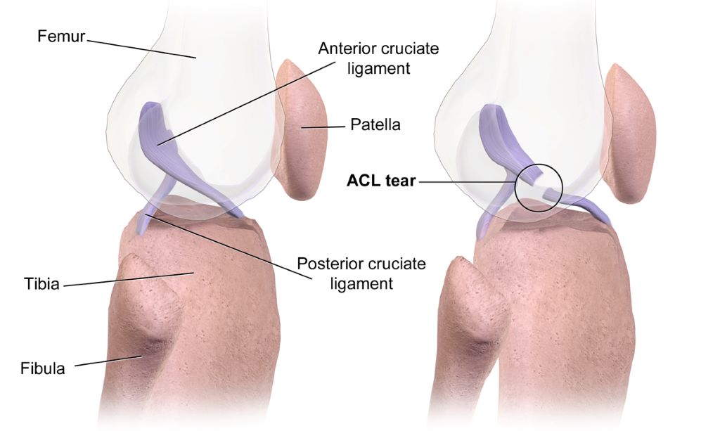
Anterior cruciate ligament injury. Credit Wikimedia/BruceBlaus CC4.0

Some individuals who have had anterior-cruciate-ligament reconstruction (ACLR), the kind of surgery often performed on athletes’ knees, may develop early-onset knee osteoarthritis. A new study in the Journal of Orthopaedic Research indicates that altered knee joint movement after ACLR could be a contributing factor. 

The study used a unique dynamic X-ray imaging system to accurately measure knee joint movement during walking in people who had undergone ACLR surgery and those with healthy knees. Compared with healthy controls, ACLR patients had a higher vertical position of the patella and a higher location of articular contact between the patella and the femur. A higher riding patella in the ACLR patients was caused by a longer-than-normal patellar tendon, the structure connecting the patella to the tibia. A surprising finding was that a higher riding patella was observed in both the ACLR knee and the uninjured contralateral knee of the ACLR patients. 

Investigators suspect that a higher riding patella may contribute to the development of knee osteoarthritis by shifting the load bearing areas between the patella and the femur to regions of cartilage unaccustomed to load and leaving previously loaded regions unloaded. 

“We don’t know whether a longer-than-normal patellar tendon that resulted in a higher riding patella existed prior to the ACL injury or resulted from the ACL injury or ACLR surgery. Further research is needed to determine the cause of a longer-than-normal patellar tendon in individuals who have undergone ACLR surgery,” said corresponding author Marcus G. Pandy, PhD, MEngSc, of the University of Melbourne, in Australia. 

Source: Wiley

Lymph Node Transfer Reduces Lymphoedema After Breast Cancer Surgery

Photo by Michelle Leman on Pexels

A multicentre study led from Finland has shown that lymph node transfer is a viable treatment for the swelling in the affected limb, a condition known as lymphoedema, after breast cancer surgery. Unfortunately, a drug to improve the outcomes of the transfer treatment was not shown to be effective. 

“I am becoming increasingly convinced that lymphoedema is not just a lymphatic problem, but is connected to an immunological factor,” says Plastic Surgeon and InFLAMES Flagship Researcher Pauliina Hartiala from the University of Turku in Finland.

The study by Hartiala and collaborators was published in the journal Plastic and Reconstructive Surgery.

Around one in four women with breast cancer undergo an axillary lymph node removal surgery. The surgery is performed if tests show that the cancer has spread from the breast tissue to the lymph nodes and is often followed by radiotherapy.

After the treatment, around 20–40% of women develop lymphoedema, a lymphatic drainage disorder in the affected arm. In 2022, about 2.3 million women were diagnosed with breast cancer worldwide. In men, the disease is rare.

Swelling can start years after treatment

“Lymphedema usually starts about six months after cancer surgery, but can also occur with a delay of several years after the cancer has been treated,” says Pauliina Hartiala.

In lymphedema, fluid accumulates in the tissue at first, but over time fat and firm connective tissue also begin to accumulate in the arm. Eventually, the upper limb becomes thick and clumsy. An elastic compression sleeve is used to try to control the problem by applying pressure to prevent the limb from swelling. However, the swelling can become so severe that the arm clearly interferes with everyday life, both at work and at leisure.

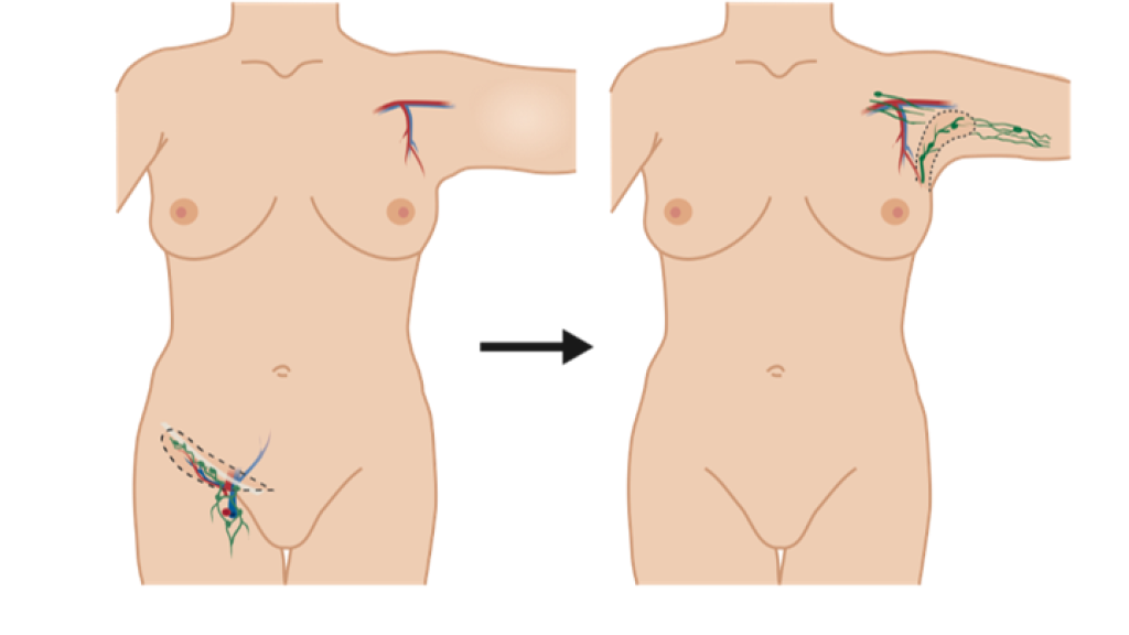
Lymphedema can be treated with surgical options including liposuction, lymphatic bypass procedure, or lymph node transfer. In the transfer surgery, the patient’s lymph nodes are transferred from the groin area to the armpit, or axilla, to replace the removed lymph nodes. The procedure involves extensive scar removal from the armpit.

Lymph node transfer is often performed at the same time as the breast operated on for cancer is reconstructed with a tissue flap taken from the patient’s abdomen. 

A lymph node transfer involves removing lymph nodes from the groin area and transferring them into the armpit. Image: Pauliina Hartiala

Additional benefits were expected from a growth factor

Pauliina Hartiala was one of the leaders of a multicentre study in Turku, Finland, focused on investigating whether the outcome of lymph node transfer could be improved by a growth factor, a drug called Lymfactin, that promotes growth and repair of lymphatic vessels. The study was carried out in five research centres in Finland and Sweden and builds on extensive basic research led by Finnish Professors Kari AlitaloSeppo Ylä-Herttuala and Anne Saarikko. Lymfactin is a research product from the Finnish pharmaceutical company Herantis Pharma.

The study tested whether the growth factor could improve lymph node flap function compared to a lymph node transfer conducted without the drug. The study involved 39 women. Of these, 20 underwent a transfer procedure where the tissue flap was injected with the lymphatic growth factor before it was transferred. For the second group, the transfer was carried out by adding only saline (placebo) to the tissue flap.

“Even though the drug therapy had worked well in combination with lymph node transfer in the animal model, it did not provide sufficient additional benefit to surgery in humans,” says Pauliina Hartiala.

Although Lymfactin did not work as expected in humans, Pauliina Hartiala is pleased with the other results of the study. In both study groups, the excess arm volume reduced during follow-up. In addition, the patients treated with Lymfactin had a significantly greater reduction in skin interstitial fluid than the placebo group.

“We are the first to show, with a double-blind study, that lymph node transfer is a viable treatment for some patients with lymphoedema after breast cancer surgery. One of the results of our study was the fact that the operation significantly improved women’s quality of life, which is an important finding.”

Pauliina Hartiala works as a Plastic Surgeon at Turku University Hospital, alongside her research work. She now believes that besides a lymphatic problem, lymphedema is linked to an immunological factor. It may be one or more of the immune cells that are involved in the accumulation of connective tissue and fat in the lymphoedema.   

“If this is the case, further research will allow us to investigate whether regulating the functions of this cell population could reduce fat accumulation in the limb,” concludes Hartiala.

Source: University of Turku

Engineered Cartilage from Nasal Septum Cells helps Treat Complex Knee Injuries

Researchers grow cartilage replacements from cells of the nasal septum to repair cartilage injuries in the knee. (Photo: University of Basel, Christian Flierl)

An unlucky fall while skiing or playing football can spell the end of sports activities. Damage to articular cartilage does not heal by itself and increases the risk of osteoarthritis. Researchers at the University of Basel and the University Hospital Basel have now shown that even complex cartilage injuries can be repaired with replacement cartilage engineered from cells taken from the nasal septum.

A team at the Department of Biomedicine led by Professor Ivan Martin, Dr Marcus Mumme and Professor Andrea Barbero has been developing this method for several years. It involves extracting the cells from a tiny piece of the patient’s nasal septum cartilage and then allowing them to multiply in the laboratory on a scaffold made of soft fibres. Finally, the newly grown cartilage is cut into the required shape and implanted into the knee joint.

Earlier studies have already shown promising results. “Nasal septum cartilage cells have particular characteristics that are ideally suited to cartilage regeneration,” explains Professor Martin. For example, it has emerged that these cells can counteract inflammation in the joints.

More mature cartilage shows better results

In a clinical trial involving 98 participants at clinics in four countries, the researchers compared two experimental approaches. One group received cartilage grafts that had matured in the lab for only two days before implantation – similar to other cartilage replacement products. For the other group, the grafts were allowed to mature for two weeks. During this time, the tissue acquires characteristics similar to native cartilage.

For 24 months after the procedure, the participants self-assessed their well-being and the functionality of the treated knee through questionnaires. The results, published in the scientific journal Science Translational Medicine, showed a clear improvement in both groups. However, patients who received more mature engineered cartilage continued to improve even in the second year following the procedure, overtaking the group with less mature cartilage grafts.

Magnetic resonance imaging (MRI) further revealed that the more mature cartilage grafts resulted in better tissue composition at the site of the implant, and even of the neighbouring cartilage. “The longer period of prior maturation is worthwhile,” emphasizes Anke Wixmerten, co-lead author of the study. The additional maturation time of the implant, she points out, only requires a slight increase in effort and manufacturing costs, and gives much better results.

Particularly suited to larger and more complex cartilage injuries

“It is noteworthy that patients with larger injuries benefit from cartilage grafts with longer prior maturation periods,” says Professor Barbero. This also applies, he says, to cases in which previous cartilage treatments with other techniques have been unsuccessful.

“Our study did not include a direct comparison with current treatments,” admits Professor Martin. “However, if we look at the results from standard questionnaires, patients treated with our approach achieved far higher long-term scores in joint functionality and quality of life.”

Based on these and earlier findings, the researchers now plan to test this method for treating osteoarthritis – an inflammatory disease that causes joint cartilage degeneration, resulting in chronic pain and disability.

Two large-scale clinical studies, funded by the Swiss National Science Foundation and the EU research framework programme Horizon Europe, are about to begin. These studies will explore the technique’s effectiveness in treating a specific form of osteoarthritis affecting the kneecaps (ie, patellofemoral osteoarthritis). The activities will further develop in Basel the field of cellular therapies, strategically defined as a priority area for research and innovation at the University of Basel and University Hospital Basel.

Source: University of Basel