Category: Surgeries & Procedures

Progressive Exercise Programme Improves Outcomes after Hip Surgery

Carers help an old man to walk
Photo by Kampus Productions on Pexels

A study published in the Journal of the American Geriatrics Society shows that a 12-month home-based supervised exercise programme can help to improve physical performance and functioning after patients undergo hip fracture surgery.

Hip fracture is a major health problem among older people, often resulting in long-term, sometimes persistent, functional impairments such as poor mobility and reduced independence in daily activities. Sedentary behaviour and low level of physical activity are also common among patients recovering from surgical repair of a hip fracture.

Standard care post-discharge care does not seem to meet the requirements of effective rehabilitation, as many patients with hip fractures do not reach their pre-fracture level of functioning. Growing evidence shows that multidisciplinary and well-coordinated rehabilitation started at the hospital and continued after discharge enhances the recovery of patients with hip fractures. Multicomponent rehabilitation in particular, which includes individualised and progressive resistance training, has improved functioning and mobility and decreased dependency in everyday activities. Longer lasting exercise programs of 6 to 12 months duration have reduced or reversed incident disability after hip fractures.

For the study, 121 patients aged 60 years and older were randomised to either an exercise group or a usual care group as a control. Home-based exercise sessions were delivered by physiotherapists twice a week and included strength, balance, mobility, and functional components as well as brief counselling on physical activity and nutrition.

Compared with patients in the usual care group, patients in the exercise group saw more improvements over the course of a year in their physical performance, their handgrip strength, and their ability to complete certain activities of daily living.

“It is worthwhile to invest in rehabilitation exercise for older people after hip fracture. Better functioning benefits the individual and also society,” said lead author Paula K. Soukkio, MSc, of the South Karelia Social and Health Care District (Eksote), in Finland.

Source: Wiley
Earlier Valve Replacement Surgery Recommended in Aortic Stenosis

Source: Pixabay CC0

Valve replacement surgery should be performed earlier than conventionally thought for people with aortic stenosis, as shown by new research published in the journal Open Heart.

Aortic stenosis is a common valvular disorder, especially in the elderly population, causing left ventricular outflow obstruction. Aetiologies include congenital (bicuspid/unicuspid), calcific, and rheumatic disease. Symptoms such as exertional dyspnoea or fatigue gradually develop after a long asymptomatic latent period of about 10 to 20 years. But many patients with aortic stenosis do not have symptoms even when they have severe narrowing of the valve and are thus not eligible for valve replacement.

The findings from this study show that these patients would benefit by undergoing a valve replacement – before they suffer irreversible heart muscle damage.

Lead researcher Prof Vassilios Vassiliou, from UEA’s Norwich Medical School, said: “The heart has four valves, which allow the blood to flow in one direction efficiently. With increasing age, one of the valves, the aortic valve, becomes increasingly narrowed or ‘stenosed’.

“A lot of patients with severe aortic stenosis do not have symptoms and therefore are not eligible for valve replacement according to the current guidelines.

“For these patients without symptoms, the guidelines suggest a ‘watchful waiting’ approach and intervention is recommended only when they show symptoms or develop pump failure.

“We wanted to know if it would be better to perform surgery and replace the valve sooner rather than later.”

In a systematic review, researchers compared early intervention versus conservative management in patients with asymptomatic severe aortic stenosis.

They then analysed data from all the available studies which involved a total of 3798 patients, out of which 302 were included in the two largest randomised controlled trials and 3496 in the observational studies.

Prof Vassiliou said: “We found that early intervention, before patients have symptoms, is associated with lower risk of death and hospitalisation for heart failure.

“By the time the patients develop symptoms, there has likely been irreversible damage to the muscle of the heart. This in turn may preclude a worse prognosis and adverse outcomes even after successful intervention.

“The timing of aortic valve intervention is crucial.

“We hope that our findings may herald the beginning of a change in the management of aortic stenosis patients, enabling the intervention to take place more commonly whilst the patients are asymptomatic.

“Ongoing trials investigating this high-risk population are anticipated to shed more light into the matter and in the identification of the optimal time of intervention,” he added.

Source: University of East Anglia

20 Years of Data Proves Safety of Islet Cell Transplantation

A 3D map of the islet density routes throughout the healthy human pancreas. Credit: MariusOrion/Wikimedia Commons

In a paper published in The Lancet Diabetes & Endocrinology, researchers report that their long-running islet cell transplant programme has shown that is safe and helps control diabetes for up to 20 years.

The researchers reported on patient survival, graft survival, insulin independence and protection from life-threatening hypoglycaemia for 255 patients who have received a total of more than 700 infusions of islets at the University of Alberta Hospital over the past two decades.

“We’ve shown very clearly that islet transplantation is an effective therapy for patients with difficult-to-control Type 1 diabetes,” said Professor James Shapiro at the University of Alberta. “This long-term safety data gives us confidence that we are doing the right thing.”

In Type 1 diabetes, the immune system mistakenly destroys the cells within the insulin-producing islets so patients have to take insulin by injection. Patients with hard-to-control diabetes face dangerous hypo- or hyperglycaemia and long-term complications.

Between March 1999 and October 2019, 255 patients received islet transplants by infusion into their livers. Seventy per cent of the grafts survived for a median time of nearly six years. The researchers reported that a combination of two anti-inflammatory medications given during the first two weeks following transplant significantly increased long-term islet function.

The transplant recipients have to take lifelong immunosuppression drugs, which in some cases lead to skin cancer or infection, but most such complications were not fatal during the study period.

After two or more islet infusions and a median time of 95 days following the first transplant, 79% of the recipients could go off insulin. A year later, 61% remained insulin-independent, 32% at five years and 8% after 20 years, the researchers reported. Even though most patients had to start taking insulin again, doses were generally much smaller and diabetes control was improved.

“Being completely free of insulin is not the main goal,” said Prof Shapiro. “It’s a big bonus, obviously, but the biggest goal for the patient — when their life has been incapacitated by wild, inadequate control of blood sugar and dangerous lows and highs — is being able to stabilise. It is transformational.”

With trials ongoing in other countries, Prof Shapiro will continue to focus on finding a more plentiful supply of islet cells to replace the current reliance on deceased donors. Human trials have already shown success using stem cells programmed to produce insulin. Trials have just started to transplant cells that have been gene edited to make them invisible to the immune system.

“Islet transplant as it exists today isn’t suitable for everybody, but it shows very clear proof of concept that if we can fix the supply problem and minimize or eliminate the anti-rejection drugs, we will be able to move this treatment forward and make it far more available for children and adults with Type 1 and Type 2 diabetes in the future,” said Shapiro.

Source: University of Alberta

In Finger Osteoarthritis, Lipofilling Reduces Pain, Improves Function

Hand osteoarthritis
Source: Pixabay CC0

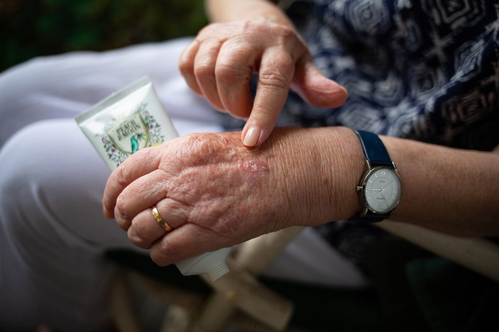
For patients with painful finger osteoarthritis, lipofilling  a nonsurgical procedure where a patient’s fat is transferred into the arthritic joints, may result in lasting improvements in hand function and especially pain, according to a study in Plastic and Reconstructive Surgery.

Researchers reported their experience with 18 patients undergoing fat transfer procedures for finger osteoarthritis.

“Even over a long-term follow-up, the transfer of fatty tissue to arthritic fingers joints appears to provide a safe and minimally invasive alternative to conventional surgery for patients with osteoarthritis,” commented the study’s lead author Max Meyer-Marcotty, MD, PhD.

In the lipofilling procedure, a sample of the patient’s own fatty tissue was obtained by liposuction from another part of the body: the upper thigh or hip area. Tiny volumes of lipid cells (< 1mL) were injected into the arthritic finger joints. For recovery, patients wore a splint around the treated finger and took pain relievers for a week. No infections or other complications were recorded.

The researchers analysed follow-up outcomes in a total of 25 finger joints treated by lipofilling. Hand function, pain scores, and patient satisfaction were evaluated an average of 44 months (maximum 50 months) after treatment.

Assessment showed a “highly significant clear improvement” in pain score: from a median of 6 points (on a 10-point scale) before treatment to 0.5 points at follow-up. “We believe that for our patients the reduction of pain represents the most striking and important result, which also has the most pronounced and highly significant effect,” Dr Meyer-Marcotty et al. wrote.

On functional evaluation, pinch grip strength of the treated fingers increased from a median of 2.0kg before lipofilling to 4.3kg at follow-up. Non-significant improvements were seen in fist closure force and score on a standard assessment of hand function during everyday tasks.

In severe cases of osteoarthritis, surgery is effective in relieving arthritis pain, but is associated with potential complications and lengthy recovery time.

Fat transfer procedures have been introduced in recent years for a growing range of purposes in plastic and reconstructive surgery. Animal studies have suggested that mesenchymal stromal cells found in fatty tissues can regenerate tissue in arthritic joints.

“The chance to preserve the joint with a minimally invasive procedure is of particular interest in the early, albeit painful, phases of finger osteoarthritis,” Dr. Meyer-Marcotty added. “Since the lipofilling procedure is nondestructive, conventional joint surgery can still be performed later, if needed.”

Larger long-term follow-up studies are needed to further corroborate these initial positive findings, the researchers said.

Source: EurekAlert!

Smartphone Pics of Post-surgical Wounds for Spotting Infections

Photo by Daniel Romero on Unsplash

Researchers have shown that smartphone pictures of post-surgical wounds taken by patients and then assessed by clinicians help spot infections early on.

These ‘surgery selfies’ were associated with a reduced number of GP visits and improved access to advice among patients who took them. This practice could help manage surgical patients’ care while they recover.

Death within 30 days of surgery is the third largest cause of mortality globally. More than a third of postoperative deaths are associated with surgical wound infections.

In the study, published in NPJ Digital Medicine, University of Edinburgh researchers conducted a randomised clinical trial involving 492 emergency abdominal surgery patients to determine if photos from smartphones and questions on symptoms of infection could be used to diagnose wound infections early.

One group of 223 patients were contacted on days three, seven and 15 after surgery and directed to an online survey, where they were asked about their wound and any symptoms they were experiencing. Then they were asked to take a picture of their wound and upload it.

A surgical team member assessed the photographs and patients’ responses were assessed for signs of wound infection. They followed up with patients 30 days after surgery to find out if they had been subsequently diagnosed with an infection.

A second group of 269 received standard care and were contacted 30 days after surgery to find out if they had been diagnosed with an infection.

No significant difference between groups was seen in the overall time it took to diagnose wound infections in the 30-days after surgery.

However, the smartphone group was nearly four times more likely to have their wound infection diagnosed within seven days of their surgery compared to the routine care group. They also had fewer GP visits and reported a better experience of trying to access post-operative care.

The research team is now conducting a follow-up study to determine how this can be best put into practice for surgical patients around the country. Artificial intelligence will also be used to help the clinical team in assessing the possibility of wound infection.

Professor Ewen Harrison, Professor of Surgery and Data Science, who led the research said: “Our study shows the benefits of using mobile technology for follow-up after surgery. Recovery can be an anxious time for everybody. These approaches provide reassurance – after all, most of us don’t know what a normally healing wound looks like a few weeks after surgery. We hope that picking up wound problems early can result in treatments that limit complications.”

Dr. Kenneth McLean, who co-led the research said: “Since the COVID-19 pandemic started, there have been big changes in how care after surgery is delivered. Patients and staff have become used to having remote consultations, and we’ve shown we can effectively and safely monitor wounds after surgery while patients recover at home – this is likely to become the new normal.”

Source: University of Edinburgh