Category: Skeletal System

Flipping the Switch on Osteoporosis with Epigenetic Discovery

Photo by Mehmet Turgut Kirkgoz on Unsplash

Van Andel Institute scientists have pinpointed a key driver of low bone density, a discovery that may lead to improved treatments with fewer side effects for women with osteoporosis. Their findings appear in the journal Science Advances.

Their research reveals that loss of an epigenetic modulator, KDM5C, preserves bone mass in mice. KDM5C works by altering epigenetic ‘marks’, switches that ensure the instructions written in DNA are read in the right time and place.

Several medications are approved to treat osteoporosis but fears of rare, severe side effects often are a barrier for their use. Treatments that leverage the hormone oestrogen also are available, but are only recommended for low-dose, short-term use due in part to associations with cancer risk.

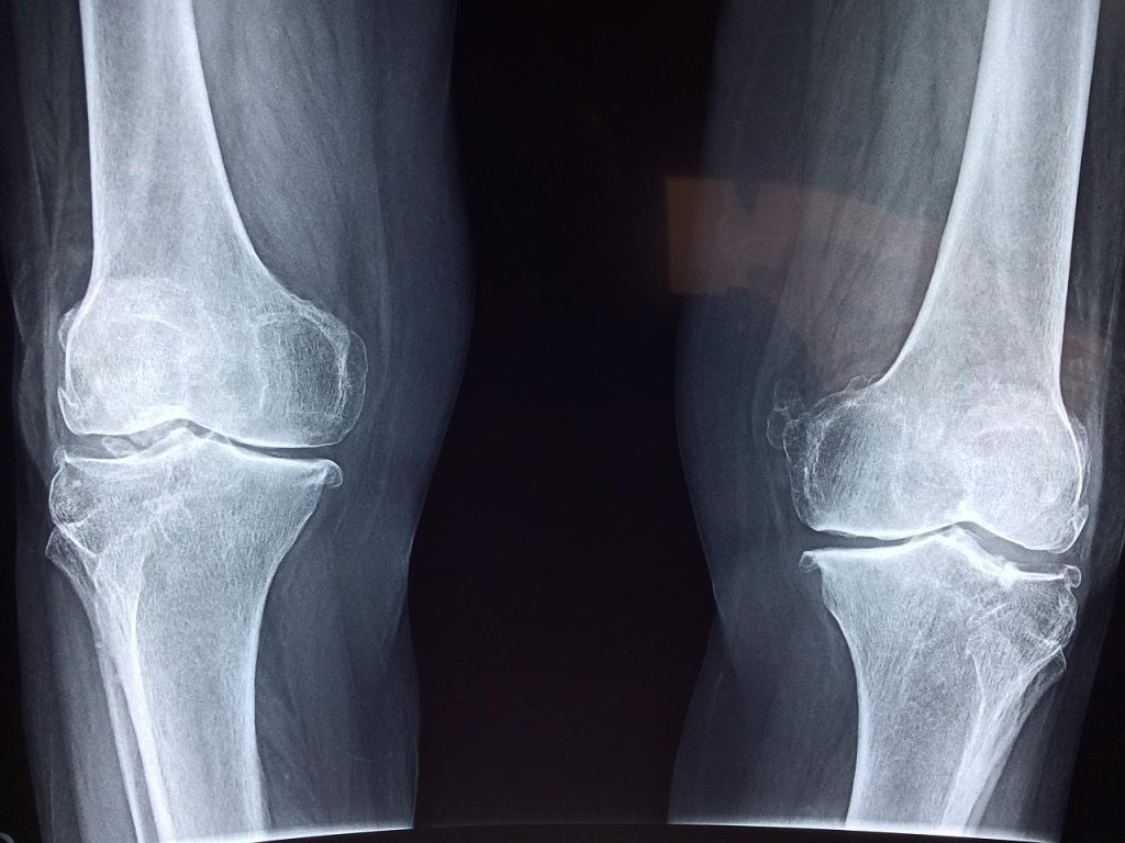
It is well-established that women experience disproportionately lower bone mass than men throughout their lives. Loss of bone mass accelerates with menopause, increasing the risk of osteoporosis and associated fractures for women as they age.

To figure out why this happens, VAI Associate Professors Connie M. Krawczyk, PhD, and Tao Yang, PhD, and their teams looked at the differences in the ways bone is regulated in male and female mice, which share many similarities with humans and are important models for studying health and disease. They focused on osteoclasts, which help maintain bone health by breaking down and recycling old bone.

“Osteoporosis is a common disease that can have debilitating outcomes,” Yang said. “KDM5C is a promising target to treat low bone mass in women because it is highly specific. We’re hopeful that our findings will contribute to improved therapies.”

The researchers found reducing KDM5C disrupted cellular energy production in osteoclasts, which slowed down the recycling process and preserved bone mass. Importantly, KDM5C is linked to X chromosomes, which means it is more active in females than in males.

“Lowering KDM5C levels is like flipping a switch to stop an overactive recycling process. The result is more bone mass, which ultimately means stronger bones,” Krawczyk said. “We’re very excited about this work and look forward to carrying out future studies to refine our findings. At the end of the day, we hope these insights make a difference for people with osteoporosis.”

Source: Van Andel Research Institute

Changes in Hyaluronic Acid Properties Drive Osteoarthritis

Source: CC0

The composition of synovial fluid changes significantly in osteoarthritis: The concentration and molecular weight of hyaluronic acid tends to decrease and is commonly used to diagnose the disease. An international group of researchers explored the disease-driven breakdown of hyaluronan and the mechanistic implications of these changes on the lubrication and subsequent wear of joints.

“One of the most important properties of the synovial fluid is its viscosity,” said Rosa Maria Espinosa-Marzal, co-author of the study published in the journal Biointerphases. “Viscosity is a measure of the internal frictional force between adjacent layers of a fluid in relative motion, or, more simply, a fluid’s resistance to flow. Large, high molecular weight polymers such as hyaluronic acid play a significant role in maintaining a high viscosity of the synovial fluid, which helps maintain a fluid film and reduces friction between articulating surfaces during motion.”

Through analysis with neutron and light scattering, the team determined that the structure of the lipid-hyaluronic-acid complexes in the bulk solution is a function of concentration and its molecular weight.

The researchers found the hyaluronic acid’s concentration and molecular weight both play a role in how the lubricant reacts with different surfaces.

“Our results show low molecular weight hyaluronic acid, which mimics osteoarthritis-diseased joints, hinders the adsorption of the hyaluronic-acid-lipid complex,” said Espinosa-Marzal, of the University of Illinois Urbana-Champaign. “The lack of the formation of an amorphous film on the surface may reflect a consequence of osteoarthritis, since this film should help reduce friction and wear.”

Their hypothesis is that this film’s absence may increase wear of the cartilage surface. In contrast, high molecular weight hyaluronic-acid-lipid complexes form an amorphous film, which presumably helps maintain the mechanical integrity and longevity of efficient lubrication in healthy cartilage.

Studies on hyaluronic acid itself and hyaluronic-acid-lipid complexes “do not entirely support hyaluronic acid’s role in providing high lubricity to the cartilage’s articular surface, which is still a bit controversial,” Espinosa-Marzal said. “Our results indicate that for low molecular weight hyaluronic acid, this is likely the case.”

By exploring the complex interplay between phospholipid and hyaluronic acid self-assembly, and the role of molecular weight on surface affinity, “our study illuminates a mechanism whereby the ‘vicious circle’ of osteoarthritis can be explained,” said co-author Mark Rutland, from KTH Royal Institute of Technology.

Source: American Institute of Physics

Scientists Close in on the Genetic Determinants of Height

Pexels Photo by Monstera

Human height is dictated by the sealing of the growth plates at the ends of bones that harden as a child develops. Along with diet and disease, heritability has long been known to be a factor determining height. Now, researchers report in Cell Genomics that cells in these plates determine the length and shape of bones and may partly predict final stature. The study identified potential “height genes” and found that genetic changes affecting cartilage cell maturation may strongly influence adult height.

“The study is really understanding the genetics of skeleton,” says paediatric endocrinologist and senior author Nora Renthal of Boston Children’s Hospital and Harvard University. “Height is a good starting point to understand the relationship between genes, growth plates, and skeletal growth because we can measure the height of every human being.”

To pinpoint height-associated genes, the team screened 600 million mouse cartilage cells to identify genes that, when deleted, can alter cell growth and maturation. These types of cellular changes in the growth plate are known to lead to variations in human height. The search turned up 145 genes mostly linked to skeletal disorders and are crucial for growth plate maturation and bone formation.

The team then compared these genes with data from genome-wide association studies (GWAS) of human height, which located “hotspots” along the entire human genome where “height genes” are located. But these regions can contain multiple genes, making it hard for researchers to track down and study an individual target.

“That’s kind of like looking for your friend’s house, but you only know the zip code,” says Renthal. “It’s difficult.”

The comparison revealed that genes affecting cartilage cells overlap with hotspots from human height GWAS, precisely locating genes in our DNA that likely play a role in determining our stature. Renthal and her team also discovered that many of the GWAS suggested height genes led to early maturation in cartilage cells. These findings suggest that genetic changes affecting cartilage cell maturation may influence height more.

Renthal notes that studies in mouse cells may not fully translate to humans, and GWAS are observational studies that cannot fully illustrate the cause and effects of height. But her study provides a novel method to bridge the two methods and provide new insights into human genetics.

Next, the team plans to use the method to understand hormones’ effect on cartilage cells. They will also look into some of the 145 genes that have no known connection to skeletal growth. The investigation may reveal new genes and pathways that play a role in the bones.

“I see patients with skeletal dysplasia, where there isn’t any treatment because genetics made their bones grow this way,” says Renthal. “It’s my hope that the more we can understand about the biology of the growth plate, the more we would be able to intervene at earlier times in growing skeletons and the life of a kid.”

Source: MedicalXpress

Bone Cells Growing on Biomaterials Like Curvatures the Best

Doctor shows an X-ray of a foot
Photo by Tima Miroshnichenko on Pexels

TU Delft engineers have shown that the curvature of biomaterials inhibits or stimulates bone cells to make new tissue. Their findings are reported in Nature Communications, and their study of geometries could be an important step in research into repairing damaged tissues.

Living cells can perceive and respond to the geometry of their environment. “Cells sense and respond to the geometry of the surfaces they are exposed to. Depending on their curvature, surfaces can either encourage cells to create new tissue or prevent them from doing so,” says Amir Zadpoor, professor of Biomaterials and Tissue Biomechanics, supervisor of the study. “Stimulating curvatures made by a 3D printer are an easy and safe way to promote tissue growth. As compared to drugs, they are also much cheaper.”

The researchers grew bone cells in vitro surrounded by small moulds made from biomaterials with which the researchers have experience. Depending on the curvatures in the moulds, the cells tended to grow, divide, and form tissue to different extents.

Cells like a saddle shape

Although curved shapes seem to exist in endless variations, they always fall roughly into one of these three categories: a ball that has a convex curvature, a saddle that has a concave curvature, and a plate that is flat. One of the authors, assistant professor of Biomaterials Lidy Fratila-Apachitei: “Cells prefer a saddle shape. If they perceive a saddle shape nearby, growth is stimulated. The study also shows that cells prefer valleys over hills.”

Rather aligned than bent

First author Sebastien Callens did the experiments and analysis in the study. “Cells also have a skeleton, which consists of fibres that are under tension to different degrees. How tension builds up in those fibres strongly influences the behaviour of cells. Our study shows that cells collectively align their stress fibres with the curvatures they experience to minimise their need to bend. I could see that cells prefer to align than to bend.”

Budget of saddle curvature

You can’t have only saddle curves around cells. Just as the three angles of a triangle always add up to 180 degrees, the sum of all curvatures must also equal some fundamental numbers. “You always have a limited budget of saddle shapes,” says Zadpoor. “If you use too much negative curvature somewhere, you must use positive curvatures somewhere else to keep the sum constant. You need to use your budget wisely to encourage maximum tissue regeneration.”

New biomaterials

The study provides guidance on the optimal geometry of biomaterials and implants to maximise tissue regeneration. The complex geometric designs required are made using high-precision 3D printing techniques to make the shapes so small that they are perceptible to cells. Callens: “We have now discovered new playing rules by which biomaterials can stimulate tissue growth. In follow-up research, we will try to apply those rules optimally.”

Source: Delft University of Technology

Bone Matrix Protein Discovery could Yield New Osteosarcoma Treatments

Doctor shows an X-ray of a foot
Photo by Tima Miroshnichenko on Pexels

A study published in the Journal of Orthopaedic Research has identified a bone matrix protein called Secreted phosphoprotein 24 kD (Spp24) that may help to treat osteosarcoma, the most common type of bone cancer.

In experiments conducted in cells and mice, investigators found that Spp24 inhibits the proliferation and invasiveness of osteosarcoma tumour cells and promotes their apoptosis, or death. Mechanistically, Spp24 binds to and neutralises a protein called bone morphogenetic protein 2, which has tumour-enhancing properties.

“Spp24 and its proteolytic products have a number of effects on bone metabolism that have been elucidated to various degrees. They have the potential to be engineered into bone therapeutics, and this anti-tumour effect through bone morphogenetic protein 2 sequestration is only one such example,” said co–corresponding author Haijun Tian, MD, PhD, of Shanghai Jiao Tong University School of Medicine. “Like many other bone matrix proteins, the more we look into the function of Spp24, the more surprising roles we find even though the primary function of Spp24 remains uncertain.”

Source: Wiley

Bone Tissue has Lymphatic Vessels – and They Aid Healing

Doctor shows an X-ray of a foot
Photo by Tima Miroshnichenko on Pexels

To date, it has been assumed that bones lack lymphatic vessels, but new research published in the journal Cell not only mapped them within bone tissue, but demonstrated their role in bone and blood cell regeneration and reveals changes associated with ageing.

The network of vessels that form the lymphatic system plays an important role in draining excess fluid from tissues, clearing waste products and supporting immune responses.

The fine network of lymph vessels extends throughout the body, but a small number of sites such as the brain, eye and bone were previously assumed to lack lymph tissue. The hard tissue of bone in particular has traditionally made studying the distribution and role of blood and lymph more difficult.

Researchers used light-sheet imaging to identify and visualise the lymphatic vessels of bone in high-resolution 3D, revealing an active network of lymph vessels within bone. The researchers further identified some of the key signals happening between lymph vessels, blood stem cells and bone stem cells.

Dr Lincoln Biswas, co-first author of this study, said: ‘Interestingly after injury, lymphatic vessels in bone show dynamic crosstalk with blood stem cells and with specialised perivascular cells in order to accelerate bone healing. Such interactions between lymphatics and bone stem cells can harnessed to promote bone healing such as in fracture repair.’

The researchers found that lymphatic vessels in bone increase during injury via a signalling molecule called IL6, and trigger expansion of bone progenitor cells by secreting a different signal, called CXCL12. Dr Junyu Chen, a co-first author of the study now based at Sichuan University said: “Ageing is associated with diminished capacity for bone repair, and our findings show that lymphatic signalling is impaired in aged bones. Remarkably, the administration of young lymphatic endothelial cells restores healing of aged bones, thus providing a future direction to promote bone healing in elderly.”

Dr Anjali Kusumbe, who led the research said: “I am very excited as these findings not only demonstrate that lymphatic vessels do exist in bone but also reveal their critical interactions with blood stem cells and perivascular bone stem cells after injury to promote healing, thereby presenting lymphatics as a therapeutic avenue to stimulate bone and blood regeneration. Further, these findings are very fundamental, opening doors for understanding the impact of bone lymphatics on the immune system and their role in bone and blood diseases.”

Source: Oxford University

Excessive Physical Activity May Impact Teens’ Leg Development

Doctor shows an X-ray of a foot
Photo by Tima Miroshnichenko on Pexels

A study published in the Journal of Orthopaedic Research showed that physical activity levels may impact adolescents’ and young adults’ leg alignment during development, as measured by magnetic resonance imaging (MRI) scans.

The study included 57 elite male soccer players compared with 34 male and 34 female controls aged 11–21 years. Outcome measures were the hip knee angle (HKA), medial proximal tibial physeal angle (MPTPhyA), lateral distal femoral physeal angle (LDFPhyA) on full leg length MRI scans, and a physical activity questionnaire score.

Using magnetic resonance imaging scans, physical activity levels were associated with the development of varsus or ‘knock knees’, an abnormal leg alignment, which may represent a physiological adaptation to load where the shin bone meets the knee.

“Our study suggests that abnormal leg alignment, a risk factor for future injury and osteoarthritis, develops in early adolescence due to high activity levels,” said corresponding author Scott Fernquest, DPhil, of the University of Oxford. “Modifying activity levels during this critical period of growth may prevent the development of abnormal leg alignment. We hope our findings lead to further research investigating this possibility.”

Source: Wiley

Weight Loss Protects against Structural Defects of Knee Osteoarthritis

Researchers conducting long-term follow ups of adults given knee X-rays found that a decrease in body mass index (BMI) was associated with both a lower incidence of the structural defects of knee osteoarthritis. Published in Arthritis & Rheumatology, also found reduced odds of these defects progressing.

In the study radiographic analyses were conducted of adults’ knees with and without the structural defects of knee osteoarthritis at baseline and at 4 to 5 years’ follow up from adults. A total of 9683 knees (from 5774 individuals) were assessed in an ‘incidence cohort’ along with 6075 knees (from 3988 individuals) in a ‘progression cohort.’

The researchers observed that a 1-unit drop in BMI was linked to a 4.76% reduction in odds of the incidence and progression of knee osteoarthritis. A 5-unit BMI drop, which can downgrade a BMI category (eg, from overweight to normal), reduced the odds of incidence and progression by 21.65%.

“These findings could be empowering for people with or at risk of knee osteoarthritis,” said lead author Zubeyir Salis, BEng, and a PhD student for Public Health at the University of New South Wales in Australia. “The current prevailing view is that knee osteoarthritis is part of ageing and that we have no control over it. However, my analyses suggest that some people could potentially prevent, slow or delay knee osteoarthritis by losing weight.”

Source: Wiley

Osteoporosis in Men is Often Overlooked

Photo by Kampus Production on Pexels

Osteoporosis in men is often overlooked by health care professionals, found the authors of a review published in The Lancet Diabetes and Endocrinology. There is a desperate need for raising awareness of the condition in men to help improve outcomes for patients, the authors said.

Women are generally at higher risk of developing osteoporosis, as their bone density declines more rapidly than men at an earlier age, especially post-menopause. In most populations, men have larger and stronger bone and joint surfaces, so they can be overlooked when diagnosing the condition.

Reviewing available data on the condition in men, researchers found that they are generally diagnosed later, comply with treatment less and present to hospital in older ages than women. With fatality rates from hospitalisations with fragility fractures, like a broken hip, being higher than women.

The review’s author, Dr Tatiane Vilaca, said: “Generally diagnosis of osteoporosis happens when a patient presents at hospital with some kind of fragility fracture in older age, for example falling from standing height, and breaking a hip, wrist or spine.

“Research suggests men hospitalised with hip fractures tend to be older than women, which could be because the condition develops more slowly in men. As older people are usually slightly frailer, with poorer states of overall health, this could explain the slightly higher levels of disability and mortality associated in men with osteoporosis who are hospitalised following a fracture.”

The review found that although there is a lack of research about which treatment options are most effective in men, diagnosis and treatment options are effective.

The team believe further research specifically tailored to osteoporosis in male patients will help improve current diagnosis systems, helping clinicians with earlier diagnosis, and a focus on education for patients will support compliance with drug treatment programs, all improving outcomes for men living with osteoporosis.

Dr. Richard Eastell, Professor of Bone Metabolism at the Department of Oncology and Metabolism, said: “As women make up larger numbers of people living with osteoporosis, the data we have on the progression of the condition in men is currently not as robust. This updated review shows that further studies of male patients could help improve current diagnosis systems, as well as resources for the education of primary care clinicians and the general public on the early warning signs of osteoporosis in men.”

Dr. Vilaca added: “Despite the current gap in knowledge, men can still easily be screened for osteoporosis at their general practitioner surgery.

“Anyone with a family history of osteoporosis, broken bones, or fractures, those with acute back pain or a loss of height should be encouraged to have a check-up.

“These are all early warning signs of the condition in both men and women, and early preventative treatment is the best way to ensure a slower disease progression and longer, healthier life without a fracture.”

Source: University of Sheffield

Prunes Also Protect Bone Health in Men

Photo by Nino Liverani on Unsplash

New research published in the Journal of Food and Medicine reports that daily prunes consumption protects bone health in men over 50. This study is the first of its kind to examine the beneficial prune effect on bones in men. 

Some 2 million men are estimated to be battling osteoporosis and another 16.1 million men have osteopenia, or low bone mass. Despite these numbers, bone disease in men is often overlooked.

“We’ve already seen significant evidence that prunes have a positive effect on bone health in women, so it’s particularly exciting to find that prunes can also play a beneficial role in men’s bone health. We look forward to continuing to study the ‘prune effect’ on bone and other health outcomes in men,” said lead researcher Professor Shirin Hooshmand at San Diego State University.

In this study, 57 healthy men aged 50-79 years old were randomised to either consume 100 grams of prunes every day or no prunes for twelve months. After a year, the prune consumers showed significant decreases in biomarkers of bone breakdown, while no changes were observed in the control group. The study authors also reported the men who ate prunes showed improvements in bone geometry indicating greater bone strength.

Historically, research has focused on osteoporosis and bone health in women, already indicating a favorable bone response to prunes specifically among postmenopausal women. Several studies have suggested that eating 50 to 100 grams of prunes everyday could lead to increased bone mass and decreased bone breakdown. Moreover, a recent case study earlier this year reported that total bone mineral density increased in a postmenopausal woman with osteopenia after she consumed 50 grams of prunes daily for 16 months.

“Bone health is not just a concern for women. Men need to think about how to protect their bones as well,” said Leslie Bonci, MPH, RDN and consultant with the California Prune Board. “Prunes are a shelf-stable and nutrient-packed food that provide a preventive, proactive, palatable option for men to optimize their bone health.”

While San Diego State University’s newest research is an exciting addition to existing prune-focused literature, more work on the effect of prunes on human bone health is currently underway. An upcoming study from Pennsylvania State University examines how consuming different amounts of prunes affects health outcomes in postmenopausal women over a one-year period. The study not only explores the impact of prunes on bone health, but it will also look at the prune-effect on inflammation and gut health.

Source: PR Newswire