Category: Skeletal System

Walking Fitness can Predict Fracture Risk in Older Adults

Photo by Teona Swift on Unsplash

The ability to walk one kilometre comfortably can help predict fracture risk, according to researchers at the Garvan Institute of Medical Research. The findings, published in JAMA Network Open, suggest that simply asking a patient about walking limitation could allow clinicians to identify those in need of further bone health screening and prescribe interventions that could prevent fractures from occurring.

“We’ve discovered that trouble walking even short distances appears closely tied to higher fracture risk over the following five years,” says lead author of the study, Professor Jacqueline Center, Head of Garvan’s Clinical Studies and Epidemiology Lab.

“Just a few simple questions about how far someone can walk could give doctors an early warning sign to check bone health.”

The researchers examined data on nearly 267 000 adults aged 45 and older from the Sax Institute’s 45 and Up Study, a major ongoing research initiative that has been tracking health outcomes in adults in the Australian state of New South Wales for more than 15 years.

Participants were asked if health issues limited their ability to walk various distances, with answer options of ‘not at all,’ ‘a little,’ or ‘a lot’. The group was then followed for five years to track fracture outcomes.

The researchers found that one in five adults reported some walking limitation at the beginning of the study.

Those with more difficulty walking were significantly more likely to experience a fracture during follow-up. For example, women who said they were limited ‘a lot’ in walking one kilometre had a 60% higher fracture risk than women with no limitation.

For men, the increased risk was over 100%.

“We saw a clear ‘dose-response’ pattern, where greater walking limitation meant higher fracture risk. This suggests a direct relationship between low walking ability and weaker bones,” says first author of the study Dr Dana Bliuc, Senior Research Officer at Garvan.

Approximately 60% of all fractures in the study were attributable to some level of walking limitation.

The link remained strong even after accounting for other factors like age, falls, prior fractures, and weight, and the findings were consistent across different fracture sites like hips, vertebrae, arms, and legs.

“In this generally healthy community-based population, we still found one in five people had trouble walking a kilometre,” says Professor Center.

“We think this simple assessment could help identify many more at-risk individuals who may benefit from bone density screening or preventative treatment.”

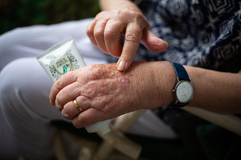
Osteoporosis medications, lifestyle changes, and other interventions are available to improve bone strength and avoid first or repeat fractures.

However, screening rates currently remain low, meaning many miss out on fracture risk assessments.

Finding easy but accurate ways to detect at-risk people is an important target for research.

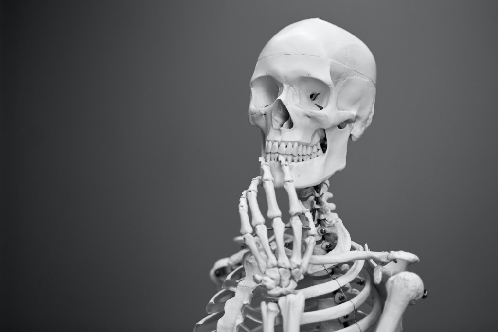
“Fracture risk assessment generally relies on a bone density test, which many people have not had when seeing their doctor,” says Professor Center.

“Asking about walking ability takes just seconds and could be a free, non-invasive way to tell if someone needs their bones checked.”

The researchers stress that walking limitation may have many causes beyond weak bones, from heart disease to arthritis.

However, a difficulty in walking even short distances appears closely tied to fracture risk independently.

“We hope these findings will encourage clinicians to consider walking ability as a red flag for possible bone health issues. For patients, if you can’t walk a full kilometre comfortably, it may be wise to ask your doctor about getting your bones checked,” says Dr Bliuc.

Source: Garvan Institute of Medical Research

Topical Corticosteroids Linked to Osteoporosis Risk

Source: Pixabay

New research indicates that higher doses of topical corticosteroids, commonly used to treat inflammatory skin conditions, are linked with elevated risks of osteoporosis and bone fractures associated with osteoporosis. The findings are published in the Journal of the European Academy of Dermatology and Venereology.

Drawing on the Taiwan National Health Insurance Research Database, the study’s investigators selected 129 682 osteoporosis cases and 34 999 major osteoporotic fracture (MOF) cases and matched them with 518 728 and 139 996 controls (without osteoporosis or MOF) by sex and age.

The team found clear dose–response relationships between long-term use of topical corticosteroids and osteoporosis and MOF, as well as differences in sex and age.

All topical corticosteroids prescriptions were converted to prednisolone equivalents (mg) according to their anti-inflammatory potency. Effects were not clear in exposure periods of six or 12 months, but effects were seen when analysis was extended to the longer term (three to five years). Compared with no doses, low, medium, and high cumulative of doses topical corticosteroids were associated with 1.22-, 1.26-, and 1.34-times higher odds of developing osteoporosis over five years. These respective doses were linked with 1.12-, 1.19-, and 1.29-times higher odds of experiencing MOF. Women had higher risks of osteoporosis and MOF than men. Also, younger people (under the age of 50 years) had a higher risk of osteoporosis compared with other age groups.

“This study emphasises that using topical corticosteroids to treat inflammatory skin conditions should be done very carefully and clinicians should be aware of these potential side effects,” said corresponding author Chia-Yu Chu, MD PhD, of National Taiwan University Hospital and National Taiwan University College of Medicine.

Source: Wiley

AI Finds that an Antimalarial Drug might Treat Osteoporosis Too

Photo by Ekamelev on Unsplash

Using a deep learning algorithm, which is a kind of artificial intelligence (AI), researchers reporting have found that dihydroartemisinin (DHA), an antimalarial drug and derivative of a traditional Chinese medicine, could treat osteoporosis as well. Publishing their findings in ACS Central Science, the team showed that in mice, DHA effectively reversed osteoporosis-related bone loss.

In healthy people, there is a balance between the osteoblasts that build new bone and osteoclasts that break it down. Current treatments for osteoporosis primarily focus on slowing the activity of the ‘wrecking crew’ of osteoclasts. But osteoblasts, or more specifically, their precursors known as bone marrow mesenchymal stem cells (BMMSCs), could be the basis for a different approach. During osteoporosis, these multipotent cells tend to turn into fat-creating cells instead, but they could be reprogrammed to help treat the disease. Previously, Zhengwei Xie and colleagues developed a deep learning algorithm that could predict how effectively certain small-molecule drugs reversed changes to gene expression associated with the disease. This time, joined by Yan Liu and Weiran Li, they wanted to use the algorithm to find a new treatment strategy for osteoporosis that focused on BMMSCs.

The team ran their program on a profile of differently expressed genes in newborn and adult mice. One of the top-ranked compounds identified was DHA, a derivative of artemisinin and a key component of malaria treatments. Administering DHA extract for six weeks to mice with induced osteoporosis significantly reduced bone loss in their femurs and nearly completely preserved bone structure. To improve delivery, the team designed a more robust system using injected, DHA-loaded nanoparticles. Bones of mice with osteoporosis that received the treatment were similar to those of the control group, and the treatment showed no evidence of toxicity. In further tests, the team determined that DHA interacted with BMMSCs to maintain their stemness and ultimately produce more osteoblasts. The researchers say that this work demonstrates that DHA is a promising therapeutic agent for osteoporosis.

Source: American Chemical Society

Newly Discovered Bone Stem Cell Drives Premature Skull Fusion

Photo by Mathew Schwartz on Unsplash

Craniosynostosis, the premature fusion of the top of the skull in infants, is caused by an abnormal excess of a previously unknown type of bone-forming stem cell, according to a preclinical study published in Nature.

Occurring in one in 2500 babies, craniosynostosis arises from one of several possible gene mutations. By constricting brain growth, it can lead to abnormal brain development if not corrected surgically. In complex cases, multiple surgeries are needed.

Led by researchers at and led by researchers at Weill Cornell Medicine, the team focused on what happens in the skull of mice with one of the most common mutations found in human craniosynostosis. They found that the mutation drives premature skull fusion by inducing the abnormal proliferation of a type of bone-making stem cell, the DDR2+ stem cell, that had never been described before.

“We can now start to think about treating craniosynostosis not just with surgery but also by blocking this abnormal stem cell activity,” said study co-senior author Dr Matt Greenblatt, an associate professor of pathology and laboratory medicine at Weill Cornell Medicine and a pathologist at NewYork-Presbyterian/Weill Cornell Medical Center.

Histology image with stem cells labeled in red and skull region in green

A new stem cell driving disorders of premature skull fusion was transplanted (red), showing that it makes the cartilage seen at sites of skull fusion (green). Credit: Greenblatt lab.

In a study published in Nature in 2018, Dr Greenblatt, study co-senior author Dr Shawon Debnath and their colleagues, described the discovery of a type of bone-forming stem cell they called the CTSK+ stem cell. Because this type of cell is present in the top of the skull, or “calvarium,” in mice, they suspected that it has a role in causing craniosynostosis.

For the new study, they knocked out genes associated with craniosynostosis in CSTK+ stem cells in mice. They expected that the gene deletion somehow would induce these calvarial stem cells to go into bone-making overdrive. This new bone would fuse the flexible, fibrous material called sutures in the skull that normally allow it to expand in infants.

“We were surprised to find that, instead of the mutation in CTSK+ stem cells leading to these stem cells being activated to fuse the bony plates in the skull as we expected, mutations in the CTSK+ stem cells instead led to the depletion of these stem cells at the sutures – and the greater the depletion, the more complete the fusion of the sutures,” Dr Debnath said.

The unexpected finding led the team to hypothesise that another type of bone-forming stem cell was driving the abnormal suture fusion. After further experiments, and a detailed analysis of the cells present at fusing sutures, they identified the culprit: the DDR2+ stem cell, whose daughter cells make bone using a different process than that utilised by CTSK+ cells.

The team found that CTSK+ stem cells normally suppress the production of the DDR2+ stem cells. But the craniosynostosis gene mutation causes the CTSK+ stem cells to die off, allowing the DDR2+ cells to proliferate abnormally.

Collaborating with other researchers, they found the human versions of DDR2+ stem cells and CTSK+ stem cells in calvarial samples from craniosynostosis surgeries—underscoring the likely clinical relevance of their findings in mice.

The findings suggest that inappropriate DDR2+ stem cell proliferation in the calvarium, in infants with craniosynostosis-linked gene mutations, could be treated by suppressing this stem cell population, through mimicking the methods that CTSK+ stem cells normally use to prevent expansion of DDR2+stem cells. The researchers found that the CTSK+ stem cells achieve this suppression by secreting a growth factor protein called IGF-1, and possibly other regulatory proteins.

“We observed that we could partly prevent calvarial fusion by injecting IGF-1 over the calvarium,” said study first author Dr Seoyeon Bok, a postdoctoral researcher in the Greenblatt laboratory.

“I can imagine DDR2+ stem cell-suppressing drug treatments being used along with surgical management, essentially to limit the number of surgeries needed or enhance outcomes,” Dr. Greenblatt said.

In addition to treatment-oriented research, he and his colleagues now are looking for other bone-forming stem cell populations in the skull.

“This work has uncovered much more complexity in the skull than we ever imagined, and we suspect the complexity doesn’t end with these two stem cell types,” Dr Greenblatt said.

Source: Weill Cornell Medicine

Why Tumours so Often Metastasise to the Spine

The vertebral bones that constitute the spine are derived from a distinct type of stem cell that secretes a protein favouring tumour metastases, according to a study led by researchers at Weill Cornell Medicine. The discovery, published in Nature, opens up a new line of research on spinal disorders and helps explain why solid tumours so often spread to the spine, and could lead to new orthopaedic and cancer treatments.

Vertebral bone was found to be derived from a stem cell that is different from other bone-making stem cells. Using bone-like “organoids” made from vertebral stem cells, they showed that the known tendency of tumours to spread to the spine rather than long bones is due largely to a protein called MFGE8, secreted by these stem cells.

“We suspect that many bone diseases preferentially involving the spine are attributable to the distinct properties of vertebral bone stem cells,” said study senior author Dr Matthew Greenblatt.

In recent years, Dr Greenblatt and other scientists have found that different types of bone are derived from different types of bone stem cells. Since vertebrae develop along a different pathway early in life, and also appear to have had a distinct evolutionary trajectory, Dr Greenblatt and his team hypothesised that a distinct vertebral stem cell probably exists.

The researchers started out by isolating what are broadly known as skeletal stem cells, which give rise to all bone and cartilage, from different bones in lab mice based on known surface protein markers of such cells. They then analysed gene activity in these cells to see if they could find a distinct pattern for the ones associated with vertebral bone.

This effort yielded two key findings. The first was a new and more accurate surface-marker-based definition of skeletal stem cells as a whole. This new definition excluded a set of cells that are not stem cells that had been included in the old stem cell definition, thus clouding some prior research in this area.

The second finding was that skeletal stem cells from different bones do indeed vary systematically in their gene activity. From this analysis, the team identified a distinct set of markers for vertebral stem cells, and confirmed these cells’ functional roles to form spinal bone in further experiments in mice and in lab-dish cell culture systems.

The researchers next investigated the phenomenon of the spine’s relative attraction for tumour metastases, including breast, prostate and lung tumours, compared to other types of bone. The traditional theory, dating to the 1940s, is that this “spinal tropism” relates to patterns of blood flow that preferentially convey metastases to the spine versus long bones. But when the researchers reproduced the spinal tropism phenomenon in animal models, they found evidence that blood flow isn’t the explanation, finding instead a clue pointing to vertebral stem cells as the possible culprits.

“We observed that the site of initial seeding of metastatic tumour cells was predominantly in an area of marrow where vertebral stem cells and their progeny cells would be located,” said study first author Dr Jun Sun, a postdoctoral researcher in the Greenblatt laboratory.

Subsequently, the team found that removing vertebral stem cells eliminated the difference in metastasis rates between spine bones and long bones. Ultimately, they determined that MFGE8, a protein secreted in higher amounts by vertebral compared to long bone stem cells, is a major contributor to spinal tropism. To confirm the relevance of the findings in humans, the team collaborated with investigators at Hospital for Special Surgery to identify the human counterparts of the mouse vertebral stem cells and characterise their properties.

The researchers are now exploring methods for blocking MFGE8 to reduce the risk of spinal metastasis in cancer patients. More generally, said Dr Greenblatt, they are studying how the distinctive properties of vertebral stem cells contribute to spinal disorders.

“There’s a subdiscipline in orthopaedics called spinal orthopaedics, and we think that most of the conditions in that clinical category have to do with this stem cell we’ve just identified,” Dr Greenblatt said.

Source: Weill Cornell Medicine

Does Low Bone Density Predict an Increased Risk of Dementia?

Photo by Mathew Schwartz on Unsplash

People who have low bone density may have an increased risk of developing dementia compared to people who have higher bone density, according to a study of over 3500 people published in Neurology. As an observational study, it only shows an association and cannot prove that low bone density causes dementia.

“Low bone density and dementia are two conditions that commonly affect older people simultaneously, especially as bone loss often increases due to physical inactivity and poor nutrition during dementia,” said study author Mohammad Arfan Ikram, MD, PhD, of the Erasmus University Medical Center in Rotterdam, Netherlands. “However, little is known about bone loss that occurs in the period leading up to dementia. Our study found that bone loss indeed already occurs before dementia and thus is linked to a higher risk of dementia.”

The study involved 3651 people in the Netherlands with an average age of 72 who did not have dementia at the start of the study. Over an average of 11 years of follow-up, 688 people or 19% developed dementia.

X-rays were used to identify bone density, and participants were interviewed every four to five years and completed physical tests such as bone scans and tests for dementia.

Of the 1211 people with the lowest total body bone density, 90 people developed dementia within 10 years, compared to 57 of the 1211 people with the highest bone density.

After adjusting for factors such as age, sex, education, other illnesses and medication use, and a family history of dementia, researchers found that within 10 years, people with the lowest total body bone density were 42% more likely to develop dementia than people in the highest group.

“Previous research has found factors like diet and exercise may impact bones differently as well as the risk of dementia,” Ikram added. “Our research has found a link between bone loss and dementia, but further studies are needed to better understand this connection between bone density and memory loss. It’s possible that bone loss may occur already in the earliest phases of dementia, years before any clinical symptoms manifest themselves. If that were the case, bone loss could be an indicator of risk for dementia and people with bone loss could be targeted for screening and improved care.”

A limitation of the study is that participants were primarily of European origin and age 70 or older at the start of the study, so these findings may vary in different races, ethnicities, and younger age groups.

Source: American Academy of Neurology

Intermittent Corticosteroid Use is Less Likely to Need Fracture Prevention Care

Photo by Mehmet Turgut Kirkgoz on Unsplash

Prolonged use of corticosteroids, such as prednisolone, has been shown to cause osteoporosis increase fracture risk. The damage can increase the more corticosteroids are taken. But an analysis of prescribing data showed that for those taking intermittent doses of corticosteroids, there was less fracture risk.

Fracture preventive measures are recommended in cases of prolonged corticosteroid use, especially in older age. These can include referrals to specialist osteoporosis clinics or prescribing bisphosphonates.

In a study published in JAMA Dermatology, a team of researchers analysed data to determine whether corticosteroid prescription patterns may affect the likelihood that fracture prevention is considered. The authors, including researchers from the London School of Hygiene & Tropical Medicine (LSHTM), looked at data from across the UK and Ontario, Canada.

Dr Julian Matthewman, lead study author and Research Fellow at LSHTM, said, “Despite well understood benefits of fracture preventive care, including the use of bisphosphonates, previous research suggests that it is under-prescribed. One reason for this could be that doctors are not made aware when some patients have been prescribed an amount of corticosteroids that can damage the bones, such as when they are prescribed gradually or intermittently over multiple prescriptions, potentially even by several doctors.

“In our study, we focused on people aged 66 or older that were prescribed corticosteroids at a level where fracture preventive care should be considered. We used data from GP practices and hospitals across the UK and Ontario, Canada, including information on both corticosteroid and bisphosphonate prescriptions.

“We found that patients prescribed gradual or intermittent corticosteroids were indeed less likely to receive fracture preventive care as compared to patients prescribed corticosteroids in fewer but higher doses or longer-lasting prescriptions. In the UK, the former were about half as likely to receive fracture preventive care. In Ontario, they are about one third less likely.

“Fractures in older age can be dangerous, even deadly, cause disability and incur high costs for health care systems. Hip fractures alone cost the UK around £2 billion, and account for 1.8 million days spent in hospitals each year, according to the Office of Health Improvement & Disparities. Better recognizing patients who can benefit from proactive care has the potential to prevent fractures and their consequences.”

Source: London School of Hygiene & Tropical Medicine

A Quick and Inexpensive Test for Osteoporosis

Photo by Mehmet Turgut Kirkgoz on Unsplash

In osteoporosis, treatment would be most effective with early detection – something not yet possible with current X-ray based osteoporosis diagnostic tests, which lack the requisite sensitivity. Now, researchers reporting in ACS Central Science have developed a biosensor that could someday help identify those most at risk for osteoporosis using less than a drop of blood.

Early intervention is critical to reducing the morbidity and mortality associated with osteoporosis. The most common technique used to measure changes in bone mineral density (BMD) – dual-energy X-ray absorptiometry – is not sensitive enough to detect BMD loss until a significant amount of damage has already occurred. Several genomic studies, however, have reported genetic variations known as single nucleotide polymorphisms (SNPs) that are associated with increased risk for osteoporosis. Using this information, Ciara K. O’Sullivan and colleagues wanted to develop a portable electrochemical device that would allow them to quickly detect five of these SNPs in finger-prick blood samples in a step toward early diagnosis.

The device involves an electrode array to which DNA fragments for each SNP are attached. When lysed whole blood is applied to the array, any DNA matching the SNPs binds the sequences and is amplified with recombinase polymerase that incorporates ferrocene, a label that facilitates electrochemical detection. Using this platform, the researchers detected osteoporosis-associated SNPs in 15 human blood samples, confirming their results with other methods.

As the DNA does not have to be purified from the blood, the analysis can be performed quickly (about 15 minutes) and inexpensively (< $0.5 per SNP). Furthermore, because the equipment and reagents are readily accessible and portable, the researchers say that the device offers great potential for use at point-of-care settings, rather than being limited to a centralised laboratory. The technology is also versatile and can be readily adapted to detect other SNPs, as the researchers showed previously when identifying drug resistance in Tuberculosis mycobacterium from sputum and cardiomyopathy risk from blood. Although the device does not diagnose osteoporosis itself, it might help physicians identify people whom they should monitor more closely.

Source: Chemical Society

Understanding Mechanisms Driving Bone Density Loss in Amputee Soldiers

Photo by MART PRODUCTION

Combat-related injuries to bone are common in military personnel and can lead to pain and disability. Results from a new study in the Journal of Bone and Mineral Research suggest that amputations for such injuries may negatively affect bone mass.

Traumatic amputation from combat injuries has the potential to lead to osteoporosis through not only systemic inflammation and hormonal changes but also altered loading. Although a documented long-term complication of lower limb amputation is osteoporosis, this is often observed in older less active subjects with comorbidities, thus it is unknown whether this is secondary to systemic changes or changes to the loading environment.

In the study of 575 male adult UK military personnel with combat-related traumatic injuries and 562 without such injuries, veterans who sustained traumatic amputations often had low bone density in the hip region. Changes in bone health appeared to be mechanically driven rather than systemic and were only evident in those with lower limb amputations.

“We hope these results will drive further research into ways to reverse bone mineral density changes,” said co-author Group Captain Alex Bennett, Defence Professor of Rehabilitation, Defence Medical Rehabilitation Centre. “We need to investigate the role of prosthetics and exercise in reversing bone mineral density loss to reduce the longer-term risk of hip fracture. Because systemic treatments like bisphosphonates are not indicated in this young population with bone mineral density loss, it is important to understand other ways to reduce their hip fracture risk.”

Source: Wiley

Gastric Surgery for Weight Loss Harms Adolescents’ Bone Development

In sleeve gastrectomy (SG), about 80% of the stomach is removed to reduce obesity and its complications. It has been observed to be associated with bone loss in adolescents, prompting a prospective study published in the Journal of Bone and Mineral Research, that revealed through imaging tests that SG decreases strength and bone mineral density of the lumbar spine in adolescents and young adults.

The researchers followed 29 adolescents and young adults with obesity underwent SG and 30 were without surgery over 12 months. At baseline and 12 months, participants underwent computed tomography of the lumbar spine for bone assessments and magnetic resonance imaging of the abdomen and thigh for body composition assessments.

Participants in the SG group lost an average of 34.3 kg 12 months after surgery, whereas weight was unchanged in controls. There were significant reductions in abdominal fat tissue and thigh muscle in the SG group compared with controls. Also, bone strength and bone mineral density decreased in the SG group compared with controls. Reductions in bone strength and bone mineral density were associated with reductions in body mass index, abdominal fat tissue, and muscle.

“Weight loss surgery is very effective in treating obesity and obesity-associated comorbidities in adolescents and young adults with obesity; however, it can cause loss of bone density and strength. We hope that our study raises awareness of the importance of bone health after weight loss surgery, so physicians can make sure that children eat a healthy diet with enough calcium and vitamin D and engage in weight-bearing activity to build up muscle mass, which is good for bones,” said corresponding author Miriam A. Bredella, MD, of Massachusetts General Hospital. 

Source: Wiley