Category: Respiratory Diseases

Emphysema Found to be More Common in Marijuana Smokers
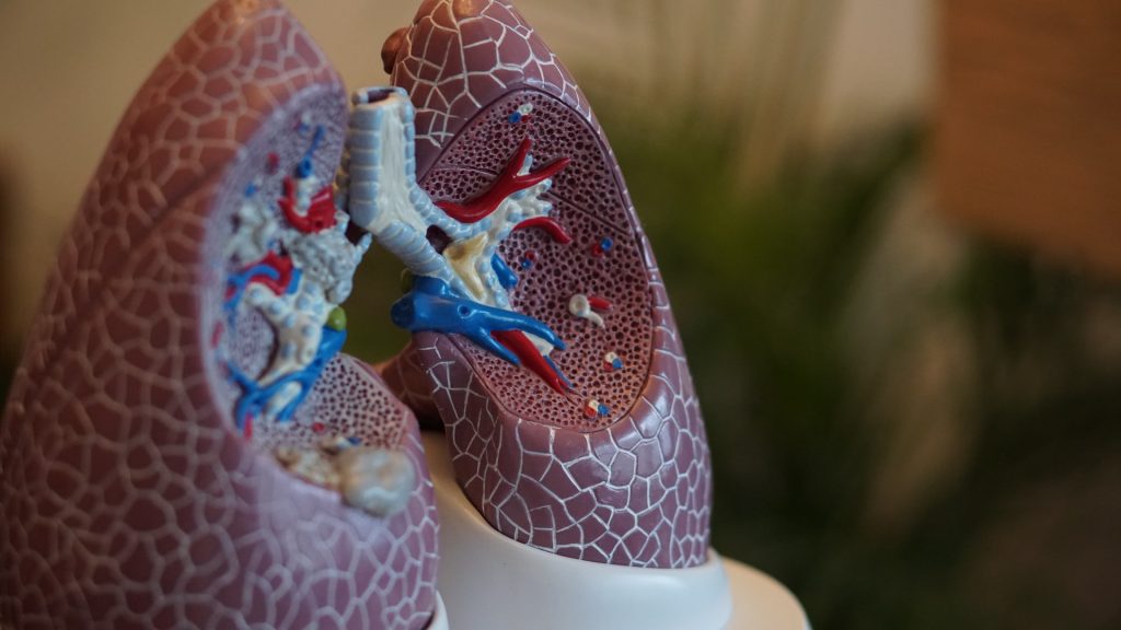
Anatomical model of lungs
Photo by Robina Weermeijer on Unsplash

Airway inflammation and emphysema are more common in marijuana smokers than cigarette smokers, according to a study published in Radiology. Researchers said the difference may be due to the way that marijuana is smoked, which is usually inhaled more deeply and without a filter.

Marijuana is one of the most widely used psychoactive substances in the world and the most-commonly smoked substance after tobacco. Its use has increased in recent years amid legalisation of recreational marijuana in many countries. The growing use has created an urgent need for information on marijuana’s effects on the lungs, something that is currently lacking.

“We know what cigarettes do to the lungs,” said study author Giselle Revah, MD, a cardiothoracic radiologist and assistant professor at the University of Ottawa. “There are well researched and established findings of cigarette smoking on the lungs. Marijuana we know very little about.” 

To find out more, Dr Revah and colleagues compared chest CT results from 56 marijuana smokers with those of 57 non-smoking controls and 33 tobacco-only smokers.
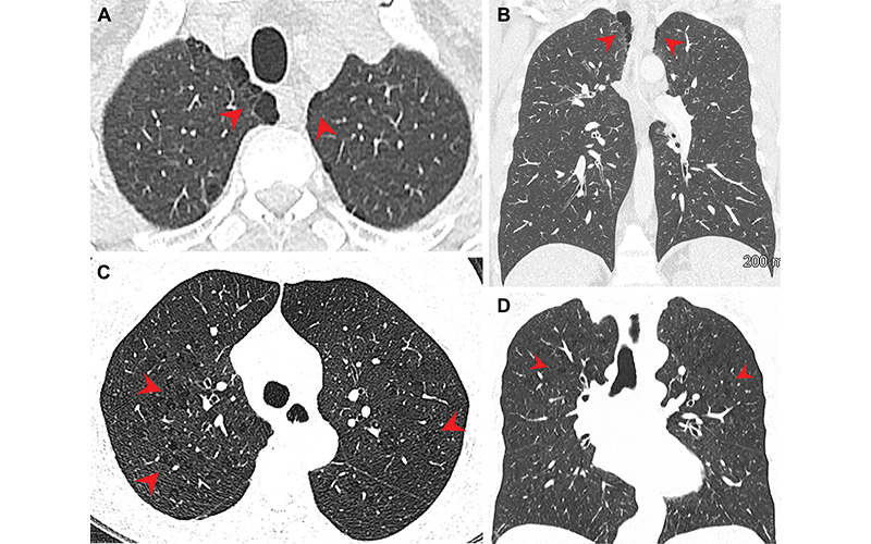
Pulmonary emphysema in (A, B) marijuana and (C, D) tobacco smokers. (A) Axial and (B) coronal CT images in a 44-year-old male marijuana smoker show paraseptal emphysema (arrowheads) in bilateral upper lobes. (C) Axial and (D) coronal CT images in a 66-year-old female tobacco smoker with centrilobular emphysema represented by areas of centrilobular lucency (arrowheads). (Murtha, et al.)

Lack of filtering partly to blame

Three-quarters of the marijuana smokers had emphysema, a lung disease that causes difficulty with breathing, compared with 67% of the tobacco-only smokers. Only 5% of the non-smokers had emphysema. Paraseptal emphysema, which damages the tiny ducts that connect to the air sacs in the lungs, was the predominant emphysema subtype in marijuana smokers compared to the tobacco-only group.

Airway inflammation was also more common in marijuana smokers than non-smokers and tobacco-only smokers, as was gynecomastia, enlarged male breast tissue due to a hormone imbalance. Gynecomastia was found in 38% of the marijuana smokers, compared with 11% of the tobacco-only smokers and 16% of the controls. 

The researchers found similar results among age-matched subgroups, where the rates of emphysema and airway inflammation were again higher in the marijuana smokers than the tobacco-only smokers.

There was no difference in coronary artery calcification between age-matched marijuana and tobacco-only groups.

Dr. Revah said the results were surprising, especially considering that the patients in the tobacco-only group had an extensive smoking history.

“The fact that our marijuana smokers – some of whom also smoked tobacco – had additional findings of airway inflammation/chronic bronchitis suggests that marijuana has additional synergistic effects on the lungs above tobacco,” she said. “In addition, our results were still significant when we compared the non-age-matched groups, including younger patients who smoked marijuana and who presumably had less lifetime exposure to cigarette smoke.” 

The reasons for the differences between the two groups is likely due to several factors. Marijuana is smoked unfiltered, Dr Revah noted, while tobacco cigarettes are usually filtered. This results in more particulates reaching the airways from smoking marijuana.

In addition, marijuana is inhaled with a longer breath hold and puff volume than tobacco smoke.

“It has been suggested that smoking a marijuana joint deposits four times more particulates in the lung than an average tobacco cigarette,” Dr Revah said. “These particulates are likely airway irritants.”

The higher incidence of emphysema may also be due to the way that marijuana is smoked. Full inhalation with a sustained Valsalva manoeuvre, an attempt at exhalation against a closed airway, may lead to trauma and peripheral airspace changes. 

More research is needed, Dr Revah said, with larger groups of people and more data on how much and how often people are smoking. Future research could also look at the impact of different inhalation techniques, such as through a bong, a joint or a pipe.

“It would be interesting to see if the inhalation method makes a difference,” Dr Revah said.

For More Information

Read the Radiology study, “Chest CT Findings in Marijuana Smokers,” and the related editorial.

Source: Radiological Society of North America

Modern Ventilators Shown to Overstretch Lung Tissue

Source: Pixabay CC0

In pulmonary medicine, it has long been debated as to whether ventilator overstretches lung tissue, and now new research published in the American Journal of Respiratory and Critical Care Medicine has proven that they do in fact cause overstretching.

The University of California Riverside researchers showed that there were major differences between natural breathing versus the forced breathing from ventilators. These results are critical, particularly in context of the COVID pandemic and the rush to build ventilators.

“Using novel techniques, we observed that ventilators can overextend certain regions of the lungs,” said Mona Eskandari, assistant professor of mechanical engineering, who led the research. These results may explain why lung health declines for patients the longer they spend on the machines, especially in the case of disease.

Eskandari’s bMECH lab pioneered a technique to study lungs as they are made to breathe. On a custom-built ventilator designed in their lab, the researchers imitated both natural and artificial breathing. Then, they observed isolated lungs involved in both types of breathing using multiple cameras collecting fast, high-resolution images, a method called digital image correlation.

“Our setup allows us to imitate both physiological and artificial breathing on the same lung with the switch of a button,” Eskandari said. “The unique combination of our ventilator with digital image correlation gives us unprecedented insights into the way specific regions of the lungs work in concert with the whole.”

Using their innovative method to interface these two systems, UCR researchers collected evidence demonstrating that natural breathing stretches certain parts of the lung as little as 25% while those same regions stretch to as much as 60% when on a ventilator.

Scholars traditionally model the lungs like balloons, or what they refer to as thin-walled pressure vessels, where pushing air in and pulling air out are understood to be mechanically equivalent.

To explain what they observed in this study, the researchers propose moving away from thin-walled pressure vessel models and instead towards thick-walled models. Unlike thin-walled pressure vessels theory, a thick-walled model accounts for the differing levels of stress in airways resulting from ventilators pushing air in versus natural breathing, which pulls air in. This helps to explain how airways are more engaged and air is more evenly distributed in the lung during physiological breathing.

Iron lungs, the gigantic ventilators used during the late 1940s polio outbreak, acted more like a human chest cavity, expanding the lung as it naturally would. This creates a vacuum effect that pulls air into the lungs. Though this action is gentler for the lungs, these bulky systems prevented easy access to monitoring other organs in hospital care.

By contrast, modern ventilators are more portable and easier for caretakers to work with. However, they push air into the lungs that is not evenly distributed, overstretching some parts and causing a decline in lung health over time.

While it is unlikely that hospitals will return to the iron lung models, it is possible that modern machines can be altered to reduce injury.

“Now that we know about excessive strain when air is delivered to the lungs, the question for us becomes about how we can improve ventilation strategies by emulating natural breathing,” Eskandari said.

Source: University of California – Riverside

Scientists Witness the Creation of a Hybrid Virus

In a world first, scientists have witnessed the fusion two viruses, influenza A virus (IAV) and respiratory syncytial virus (RSV), forming a single, hybrid virus particle (HVP). The discovery was published in Nature Microbiology.

Viruses often share tropism for the same system, such as respiratory viruses preferentially infecting the respiratory system. Coinfections by more than one virus represent between ~10–30% of all respiratory viral infections and are common among children. The clinical impact of viral coinfections is unclear: while some studies indicate that coinfections do not alter the outcome of disease, others report increased incidence of viral pneumonia.

Though evidence suggests virus–virus interactions play an important role in virus dynamics and transmission, viruses are typically studied in isolation. Recent work showed that interactions among respiratory viruses occur and have impacts at multiple levels, from populations, to individuals and tissues. However, studies characterising direct virus–virus interactions within cells are scarce. Here we report previously unknown interactions between IAV and RSV, two clinically important respiratory viruses that belong to different taxonomical families.

To investigate virus–virus interactions, the researchers infected human lung cells with both influenza A virus (IAV) and respiratory syncytial virus (RSV). Using super-resolution microscopy, live-cell imaging, scanning electron microscopy and cryo-electron tomography, the researchers found extracellular and membrane-associated filamentous structures consistent with hybrid viral particles (HVPs).

The researchers found that HVPs harbour surface glycoproteins and ribonucleoproteins of IAV and RSV. HVPs use the RSV fusion glycoprotein to evade anti-IAV neutralising antibodies and infect and spread among cells lacking IAV receptors. Finally, we show that IAV and RSV coinfection in primary cells of the bronchial epithelium results in viral proteins from both viruses latching on together at the apical cell surface.

“Our observations define a previously unknown interaction between respiratory viruses that might affect virus pathogenesis by expanding virus tropism and enabling immune evasion,” the researchers wrote.

“This kind of hybrid virus has never been described before,” virologist and senior author Pablo Murcia told The Guardian. “We are talking about viruses from two completely different families combining together with the genomes and the external proteins of both viruses. It is a new type of virus pathogen.”

When IAV and RSV coinfect, IAV becomes more infectious, infecting a wider array of human cells. Carrying the RSV surface proteins, IAV was able to better evade the immune system. The HVP also spread into cells lacking influenza receptors, letting it progress further down the respiratory tract.

The relationship is not mutually beneficial for the viruses as RSV loses potency. Overall though, pilfering another virus’s tools could play a role in viral pneumonia.

“RSV tends to go lower down into the lung than the seasonal flu virus, and you’re more likely to get more severe disease the further down the infection goes,” said Dr Stephen Griffin, a virologist at the University of Leeds who was not involved in the study.

“It is another reason to avoid getting infected with multiple viruses, because this [hybridisation] is likely to happen all the more if we don’t take precautions to protect our health,” he added.

The researchers also found that the combination of viruses was important; IAV did not form an effective hybrid with rhinovirus.

Air Pollution Worsens Lung Disease Outcomes

Photo by Kouji Tsuru on Unsplash

People with fibrotic interstitial lung disease that has no obvious cause are more likely to die if they live in areas with higher levels of air pollution composed of chemicals associated with industrial sources and vehicular traffic, according to new published today in JAMA Internal Medicine.

The University of Pittsburgh study is the first to link the chemical composition of fine particulate air pollution to worsened fibrotic interstitial lung disease (fILD) outcomes. It is also the largest study ever done to evaluate the impact of air pollution on these patients.

“Some people with these lung diseases have an expected lifespan from diagnosis to death of only a few years, and yet it’s a mystery as to why they developed the disease, why their lungs become so scarred,” said lead author Gillian Goobie, MD, doctoral candidate. “Our study points to air pollution – specifically pollutants from factories and vehicles – as potentially driving faster disease progression and premature death in these patients.”

Goobie and her team obtained data from 6,683 patients with fILDs in the U.S. and Canada and linked their home addresses with satellite and ground-monitoring air pollution data to determine air pollutant composition to an accuracy of less than half a mile.

The team specifically looked at a pollutant known as PM2.5, which refers to particulate matter that measures less than 2.5 microns across, a size invisible to the naked eye. This type of pollution is so small that it can infiltrate deep into the lungs and even cross into the blood stream, where it can contribute to other diseases outside of the lungs, such as heart disease.

“In the past, most environmental health research has focused on the simple definition of PM2.5 as anything of that size,” said co-author James Fabisiak, Ph.D., associate professor in Pitt Public Health’s Department of Environmental and Occupational Health. “But PM2.5 is chemically diverse, with a different composition depending on whether it came from a forest fire or a tailpipe. Research has lacked in determining if the type of PM2.5 matters when it comes to health effects. Our new research is a big step toward filling in that knowledge gap.”

The team found that increasing levels of PM2.5 were linked to more severe disease at diagnosis, faster disease progression as measured by lung function decline and higher likelihood of dying sooner. Pollution high in sulfate (typically produced by factories, such as the coal and steel industries), nitrate (primarily from fossil fuel combustion) and ammonium (usually produced by industry or agriculture) were associated with worse outcomes, whereas chemical signatures from more naturally occurring particulate matter such as sea salt or soil dust didn’t carry as high of an association.

After pollution leaves a smokestack or tailpipe, Goobie noted that sulfate- and nitrate-containing aerosols can be formed in the atmosphere from those and other gaseous pollutants and can be acidic, which can be very damaging to the tiny air sacs of the lungs.

The team is now doing laboratory studies looking at the impact of these pollutants on lung cells at the molecular level to better understand why they are particularly damaging to the lungs of certain people and whether exposure to the pollutants triggers changes to how certain genes work that could cause runaway scarring.

According to the team’s calculations, if exposure to industrial pollutants hadn’t occurred, most premature deaths among participants living in areas of North America with a heavier burden of industry could have been avoided. Participants of colour were disproportionately exposed to higher levels of human-made air pollutants: 13% of the high-exposure group were non-white, but only 8% of the low-exposure group, highlighting the impact of environmental injustice in these findings as well.

Co-senior author S. Mehdi Nouraie, MD, PhD, associate professor of pulmonary, allergy and critical care medicine at Pitt’s School of Medicine, said that the findings further emphasise the need for people with lung conditions that make them more vulnerable to pollution to pay attention to the air quality index and consider minimising time outdoors or in rooms without good air filtration during poor air quality days.

“Ultimately, we want to encourage a data-driven awareness,” A/Prof Nouraie said. “We want people to think about the quality of the air we breathe. Patients, health care providers and policymakers can all use the new information we’re providing to try to improve health outcomes. When you make the air safe for the most vulnerable to breathe, you’re making it safe for all of us.”

Source: University of Pittsburgh

Controlling Allergic Asthma without Compromising Flu Resistance

Young girl sneezing
Photo by Andrea Piacquadio on Unsplash

Blocking calcium signalling in immune cells suppresses allergic asthma, but without compromising the immune defence against flu viruses, according to the findings of a new study published in Science Advances.

The researchers showed that, in a mouse model, removing the gene for a certain calcium channel reduced asthmatic lung inflammation caused by house dust mite faeces, a common cause of allergic asthma. Blocking signals sent through this channel, the calcium release-activated calcium (CRAC) channel, with an investigational inhibitor drug had a similar effect.

The study revolved human cells’ use of signalling and switch-flipping ions, mainly calcium. When triggered by viral proteins or allergens, T cells open channels in their outer membranes, allowing calcium in to activate signalling pathways that control cell division and secretion of cytokine molecules.

Past work had found that CRAC channels in T cells regulate their ability to multiply into armies of cells designed to fight infections caused by viruses and other pathogens.

The new study showed that the CRAC channel inhibitor reduced allergic asthma and mucus build-up in mice without undermining their immune system’s ability to fight influenza, a main worry of researchers seeking to tailor immune-suppressing drugs for several applications.

“Our study provides evidence that a new class of drugs that target CRAC channels can be used safely to counter allergic asthma without creating vulnerability to infections,” said senior study author Stefan Feske, MD, a professor at NYU Langone Health. “Systemic application of a CRAC channel blocker specifically suppressed airway inflammation in response to allergen exposure.”

Allergic asthma, which is the most common form of the disease, is characterised by increased type 2 (T2) inflammation, which involves T helper (Th) 2 cells, the study authors noted. Th2 cells produce cytokines that play important roles in both normal immune defences, and in disease-causing inflammation that occurs in the wrong place and amount. In allergic asthma, cytokines promote the production of IgE antibodies and the recruitment to the lungs of inflammation-causing immune cells called eosinophils, the hallmarks of the disease.

In the new study, the research team found that deletion of the ORAI1 protein in T cells, which makes up the CRAC channel, or treating mice with the CRAC channel inhibitor CM4620, thoroughly suppressed Th2-driven airway inflammation in response to house dust mite allergens.

Treatment with CM4620 significantly reduced airway inflammation when compared to an inactive control substance, with the treated mice also showing much lower levels of Th2 cytokines and related gene expression. Without calcium entering through CRAC channels, T cells are unable to become Th2 cells and produce the cytokines that cause allergic asthma, the authors say.

Conversely, ORAI1 gene deletion, or interfering with CRAC channel function in T cells via the study drug, did not hinder T cell-driven antiviral immunity, as lung inflammation and immune responses were similar in mice with and without ORAI1.

“Our work demonstrates that Th2 cell-mediated airway inflammation is more dependent on CRAC channels than T cell-mediated antiviral immunity in the lung,” said study co-first author Yin-Hu Wang, PhD. “This suggests CRAC channel inhibition as a promising, potential future treatment approach for allergic airway disease.”

Source: NYU Langone Health via PRNewsWire

Asthma from Smoke Exposure Can Pass Down the Generations

Cigarette smoking
Source: Sabine R on Unsplash

Children are more likely to develop asthma if their father was exposed to secondhand smoke when he was a child, according to a study published today in the European Respiratory Journal. The researchers also found that the children have an even higher asthma risk if their father was exposed to secondhand smoke and then also became a smoker.

The researchers say their findings highlight how smoking can cause intergenerational harm, impacting even grandchildren.

The research drew on on data from the Tasmanian Longitudinal Health Study (TAHS). TAHS began in 1968 and is one of the world’s largest and longest ongoing respiratory studies.

For this study, researchers looked at 1689 children who grew up in Tasmania, and their fathers and their paternal grandparents. They compared data on whether the children had developed asthma by age 7 with data on whether the fathers grew up with parents who smoked when they were under age 15. They also included data on whether the fathers were current or former smokers.

First author Mr Jiacheng Liu said, “We found that the risk of non-allergic asthma in children increases by 59% if their fathers were exposed to secondhand smoke in childhood, compared to children whose fathers were not exposed. The risk was even higher, at 72%, if the fathers were exposed to secondhand smoke and went on to smoke themselves.”

Researcher Dr Dinh Bui said, “Our findings show how the damage caused by smoking can have an impact not only on smokers, but also their children and grandchildren. For men who were exposed to secondhand smoke as children, our study suggests that they can still lower the risk they pass on to their own children, if they avoid smoking.”

Senior author Professor Shyamali Dharmage said, “We can’t be certain of how this damage is passed on through generations, but we think it may be to do with epigenetic changes. This is where factors in our environment, such as tobacco smoke, interact with our genes to modify their expression. These changes can be inherited but may be partially reversible for each generation.

“It’s possible that tobacco smoke is creating epigenetic changes in the cells that will go on to produce sperm when boys grow up. These changes can then be passed on to their children.”

The researchers will now investigate if the increased risk of asthma persists into adult life and whether fathers who were exposed to secondhand smoke as children pass on any increase in allergies or other lung diseases to their children.

Source: University of Melbourne

‘Alveoli on a Chip’ Reveals Respiration Secrets

Schematic diagram of the alveolar chip (upper left), photograph of the chip (upper middle), CAD drawing of the multi-generation alveolar structure (upper right), and two typical flow patterns in the alveolar chip (bottom). CREDIT: Yonggang Zhu

Understanding how air and particulates through the alveoli is important to better treat respiratory disease. In Biomicrofluidics, researchers detail a model alveolar system that they built to mimic the breathing action of the human lung and allows visualisation of flow patterns within the alveoli. They observed that flow changes after the 20th branching of the alveoli.

The scientists, from Harbin Institute of Technology in China, designed a chip that includes tubes arranged like the structure of a bifurcation point in the bronchial network. The upper layer of the chip is made of a flexible polymer moulded into small tubes that mimic the alveolar structure. The lower layer is glass, which allows the authors to visualise fluid flow through the tubes.

To mimic inhalation and exhalation, the scientists devised a system in which gas was pressurised in a sinusoidal fashion and pumped around the flexible tubes. Small red polystyrene spheres were added to the fluid flowing through tubes. These spheres allowed them to photograph movement of the fluid as it was pushed through the tubes by the artificial breathing apparatus.

Subsequent branches in the bronchial network are termed ‘generations’, and the team found different flow patterns for different generations. In the human lung, alveoli appear at the 15th generation and remain present for generations up to 23. The researchers found a change in flow pattern between the 19th–20th and the 21st–22nd generations.

“The alveolar flow pattern of the 19th generation is dominated by vortex flow,” author Yonggang Zhu said. “Alveolar flow patterns in the 20th generation are similar to those in the 19th, but somewhat compressed.”

The investigators observed a change in the next generation.

“The alveolar flow pattern in the 21st generation has both vortex flow and radial flow. The vortex region is much smaller than the radial flow region. By the time the flow reaches the 22nd generation, vortex flow disappears completely, and we observe only radial flow,” Zhu said.

The authors also found evidence of chaotic behaviour near the vortex centre. They said more research is needed to fully understand this, but they felt the current study provides a good baseline for deeper investigations.

With the model, researchers will be able to study changes in flow patterns in the alveoli due to diseases such as emphysema and COPD.

Source: American Institute of Physics

Bronchodilators Don’t Ease Smoking-related Respiratory Symptoms in non-COPD Patients

Anatomical model of lungs
Photo by Robina Weermeijer on Unsplash

A study published in the New England Journal of Medicine have found that dual bronchodilators do little to help people who do not have chronic obstructive pulmonary disease (COPD), but who do have respiratory symptoms and a history of smoking.

Millions of people who smoke or used to smoke and have some symptoms of COPD have been prescribed bronchodilators.

“We’ve assumed these medications worked in patients who don’t meet lung function criteria for COPD, but we never checked,” said MeiLan K. Han, MD, a principal investigator and first author of the study. “We now know these existing medications don’t work for these patients.”

According to scientists, the implications are significant. First, they show the importance of diagnosing lung conditions through spirometry, a lung function test Dr Han noted is underutilised in clinical practice. Second, they show the need for new, effective therapies for patients without COPD.

Inhalers have long been the primary go-to treatment for these patients, she explained, because doctors either assume a patient has COPD, or else that their smoking-related symptoms could be helped by the inhalers. But while tobacco smoking causes a large spectrum of lung damage, the study showed bronchodilator therapy only helps patients with enough lung damage that would result in abnormal spirometry readings.

In the 12-week, randomised, double-blinded study, which was part of the Redefining Therapy in Early COPD for the Pulmonary Trials Cooperative (RETHINC), researchers enrolled 535 adults with symptoms of COPD, ages 40–80. Participants used an inhaler twice daily that contained either medication or a placebo.

By the end of the trial, some adults in the medication and placebo groups saw slight respiratory improvements, eg coughed less, produced less phlegm, or felt less winded, which was assessed through the St. George’s Respiratory Questionnaire. However, the researchers found no significant differences between those receiving medication or placebo. They reported 56% (128 of 227) of participants who received the medication saw respiratory symptom improvements, compared to 59% (144 of 244) of those who took the placebo.

According to Dr Han, these data underscore the need to change the standard practice, which is not doing spirometry and just treating patients with the same COPD medications and expecting to see improvement.

Antonello Punturieri, MD, PhD, program director of NHLBI’s Chronic Obstructive Pulmonary Disease/Environment Program, said spirometry testing should be used for any patient who shows signs of COPD, airflow obstruction, or who has a history of cigarette smoking. Though spirometry readings are used during about one-third of medical visits related to COPD, roughly half of patients who would meet criteria for COPD go undiagnosed.

Promoting smoking cessation a primary way to prevent COPD or COPD-like symptoms, the study noted. About one in four current or former smokers without COPD have reported having shortness of breath. In addition to encouraging smoking cessation, doctors can help patients who do not meet lung-function criteria of COPD by working with them to address any other underlying issues, such as overweight and obesity, heart failure, or other lung issues.

“In the meantime, research should be focused on finding new treatments for them,” Dr Han explained. “The next question is, can we develop more targeted therapies for these patients who are on the milder end of the spectrum?”

“Because cough and mucus production show up prominently among these patients, we believe therapies that target mucus production in the airways may be effective,” said Prescott G. Woodruff, MD, a principal investigator and senior author of the study.

Some of these therapies are already in development, and data from other studies may offer insight into the biological causes of excessive airway mucus, which could help point to additional therapies.

Source: NIH/National Heart, Lung and Blood Institute

Hospital Readmissions for Children with Asthma on The Increase

Photo by Kelly Sikkema on Unsplash

Hospital readmissions for asthma are increasing among children, likely stemming from COVID lockdowns reducing immunity to common respiratory viruses. These are the findings of a new study published in the Journal of Asthma. The finding highlights the gaps in health care for this most common of chronic paediatric illnesses.

The Australian study, led by the Murdoch Children’s Research Institute, found about one in three children, mostly pre-schoolers, are readmitted to hospital for asthma compared to one in five a decade ago.

Most asthma hospital presentations were preventable, Murdoch Children’s Dr Katherine Chen said, which emphasises the need for a holistic evaluation of each child’s asthma management to prevent future readmissions.

The study involved 767 children, aged three to 18 years, who were admitted to three hospitals in Victoria state between 2017-2018 with a diagnosis of asthma. It found that 34.3% were readmitted to hospital for asthma, with those aged three to five years accounting for 69.2%. Of the 767 participants, 20.6% were readmitted once, and 13.7% had two or more readmissions in 12 months. 

“Our study highlighted gaps in the children’s asthma care,” Dr Chen said. Over a third of children hadn’t had a review of their inhaler technique, and only about a quarter were prescribed a preventer or asked to continue using it.

“Almost three quarters were discharged without a preventer medication, and over 80 per cent did not have a follow-up clinic booked at the hospital, often reserved for children with difficult-to-control asthma. Most families, therefore, need to navigate their child’s asthma follow-up with their GP.”

Recently, said Dr Chen, asthma admissions had spiked due to the rise in respiratory infections and children lacking immunity to common viruses as a result of COVID lockdowns.

Professor Harriet Hiscock at MCRI said that the findings confirmed the important role of GPs in paediatric asthma management and how targeted interventions at each hospital could reduce readmissions.

“Less than 10 per cent were readmitted within 30 days suggesting the importance of ongoing community care and longer-term asthma control,” she said. The need to regularly review overall asthma management, minimise risk factors, arrange follow-up, and support optimum care in the community are key.

“Interactive digital symptom monitoring with specialist nurse support, home-based education and a culturally tailored education program could also help.”

Prof Hiscock said linked datasets were important to objectively measure the burden of asthma cases on health services.

“Our current dataset cannot verify whether the follow-up appointment was attended, whether caregivers had arranged follow-up post-discharge and if the medications were used as prescribed,” she said. “Integrating datasets such as health services and medication use into clinical care will improve the clinician’s understanding of the child’s asthma control and medication adherence and would assist in providing targeted treatments.”

Asthma is the most common chronic paediatric illness in industrialised countries, affecting 8–10% of children.

Source: Murdoch Children’s Research Institute

Urine Metabolites Yield Clues on Severe Asthma

Asthma inhaler
Source: PIxabay/CC0

A study published in the European Respiratory Journal found severe asthmatics have a distinct metabolite profile detectable in their urine, compared to healthy individuals and those with milder asthma.

Researchers analysed urine samples from more than 600 participants as part of the U-BIOPRED study, a Europe-wide initiative to identify and better understand different sub-types of severe asthma.

The research team discovered a specific type of metabolite, called carnitines, decreased in severe asthmatics. Carnitines play an important role in cellular energy generation and immune responses. Further analyses found carnitine metabolism was lower in severe asthmatics.

These new findings will help enable researchers work towards new, more effective therapies for asthmatics.

Study leader Dr Stacey Reinke said it is vital that asthma treatment is improved.

“To identify and develop new treatment options, we first need to better understand the underlying mechanisms of the disease,” she said.

Examining the body’s chemical profile, or ‘metabolome’, provides a snapshot of a person’s current physiological state and gives useful insight into disease processes.

“In this case, we were able to use the urinary metabolome of asthmatics to identify fundamental differences in energy metabolism that may represent a target for new interventions in asthma control,” Dr Reinke said.

Dr Reinke said it can be difficult and invasive to investigate the lungs directly – but fortunately they contain a lot of blood vessels.

“Therefore, any biochemical changes in the lungs can enter the blood stream, and then be excreted through the urine,” she said.

“These are preliminary results, but we will continue to investigate carnitine metabolism to evaluate its potential as a new asthma treatment target.”

‘Urinary metabotype of severe asthma evidences decreased carnitine metabolism independent of oral corticosteroid treatment in the U-BIOPRED study’ was published in the .

Source: Edith Cowan University