Category: Pain Management

First-visit Communication with Doctor Affects Outcomes of Pain Patients

Photo by Alex Green on Pexels

Chronic pain, defined as daily or significant pain that lasts more than three month, can be complicated to diagnose and treat. Studies have shown that, since chronic pain conditions are clouded with uncertainties, patients often struggle with anxiety and depression – something challenging to for they and their doctors to discuss and manage.

A recent study of 200 chronic neck or back pain sufferers found that effective physician-patient communication during the initial consultation helps patients manage their uncertainties, including their fears, anxieties and confidence in their ability to cope with their condition.

Study leader Charee Thompson, communication professor at University of Illinois Urbana-Champaign, said: “We found that providers and patients who perceive themselves and each other as competent medical communicators during consultations can alleviate patients’ negative feelings of uncertainty such as distress and increase their positive feelings about uncertainties such as their sense of hope and beliefs in their pain-management self-efficacy. Providers and patients successfully manage patients’ uncertainty through two fundamental medical communication processes – informational and socioemotional, each of which can have important clinical implications.”

According to the study, informational competence reflects patients’ abilities to accurately describe their symptoms and verify their understanding of doctors’ explanations and instructions, as well as clinicians asking appropriate questions, providing clear explanations and confirming patients’ understanding. The extent to which doctors and patients establish a trusting relationship through open, honest communication and patients’ feelings of being emotionally supported by the physician reflects socioemotional communication competence.

Thompson and her co-authors — Manuel D. Pulido, a communication professor at California State University, Long Beach; and neurosurgery chair Dr. Paul M. Arnold and medical student Suma Ganjidi, both of the Carle Illinois College of Medicine — published their findings in the Journal of Health Communication.

The current study was based on uncertainty management theory, the hypothesis that people faced with uncertainty about a health condition appraise it and decide whether obtaining information is a benefit or a threat. For example, patients may seek information about the origins of a new symptom to mitigate their anxiety-related uncertainty — or, conversely, they might avoid information-seeking so they can maintain hopeful uncertainty about their prognosis, the team wrote.

The study was conducted at an institute in the Midwest composed of several clinics and programs that treat diseases and injuries of the brain, spinal cord and nervous system. Ranging in age from 18–75, those in the study sample had pain that included but was not limited to their neck, back, buttocks and lower extremities. About 59% of the patients were female. 

Before the consultation, the patients completed surveys rating how they experienced and managed their pain and their certainty or uncertainty about it. They and the providers also completed post-consultation surveys rating themselves and each other on their communication skills. 

The patients rated how well the provider ensured that they understood their explanations and asked questions related to their medical problem. 

To determine if patients’ levels of uncertainty changed, on the pre- and post-consultation surveys the patients ranked how certain or uncertain they felt about six aspects of their pain – including its cause, diagnosis, prognosis, the available treatment options and the risks and benefits of those. The patients also rated themselves on catastrophising – their tendency to worry that they would always be in pain and never find relief.

Patients’ feelings of distress were reduced when they and their physician mutually agreed that the other person was effective at seeking and providing medical information, and when the patients felt emotionally supported by their doctors, the team found.

“Patients’ ratings of their providers’ communication competency significantly predicted reductions in their pain-related uncertainty and in their appraisals of fear and anxiety, as well as increases in their positive uncertainty and pain self-efficacy,” Thompson said. “Providers’ reports of patients’ communication competency were likewise associated with decreases in patients’ pain-related uncertainty and marginally significant improvements in their positive appraisals of uncertainty.”

In a related study, the U. of I.-led team found that, for a subset of spinal pain patients, satisfaction, trust in and agreement with their doctor were strongly associated with the doctors exceeding patients’ expectations for shared decision-making and the quality of the provider’s history-taking and people skills. U. of I. graduate student Junhyung Han was a co-author of that paper, which was published in the journal Patient Education and Counseling

The team wrote that providers and patients need to discuss their mutual expectations for testing, medication and treatment, such as which options are worth pursuing and their potential to meet patients’ expectations for pain relief. 

Thompson said that while these studies’ findings highlight the effects that providers’ overall communication skills have on chronic pain patients’ emotions, expectations and attitudes about their condition, the patients’ communication skills matter, too. 

“I wanted to challenge the notion that pain patients are frustrated or ‘difficult’ because they have unrealistic standards,” Thompson said. “No matter how high their expectations are, what seems to matter most to conversation outcomes is the extent to which patients’ expectations are met or exceeded.

“Consultations mark what may be a long, challenging diagnostic and treatment journey for these patients, and they could benefit from learning about therapies and strategies to help them manage their pain and uncertainties,” Thompson said. “Giving them the tools and language to communicate their symptoms and concerns to providers could make their interactions more productive. Learning about the uncertain nature of pain may validate their fears and anxieties, while awareness and education about the various treatment options and therapies such as cognitive behavioural therapy could enhance their coping and dispel feelings of helplessness and fear.”

Source: University of Illinois at Urbana-Champaign

Men and Women Use Different Biological Systems to Reduce Pain

Photo by Sasun Bughdaryan on Unsplash

In a new study evaluating meditation for chronic lower back pain, researchers at University of California San Diego School of Medicine have discovered that men and women utilise different biological systems to relieve pain. While men relieve pain by releasing endogenous opioids, the body’s natural painkillers, women rely instead on other, non-opioid based pathways. The study was published in PNAS Nexus.

Synthetic opioid drugs, such as morphine and fentanyl, are the most powerful class of painkilling drugs available. Women are known to respond poorly to opioid therapies, which use synthetic opioid molecules to bind to the same receptors as naturally-occurring endogenous opioids. This aspect of opioid drugs helps explain why they are so powerful as painkillers, but also why they carry a significant risk of dependence and addiction.

“Dependence develops because people start taking more opioids when their original dosage stops working,” said Fadel Zeidan, PhD, professor of anaesthesiology and Endowed Professor in Empathy and Compassion Research at UC San Diego Sanford Institute for Empathy and Compassion. “Although speculative, our findings suggest that maybe one reason that females are more likely to become addicted to opioids is that they’re biologically less responsive to them and need to take more to experience any pain relief.”

The study combined data from two clinical trials involving a total of 98 participants, including both healthy individuals and those diagnosed with chronic lower back pain. Participants underwent a meditation training program, then practiced meditation while receiving either placebo or a high-dose of naloxone, a drug that stops both synthetic and endogenous opioids from working. At the same time, they experienced a very painful but harmless heat stimulus to the back of the leg. The researchers measured and compared how much pain relief was experienced from meditation when the opioid system was blocked versus when it was intact.

The study found:

  • Blocking the opioid system with naloxone inhibited meditation-based pain relief in men, suggesting that men rely on endogenous opioids to reduce pain.
  • Naloxone increased meditation-based pain relief in women, suggesting that women rely on non-opioid mechanisms to reduce pain.
  • In both men and women, people with chronic pain experienced more pain relief from meditation than healthy participants.

“These results underscore the need for more sex-specific pain therapies, because many of the treatments we use don’t work nearly as well for women as they do for men,” said Zeidan.

The researchers conclude that by tailoring pain treatment to an individual’s sex, it may be possible to improve patient outcomes and reduce the reliance on and misuse of opioids.

“There are clear disparities in how pain is managed between men and women, but we haven’t seen a clear biological difference in the use of their endogenous systems before now,” said Zeidan. “This study provides the first clear evidence that sex-based differences in pain processing are real and need to be taken more seriously when developing and prescribing treatment for pain.”

Source: University of California – San Diego

Ultrasound Chronic Pain Relief Device Takes a Step Closer

Photo by Pawel Czerwinski on Unsplash

Chronic pain is often caused by faulty signals emerging deep within the brain, giving false alarms about a wound that has since healed, a limb that has since been amputated, or other intricate, hard-to-explain scenarios. Effective treatment options are sorely needed; now, a new device from the University of Utah may represent a practical long-sought solution, using ultrasound to target pain centres deep inside the brain.

Researchers at the University of Utah’s John and Marcia Price College of Engineering and Spencer Fox Eccles School of Medicine have published promising findings about an experimental therapy that has given many participants relief after a single treatment session.

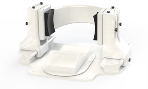
At the core of this research is Diadem, a new biomedical device that uses ultrasound to noninvasively stimulate deep brain regions, potentially disrupting the faulty signals that lead to chronic pain.

The Diadem Device

The findings of a recent clinical trial are published in the journal PainThis study constitutes a translation of two previous studies, published in Nature Communications Engineering and IEEE Transactions on Biomedical Engineering, which describe the unique features and characteristics of the device.

The study was conducted by Jan Kubanek, professor in Price’s Department of Biomedical Engineering (BME), and Thomas Riis, a postdoctoral researcher in his lab, and other collaborators.

The randomised sham-controlled study recruited 20 participants with chronic pain, who each experienced two 40-minute sessions with Diadem, receiving either real or sham ultrasound stimulation. Patients described their pain a day and a week after their sessions, with 60% of the experimental group receiving real treatment reporting a clinical meaningful reduction in symptoms at both points.

“We were not expecting such strong and immediate effects from only one treatment,” says Riis.

“The rapid onset of the pain symptom improvements as well as their sustained nature are intriguing, and open doors for applying these noninvasive treatments to the many patients who are resistant to current treatments,” Kubanek says.

Diadem’s approach is based on neuromodulation, a therapeutic technique that seeks to directly regulate the activity of certain brain circuits. Other neuromodulation approaches are based on electric currents and magnetic fields, but those methods cannot selectively reach the brain structure investigated in the researchers’ recent trial: the anterior cingulate cortex.

After an initial functional MRI scan to map the target region, the researchers adjust Diadem’s ultrasound emitters to correct for the way the waves deflect off of the skull and other brain structures. This procedure was published in Nature Communications Engineering.

The team is now preparing for a Phase 3 clinical, trial which is the final step before FDA approval to use Diadem as a treatment for the general public.

Source: University of Utah

Asymmetric Placebo Effect in Response to Spicy Food

Positive expectations facilitate reward processing and negative expectations prime pain processing

Photo by Ryan Quintal on Unsplash

The expectations humans have of a pleasurable sensation asymmetrically shape neuronal responses and subjective experiences to hot sauce, according to a study published October 8th, in the open-access journal PLOS Biology by Yi Luo from East China Normal University, Kenneth Kishida from Wake Forest School of Medicine, US, and colleagues.

Expectations shape our perception, profoundly influencing how we interpret the world. Positive expectations about sensory stimuli can alleviate distress and reduce pain through what’s known as the placebo effect, while negative expectations may heighten anxiety and exacerbate pain. In the new study, Luo, Kishida, and colleagues investigated the impact of the hedonic aspect of expectations on subjective experiences.

Specifically, the researchers measured neurobehavioral responses to the taste of hot sauce among individuals with a wide range of taste preferences. In total, 47 participants completed the tasks while undergoing functional magnetic resonance imaging scanning. The researchers identified participants who liked versus those who strongly disliked spicy flavors and provided contextual cues about the spiciness of the sauce to be tasted. That way, they were able to dissociate the effects of positive and negative expectations from sensory stimuli (i.e., visual and taste stimuli), which were the same across all participants.

The results showed that positive expectations lead to modulations in the intensity of subjective experience. These modulations were accompanied by increased activity in brain regions previously linked to pleasure, information integration, and the placebo effect, including the anterior insula, dorsolateral prefrontal cortex, and dorsal anterior cingulate cortex. By contrast, negative expectations decreased hedonic experience and increased neural activity in the Neurological Pain Signature network.

Taken together, these findings demonstrate that hedonic aspects of one’s expectations asymmetrically shape how the brain processes sensory input and associated behavioral reports of one’s subjective experiences of intensity, pleasure, and pain. The results suggest a dissociable impact of hedonic information. While positive expectations facilitate higher-level information integration and reward processing, negative expectations prime lower-level processes related to pain and emotions. According to the authors, this study demonstrates the powerful role of hedonic expectations in shaping subjective reality and suggests potential avenues for consumer and therapeutic interventions targeting expectation-driven neural processes.

The authors add, “Our study highlights how hedonic expectations shape subjective experiences and neural responses, offering new insights into the mechanisms behind pain perception.”

Provided by PLOS

Sitting Less may Prevent Back Pain – Even Without Exercise

Photo by Thestandingdesk on Unsplash

A new study from the University of Turku showed that reducing daily sitting prevented back pain from worsening over six months. The results, published in BMJ Open, strengthen the current understanding of the link between activity and back pain as well as the mechanisms related to back pain.

Intuitively, it is easy to think that reducing sitting would help with back pain, but previous research data is surprisingly scarce. The study from the Turku PET Centre and UKK Institute in Finland investigated whether reducing daily sitting could prevent or relieve back pain among overweight or obese adults who spend the majority of their days sitting. The participants were able to reduce their sitting by 40 min/day, on average, during the six-month study.

“Our participants were quite normal middle-aged adults, who sat a great deal, exercised little, and had gained some extra weight. These factors not only increase the risk for cardiovascular disease but also for back pain,” says Doctoral Researcher and Physiotherapist Jooa Norha from the University of Turku in Finland.

Previous results from the same and other research groups have suggested that sitting may be detrimental for back health but the data has been preliminary.

The figure presents the change in back pain intensity on a scale from 0 to 10. The blue bars represent individuals in the intervention group that reduced sitting and the red bars represent the control participants who did not change their sitting habits. Most of the participants in the intervention group decreased their back pain whereas the back pain in the control participants tended to increase.

Robust methods for studying the mechanisms behind back pain

The researchers also examined potential mechanisms behind the prevention of back pain.

”However, we did not observe that the changes in back pain were related to changes in the fattiness or glucose metabolism of the back muscles,” Norha says.

Individuals with back pain have excessive fat deposits within the back muscles, and impaired glucose metabolism, or insulin sensitivity, can predispose to pain. Nevertheless, back pain can be prevented or relieved even if no improvements in the muscle composition or metabolism take place. The researchers  used magnetic resonance imaging (MRI) and PET imaging that is based on a radioactive tracer to measure the back muscles.

“If you have a tendency for back pain or excessive sitting and are concerned for your back health, you can try to figure out ways for reducing sitting at work or during leisure time. However, it is important to note that physical activity, such as walking or more brisk exercise, is better than simply standing up,” Norha points out.

The researchers wish to remind that switching between postures is more important than only looking for the perfect posture.

Source: University of Turku

When a Child Hurts, Validating their Pain may be the Best First Aid

Photo by cottonbro studio

Whether it’s a sore arm or a fear of injections, how a child is treated when they present with pain could significantly affect how they respond to and manage pain later in life.

In a new study published in the journal Pain, researchers say that parents and doctors should be mindful of how they talk to and treat children experiencing pain – no matter how big or small the injury – knowing that these foundational experiences can be carried forward into adulthood.

Drawing from diverse research across developmental psychology, child mental health, and pain sciences, the University of South Australia researchers say that it may be important to validate children’s pain by demonstrating that their pain-related experiences, emotions, or behaviours are acceptable, understood, and legitimate.

By validating a child’s pain, the child feels heard and believed, which reinforces their trust and connection with their parent, or with a treating doctor.

UniSA researcher Dr Sarah Wallwork says social relationships play a critical role in shaping how health is experienced throughout the lifespan.

“When a parent or doctor validates a child’s experiences in a way that matches their expressed vulnerability, it helps the child to feel accepted, builds connection and trust, and may help the child to develop critical skills in regulating their emotions,” Dr Wallwork says.

“For example, when a doctor is attentive, and responds to a child’s emotional and behavioural cues, particularly about seeking help, the clinician is telling the child their pain is real and concurrently reinforcing helpful pain management behaviours, such as attending the clinic.

“However, if these cues are missed, or the doctor questions the validity of their pain, this can have negative consequences for the child. Not only can it affect the clinician-patient relationship and trust but it can also impact future attendance at appointments and adherence to a pain management plan.

“Pain and emotion are inextricably linked, with emotion dysregulation commonly co-occurring with chronic pain.

“By validating children’s experiences of pain, they are likely to hold fewer negatively biased memories of pain and be in better position to seek help in the future, when then need it.”

In Australia, as many as one in four children experience chronic pain.

Dr Wallwork says that setting children up for success should cover all aspects of life, including pain management.

“Our research highlights an underemphasised element of child and youth pain treatment, especially for children in minoritised groups, who are systematically undertreated for pain,” Dr Wallwork says.

“People with chronic pain often report that their pain-related experiences are met with disbelief or dismissal. This can have significant consequences, including poor mental health and reduced quality of life.

“Given the significant burden of chronic pain, and the clear intersection with the rising child mental health crisis, it’s important that we better manage pain earlier on, rather than waiting until it is too late.”

Dr Wallwork says this review provides a building block for future empirical research.

Source: University of South Australia

Scientists Trace the Neural Pathway of the Placebo Effect

Photo by Danilo Alvesd on Unsplash

The placebo effect is very real. This we’ve known for decades, as seen in real-life observations and the best double-blinded randomised clinical trials researchers have devised for many diseases and conditions, especially pain. And yet, how and why the placebo effect occurs has remained a mystery. Now, neuroscientists have discovered a key piece of the placebo effect puzzle, reporting it in Nature.

Researchers at the University of North Carolina School of Medicine – with colleagues from Stanford, the Howard Hughes Medical Institute, and the Allen Institute for Brain Science – discovered a pain control pathway that links the cingulate cortex in the front of the brain, through the pons region of the brainstem, to cerebellum in the back of the brain.

The researchers, led by Greg Scherrer, PharmD, PhD, associate professor in the UNC Department of Cell Biology and Physiology, the UNC Neuroscience Center, and the UNC Department of Pharmacology, then showed that certain neurons and synapses along this pathway are highly activated when mice expect pain relief and experience pain relief, even when there is no medication involved.

“That neurons in our cerebral cortex communicate with the pons and cerebellum to adjust pain thresholds based on our expectations is both completely unexpected, given our previous understanding of the pain circuitry, and incredibly exciting,” said Scherrer. “Our results do open the possibility of activating this pathway through other therapeutic means, such as drugs or neurostimulation methods to treat pain.”

Scherrer and colleagues said research provides a new framework for investigating the brain pathways underlying other mind-body interactions and placebo effects beyond the ones involved in pain.

The Placebo Paradox

In conjunction with millennia of evolution, our brains can search for ways to alleviate the sensation of pain, in some cases quantifiably as with released chemicals, and less quantifiably through positive thinking and even prayer which have some documented benefit. And then there is the placebo effect.

In clinical research, the placebo effect is often seen in the “sham” treatment group that receives a fake pill or intervention that is supposed to be inert; no benefit is expected. Except that the brain is so powerful and individuals so desire to feel better that some experience a marked improvement in their symptoms. Some placebo effects are so strong that individuals are convinced they received a real treatment meant to help them.

In fact, it’s thought that some individuals in the “actual” treatment group also derive benefit from the placebo effect, complicating experimental design and driving larger sample sizes. One way to help scientists account for this is to first understand what precisely is happening in the brain of someone experiencing the placebo effect.

Enter the Scherrer lab

The scientific community’s understanding of the biological underpinnings of pain relief through placebo analgesia came from human brain imaging studies, which showed activity in certain brain regions. Those imaging studies did not have enough precision to show what was actually happening in those brain regions. So Scherrer’s team designed a set of meticulous, complementary, and time-consuming experiments to learn in more detail, with single nerve cell precision, what was happening in those regions.

First, the researchers created an assay that generates in mice the expectation of pain relief and then very real placebo effect of pain relief. Then the researchers used a series of experimental methods to study the intricacies of the anterior cingulate cortex (ACC), which had been previously associated with the pain placebo effect. While mice were experiencing the effect, the scientists used genetic tagging of neurons in the ACC, imaging of calcium in neurons of freely behaving mice, single-cell RNA sequencing techniques, electrophysiological recordings, and optogenetics – the use of light and fluorescent-tagged genes to manipulate cells.

These experiments helped them see and study the intricate neurobiology of the placebo effect down to the brain circuits, neurons, and synapses throughout the brain.

The scientists found that when mice expected pain relief, the rostral anterior cingulate cortex neurons projected their signals to the pontine nucleus, which had no previously established function in pain or pain relief. And they found that expectation of pain relief boosted signals along this pathway.

“There is an extraordinary abundance of opioid receptors here, supporting a role in pain modulation,” Scherrer said. “When we inhibited activity in this pathway, we realised we were disrupting placebo analgesia and decreasing pain thresholds. And then, in the absence of placebo conditioning, when we activated this pathway, we caused pain relief.

Lastly, the scientists found that Purkinje cells – a distinct class of large branch-like cells of the cerebellum – showed activity patterns similar to those of the ACC neurons during pain relief expectation. Scherrer and first author Chong Chen, MD, PhD, a postdoctoral research associate in the Scherrer lab, said that this is cellular-level evidence for the cerebellum’s role in cognitive pain modulation.

“We all know we need better ways to treat chronic pain, particularly treatments without harmful side effects and addictive properties,” Scherrer said. “We think our findings open the door to targeting this novel neural pain pathway to treat people in a different but potentially more effective way.”

Source: University of North Carolina Health Care

Radiology Helps Treat Chronic Pain

Dr Winter performing a CT-guided interventional procedure. Photo: Supploed

Radiology encompasses more than just imaging. It is a medical field that uses various imaging techniques to diagnose conditions, guide minimally invasive procedures and, much to the relief of agonised patients, treat chronic pain.

‘Traditionally, radiology is known as a modality where causes of pain are only diagnosed’, says Dr Arthur Winter, a radiologist at SCP Radiology. ‘Interventional radiology has changed this. It is a rapidly developing branch of radiology involving minimally invasive procedures.  Pain management procedures are becoming a daily part of busy radiology departments.’

Simply put, interventional radiologists can use precisely targeted injections to intervene in the body’s perception of pain.

Understanding pain

Pain is a signal from the nervous system to let you know that something is wrong in your body. It is transmitted in a complex interaction between specialised nerves, the spinal cord and the brain. It can take many forms, be localised to one part of the body or appear to come from all over.

Pain can be acute or chronic

Harvard Medical School gives an overview of the difference between the two. ‘Most acute pain comes from damage to body tissues. It results from physical trauma such as a sports or exercise injury, a broken bone, a medical procedure or an accident like stubbing your toe, cutting a finger or bumping into something. The pain can feel sharp, aching or throbbing and often heals within a few days to a few weeks.’

In comparison, chronic pain lasts at least two to three months, often long after you have recovered from the injury or illness and may even become permanent. It could also be a result of lifestyle diseases. Symptoms and severity vary and may include a dull ache, shooting, burning, stabbing or electric shock-like pain and sensations like tingling and numbness. Chronic pain can be debilitating and affect your ability to perform activities of daily living.

Interventional pain management

Although some acute pain can be managed with interventions, it is patients with chronic pain that truly benefit. ‘These patients often use high doses of opioid painkillers that may cause nausea, constipation, anorexia and addiction. Other painkillers may also irritate the stomach lining and cause kidney problems,’ says Dr Winter.

An alternative that interventional pain management offers, involves injections called nerve blocks that target very specific nerves.

‘Most of these interventions prevent nerve impulses or pain signals from being transmitted, using long-acting local anaesthetics. The effect is usually temporary but the addition of cortisone – or steroids – often brings longer-lasting relief. In some cases, it could be appropriate to follow the temporary block with neurolysis, which is a permanent disruption or destruction of the target nerves.’

Although nerve blocks and other long-acting pain injections have been done for years, the scope of procedures is evolving fast. The involvement of radiologists has also grown.

Dr Winter explains. ‘Pain management has traditionally been the responsibility of clinicians and anaesthetists. During nerve block procedures, they were typically guided by their knowledge of anatomy or a continuous X-ray technique called fluoroscopy. As ultrasound became more widely available, many anaesthetists learned to do these procedures under ultrasound guidance.

‘These specialists still provide these treatments but, thanks to the availability of specialised imaging equipment, radiologists now have the tools and skill to do procedures under sophisticated image guidance. With CT guidance, some procedures can be performed with great accuracy while avoiding blood vessels and non-target organs,’ says Dr Winter.

‘A lower dose of medication is also needed if the needle is placed accurately next to the target nerves. It is therefore not surprising that this is increasingly becoming a responsibility of interventional radiologists.’

Other procedures where radiologists are involved include targeted Botox injections to treat the symptoms of Piriformis syndrome, epidural cortisone injections for inflammation in the spine and a procedure called epidural blood patch. This is to seal spinal fluid leaks that cause low-pressure headaches.

In conclusion, Dr Winter says chronic pain may cause poor quality of life and depression, often seen in patients with underlying cancer. ‘It is especially these patients who should be considered for interventions. There are, for example, very effective procedures to manage pain caused by pancreatic and pelvic cancers.

‘Specialists like oncologists and neurologists recognise the value of interventional radiology in pain management and work closely with us to support their patients. It is a growing branch of radiology that offers a minimally invasive solution and it’s quite rewarding to see patients regain some quality of life.’

Researchers Delve into the Roots of Chronic Pain

Source: Pixabay CC0

A team of researcher have identified a new function for the PIEZO2 protein – in mediating chronic pain hypersensitivity. The research suggests a new target for analgesics and potentially explains why pain medications that target voltage gated sodium channels have been disappointing as clinical targets. The study, led by Oscar Sánchez-Carranza in Professor Gary Lewin’s lab at the Max Delbrück Center, was published in the journal Brain.

“There’s a good correlation between chronic pain and the sensitisation of pain receptors, called nociceptors, in humans,” says Lewin. “This study implicates the PIEZO2 channel as a critical mediator of sensory signals that maintain chronic pain.”

PIEZO2 protein forms an ion channel in human sensory receptors. Previous studies have shown that the ion channel is involved in communicating the sense of touch to the brain. People with “loss-of-function” mutations in the PIEZO2 gene are hypo-sensitive to gentle touch or vibration. By contrast, patients with “gain-of-function mutations” in PIEZO are often diagnosed with complex developmental disorders. But whether gain-of-function mutations are responsible for mechanical hypersensitivity had never been proven.

Mutation dramatically sensitises nociceptors

To study the connection, Sánchez-Carranza created two strains of so called “gain-of-function” mice, each carrying a different version of a mutated PIEZO2 gene. He expected to find the touch receptors of these mice to be highly sensitive. In cell biology experiments his team has found that PIEZO2 mutations have a powerful effect on the activity of the ion channel. One mutation, for example, causes the channel to open with 10 times less force compared to normal non-mutated channels.

Using electrophysiological methods developed in the Lewin lab, Sánchez-Carranza and his colleagues measured electrical activity in sensory neurons isolated from the transgenic mice. They found that in addition to sensitising touch receptors as expected, the mutations made nociceptive receptors – neurons that detect painful mechanical stimuli – dramatically more sensitive to mechanical stimuli.

Moreover, the researchers found that the nociceptors were activated by mechanical stimuli that would normally be experienced as light touch.

“You pretty much need to crush the skin to activate nociceptors,” Sánchez-Carranza explains. But the nociceptors from the transgenic mice were triggered by levels of mechanical force that would normally be perceived as a touch. They were incredibly sensitive.”

That a single mutation in PIEZO2 was enough to change the physiology of the nociceptors from one type of neuron to another, was especially surprising, says Lewin. More significantly, when the stimulus was removed, the neurons kept firing. The study is the first time that anyone has linked gain-of-function mutations in the PIEZO2 gene to pain receptors.

PIEZO2 might be involved in pain syndromes like fibromyalgia

Clinical studies have shown that in patients with chronic pain syndromes such as fibromyalgia and small fibre neuropathies, C-fibre nociceptors, which are the sensory receptors that initiate pain, are hyperactive. When researchers have recorded the activity of nociceptors in such people, they found that the they were active in the absence of any mechanical stimulus. But the mechanism was not clear.

“We show that just by changing one amino acid in PIEZO2, we can actually mimic a lot of what happens in chronic pain in the C-fibres,” says Lewin. In humans, “PIEZO2 might be involved in many of these pathologies.” Nociceptive neurons are the largest population of sensory neurons that innervate the skin – humans have four times more pain receptors in the skin than touch receptors.

Up to 20% of the adult population suffers from chronic pain, according to a 2023 study by the U.S. National Institutes of Health, which is poorly treated with existing medications. The same NIH study found that two thirds of people who reported chronic pain in 2019 were still suffering one year later.

The findings suggest that a particular aspect of the PIEZO2 channels mechanism of opening could be targeted by new pain medications. Much effort on developing new analgesics has focused on voltage gated sodium channels with limited success, says Lewin. “By addressing the root cause of nociceptor sensitisation, new drugs could provide better relief for chronic pain sufferers.”

Source: Max Delbrück Center

Researchers Figure out How Propofol Makes Patients Lose Consciousness

Photo by Anna Shvets on Pexels

There are many drugs that anaesthesiologists can use to induce unconsciousness in patients. Exactly how these drugs cause the brain to lose consciousness has been a longstanding question, but MIT neuroscientists have now answered that question for the commonly used drug propofol.

Using a novel technique for analysing neuron activity, the researchers discovered that the drug propofol induces unconsciousness by disrupting the brain’s normal balance between stability and excitability. The drug causes brain activity to become increasingly unstable, until the brain loses consciousness.

“The brain has to operate on this knife’s edge between excitability and chaos. It’s got to be excitable enough for its neurons to influence one another, but if it gets too excitable, it spins off into chaos. Propofol seems to disrupt the mechanisms that keep the brain in that narrow operating range,” says Earl K. Miller, the Picower Professor of Neuroscience and a member of MIT’s Picower Institute for Learning and Memory.

The new findings, published in Neuron, could help researchers develop better tools for monitoring patients as they undergo general anaesthesia.

Miller and Ila Fiete, a professor of brain and cognitive sciences, the director of the K. Lisa Yang Integrative Computational Neuroscience Center (ICoN), and a member of MIT’s McGovern Institute for Brain Research, are the senior authors of the new study. MIT graduate student Adam Eisen and MIT postdoc Leo Kozachkov are the lead authors of the paper.

Losing consciousness

Propofol is a drug that binds to GABA receptors in the brain, inhibiting neurons that have those receptors. Other anaesthesia drugs act on different types of receptors, and the mechanism for how all of these drugs produce unconsciousness is not fully understood.

Miller, Fiete, and their students hypothesised that propofol, and possibly other anaesthesia drugs, interfere with a brain state known as “dynamic stability.” In this state, neurons have enough excitability to respond to new input, but the brain is able to quickly regain control and prevent them from becoming overly excited.

Previous studies of how anaesthesia drugs affect this balance have found conflicting results: Some suggested that during anaesthesia, the brain shifts toward becoming too stable and unresponsive, which leads to loss of consciousness. Others found that the brain becomes too excitable, leading to a chaotic state that results in unconsciousness.

Part of the reason for these conflicting results is that it has been difficult to accurately measure dynamic stability in the brain. Measuring dynamic stability as consciousness is lost would help researchers determine if unconsciousness results from too much stability or too little stability.

In this study, the researchers analysed electrical recordings made in the brains of animals that received propofol over an hour-long period, during which they gradually lost consciousness. The recordings were made in four areas of the brain that are involved in vision, sound processing, spatial awareness, and executive function.

These recordings covered only a tiny fraction of the brain’s overall activity, so to overcome that, the researchers used a technique called delay embedding. This technique allows researchers to characterize dynamical systems from limited measurements by augmenting each measurement with measurements that were recorded previously.

Using this method, the researchers were able to quantify how the brain responds to sensory inputs, such as sounds, or to spontaneous perturbations of neural activity.

In the normal, awake state, neural activity spikes after any input, then returns to its baseline activity level. However, once propofol dosing began, the brain started taking longer to return to its baseline after these inputs, remaining in an overly excited state. This effect became more and more pronounced until the animals lost consciousness.

This suggests that propofol’s inhibition of neuron activity leads to escalating instability, which causes the brain to lose consciousness, the researchers say.

Better anesthesia control

To see if they could replicate this effect in a computational model, the researchers created a simple neural network. When they increased the inhibition of certain nodes in the network, as propofol does in the brain, network activity became destabilized, similar to the unstable activity the researchers saw in the brains of animals that received propofol.

“We looked at a simple circuit model of interconnected neurons, and when we turned up inhibition in that, we saw a destabilization. So, one of the things we’re suggesting is that an increase in inhibition can generate instability, and that is subsequently tied to loss of consciousness,” Eisen says.

As Fiete explains, “This paradoxical effect, in which boosting inhibition destabilises the network rather than silencing or stabilising it, occurs because of disinhibition. When propofol boosts the inhibitory drive, this drive inhibits other inhibitory neurons, and the result is an overall increase in brain activity.”

The researchers suspect that other anesthetic drugs, which act on different types of neurons and receptors, may converge on the same effect through different mechanisms – a possibility that they are now exploring.

If this turns out to be true, it could be helpful to the researchers’ ongoing efforts to develop ways to more precisely control the level of anaesthesia that a patient is experiencing. These systems, which Miller is working on with Emery Brown, the Edward Hood Taplin Professor of Medical Engineering at MIT, work by measuring the brain’s dynamics and then adjusting drug dosages accordingly, in real-time.

“If you find common mechanisms at work across different anaesthetics, you can make them all safer by tweaking a few knobs, instead of having to develop safety protocols for all the different anaesthetics one at a time,” Miller says. “You don’t want a different system for every anesthetic they’re going to use in the operating room. You want one that’ll do it all.”

The researchers also plan to apply their technique for measuring dynamic stability to other brain states, including neuropsychiatric disorders.

“This method is pretty powerful, and I think it’s going to be very exciting to apply it to different brain states, different types of anaesthetics, and also other neuropsychiatric conditions like depression and schizophrenia,” Fiete says.

Source: MIT