Researchers used a computer screening approach to identify two compounds that could help prevent vision loss in people with a genetic eye disease
Photoreceptor cells in the retina. Credit: Scientific Animations
Two new compounds may be able to treat retinitis pigmentosa, a group of inherited eye diseases that cause blindness. The compounds, described in a study published January 14th in the open-access journal PLOS Biologyby Beata Jastrzebska from Case Western Reserve University, US, and colleagues, were identified using a virtual screening approach.
In retinitis pigmentosa, the retina protein rhodopsin is often misfolded due to genetic mutations, causing retinal cells to die off and leading to progressive blindness. Small molecules to correct rhodopsin folding are urgently needed to treat the estimated 100 000 people in the United States with the disease. Current experimental treatments include retinoid compounds, such as synthetic vitamin A derivatives, which are sensitive to light and can be toxic, leading to several drawbacks.
In the new study, researchers utilised virtual screening to search for new drug-like molecules that bind to and stabilise the structure of rhodopsin to improve its folding and movement through the cell. Two non-retinoid compounds were identified which met these criteria and had the ability to cross the blood-brain and blood-retina barriers. The team tested the compounds in the lab and showed that they improved cell surface expression of rhodopsin in 36 of 123 genetic subtypes of retinitis pigmentosa, including the most common one. Additionally, they protected against retinal degeneration in mice with retinitis pigmentosa.
“Importantly, treatment with either compound improved the overall retina health and function in these mice by prolonging the survival of their photoreceptors,” the authors say. However, they note that additional studies of the compounds or related compounds are needed before testing the treatments in humans.
The authors add, “Inherited mutations in the rhodopsin gene cause retinitis pigmentosa (RP), a progressive and currently untreatable blinding disease. This study identifies small molecule pharmacochaperones that suppress the pathogenic effects of various rhodopsin mutants in vitro and slow photoreceptor cell death in a mouse model of RP, offering a potential new therapeutic approach to prevent vision loss.”
Could complex beliefs like paranoia have roots in something as basic as vision? A new Yale study finds evidence that they might.
When completing a visual perception task, in which participants had to identify whether one moving dot was chasing another moving dot, those with greater tendencies toward paranoid thinking (believing others intend them harm) and teleological thinking (ascribing excessive meaning and purpose to events) performed worse than their counterparts, the study found. Those individuals more often – and confidently – claimed one dot was chasing the other when it wasn’t.
The findings, published in the journal Communications Psychology, suggest that, in the future, testing for illnesses like schizophrenia could be done with a simple eye test.
“We’re really interested in how the mind is organised,” said senior author Philip Corlett, an associate professor of psychiatry at Yale School of Medicine and member of the Wu Tsai Institute. “Chasing or other intentional behaviours are what you might think of as experiences perceived at a very high-level in the brain, that someone might have to reason through and deliberate. In this study, we can see them low down in the brain, in vision, which we think is exciting and interesting – and has implications for how those mechanisms might be relevant for schizophrenia.”
Paranoia and teleological thinking are similar in that they are both misattributions of intention, but paranoia is a negative perception while teleological thinking tends to be positive. Both patterns of thinking are linked to psychosis and schizophrenia.
Hallucinations are associated with psychosis as well and are often about other people, said Corlett, suggesting there may be a social component to these visual misperceptions.
“So we wondered whether there might be something related to social perception – or misperception, what we refer to as social hallucination – that we could measure and that relate to these symptoms of psychosis,” he said.
For the task, participants were shown dots moving on a screen. Sometimes one dot was chasing another; other times there was no chase. Across different trials of the task, participants had to say whether a chase was occurring or not.
Those with higher degrees of paranoia and teleological thinking (as measured through questionnaires) were more likely than others to say with confidence that a chase was happening when one wasn’t. Essentially, they perceived a social interaction that wasn’t occurring.
In additional experiments, the researchers asked participants to identify which dot was doing the chasing and which dot was being chased. In these results, paranoia and teleological thinking began to diverge.
“People with paranoia were particularly bad at detecting which dot was being chased,” said Santiago Castiello, lead author of the study and a postdoctoral researcher in Corlett’s lab. “And people with high teleology were particularly bad at detecting which dot was doing the chasing.”
That these two types of beliefs differed in this way highlights that they are distinct and may have implications for diagnosis or treatment, said the researchers. The connection to vision may also shift thinking around how the brain gives rise to psychotic symptoms.
“Very few people with congenital blindness develop schizophrenia,” said Castiello. “Finding these social hallucinations in vision makes me wonder if schizophrenia is something that develops through errors in how people sample the visual world.”
While there are no immediate therapeutic implications from these findings, deeper understanding of these beliefs could aid in pharmacological treatment development and risk assessment.
“One thing we’re thinking about now is whether we can find eye tests that predict someone’s risk for psychosis,” said Corlett. “Maybe there is some very quick perceptual task that can identify when someone might need to talk to a clinician.”
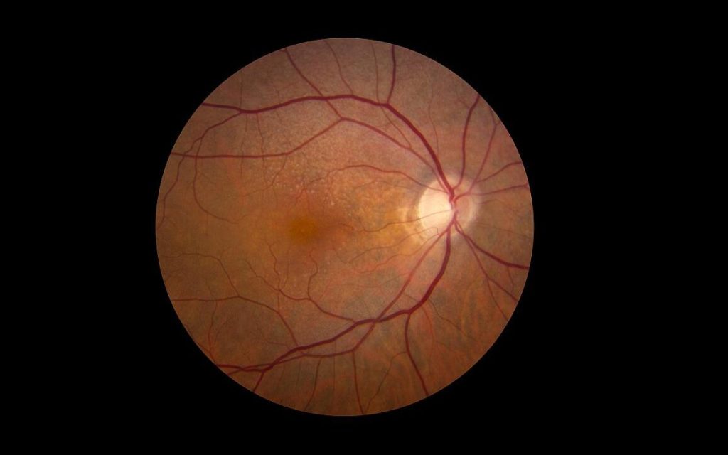
Retina showing reticular pseudodrusen. Although they can infrequently appear in individuals with no other apparent pathology, their highest rates of occurrence are in association with age-related macular degeneration (AMD), for which they hold clinical significance by being highly correlated with end-stage disease sub-types, choroidal neovascularisation and geographic atrophy. Credit: National Eye Institute
A new study from researchers at Wilmer Eye Institute, Johns Hopkins Medicine explains not only why some patients with wet age-related macular degeneration (or “wet” AMD) fail to have vision improvement with treatment, but also how an experimental drug could be used with existing wet AMD treatments to save vision.
Wet AMD, one of two kinds of AMD, is a progressive eye condition caused by an overgrowth of blood vessels in the retina. Such blood vessels – caused by an overexpression of a protein known as VEGF that leads to blood vessel growth – then leak fluid or bleed and damage the retina, causing vision loss.
Despite the severe vision loss often experienced by people with wet AMD, less than half of patients treated with monthly eye injections, known as anti-VEGF therapies, show any major vision improvements. Additionally, for those who do benefit with improved vision, most will lose those gains over time.
Now, in the full report published November 4 in the Proceedings of the National Academy of Sciences, the Wilmer-led team of researchers share how such anti-VEGF therapies may actually contribute to lack of vision improvements by triggering the overexpression of a second protein. Known as ANGPTL4, the protein is similar to VEGF, as it can also stimulate overproduction of abnormal blood vessels in the retina.
“We have previously reported that ANGPTL4 was increased in patients who did not respond well to anti-VEGF treatment,” says Akrit Sodhi, MD, PhD, corresponding author and associate professor of ophthalmology at the Johns Hopkins University School of Medicine and the Wilmer Eye Institute. “What we saw in this paper was a paradoxical increase of ANGPTL4 in patients that received anti-VEGF injections – the anti-VEGF therapy itself turned on expression of this protein.”
The team compared VEGF and ANGPTL4 levels in the eye fluid of 52 patients with wet AMD at various stages of anti-VEGF treatment. Prior to anti-VEGF injections, patients with wet AMD had high levels of ANGPTL4 and VEGF proteins. After treatment, their VEGF levels predictably decreased, yet ANGPTL4 levels rose higher, indicating ANGPTL4 remained active following the anti-VEGF injections and the treatments contributed to an increase in ANGPTL4. Such ANGPTL4 activity can lead to blood vessel overgrowth and lack of vision improvement.
The team then investigated ways to bridge the gap between patients with increased ANGPTL4 following anti-VEGF treatments by testing the experimental drug 32-134D in mice with wet AMD. The drug decreases levels of a third protein, HIF-1, known to be involved in wet AMD and diabetic eye disease for its role in switching on VEGF production. Researchers believed the HIF-inhibitor 32-134D would have a similar effect on ANGPTL4 following anti-VEGF treatment, since ANGPTL4 production is also turned on by HIF-1.
In mice treated with 32-134D, the team observed a decrease in HIF-1 levels and VEGF, as well as decreased levels of ANGPTL4 and blood vessel overgrowth. Mice treated only with anti-VEGF therapies corroborated the team’s findings in human patients: levels of VEGF were lower, yet ANGPTL4 levels rose, preventing anti-VEGF therapies from fully working to prevent blood vessel growth (and vision loss). Researchers also determined that combining 32-134D with anti-VEGF treatments prevented the increase in HIF-1, VEGF and ANGPTL4. This treatment combination was more effective than either drug alone, showing promise for treating wet AMD.
“This work exposes a way to improve anti-VEGF therapy for all patients and potentially help a subset of patients with wet AMD who still lose vision over time despite treatment,” Sodhi says. “Our hope is that this [project] will further the three goals we have related to wet AMD: make current therapies as effective as possible, identify new therapies, and prevent people from ever getting wet AMD.”
A Birmingham-led study has found that AI-powered models match ophthalmologists in diagnosing infectious keratitis, offering promise for global eye care improvements.
Infectious keratitis (IK) is a leading cause of corneal blindness worldwide. This new study finds that deep learning models showed similar levels of accuracy in identifying infection.
In a meta-analysis study published in eClinicalMedicine, Dr Darren Ting from the University of Birmingham conducted a review with a global team of researchers analysing 35 studies that utilised Deep Learning (DL) models to diagnose infectious keratitis.
AI models in the study matched the diagnostic accuracy of ophthalmologists, exhibiting a sensitivity of 89.2% and specificity of 93.2%, compared to ophthalmologists’ 82.2% sensitivity and 89.6% specificity.
The models in the study had analysed a combined total of more than 136 000 corneal images, and the authors say that the results further demonstrate the potential use of artificial intelligence in clinical settings.
Dr Darren Ting, Senior author of the study, Birmingham Health Partners (BHP) Fellow and Consultant Ophthalmologist, University of Birmingham said: “Our study shows that AI has the potential to provide fast, reliable diagnoses, which could revolutionise how we manage corneal infections globally. This is particularly promising for regions where access to specialist eye care is limited, and can help to reduce the burden of preventable blindness worldwide.”
The AI models also proved effective at differentiating between healthy eyes, infected corneas, and the various underlying causes of IK, such as bacterial or fungal infections.
While these results highlight the potential of DL in healthcare, the study’s authors emphasised the need for more diverse data and further external validation to increase the reliability of these models for clinical use.
Infectious keratitis, an inflammation of the cornea, affects millions, particularly in low- and middle-income countries where access to specialist eye care is limited. As AI technology continues to grow and play a pivotal role in medicine, it may soon become a key tool in preventing corneal blindness globally.
Pete Williams at Karolinska Institutet is one of the few researchers in Sweden concentrating on glaucoma. The goal is an effective treatment, something that stops the degenerative process in the nerve cells of the eye. He is the senior author of a new paper in Nature Communications on how deficiency of the enzyme NMNAT2 renders the nerve cells of the eye vulnerable to neurodegeneration and could be a key in the search for a treatment.
Glaucoma is very common. Eighty million people worldwide have the eye disease. There is no cure, but there are treatments that lower the pressure in the eye and that can slow down the progression of the disease, which otherwise leads to irreversible blindness.
Not always treatable
“Most people who have heard of glaucoma believe that it can be treated with eye drops and surgery. Unfortunately, this is not entirely true. For many of the patients, the treatment lower the eye pressure but doesn’t prevent further vision loss,” says Pete Williams.
Knowledge about glaucoma has taken time to develop because the disease progresses slowly. This means that in the past, it took many years before researchers could see if a particular treatment had any effect. However, in the last decade or so, the availability of instruments that measure changes in the eye much earlier than the patient experiences them has given new impetus to research into the eye disease.
The importance of NAD
In the 1980s, research into neurodegenerative diseases discovered a link with NAD, a co-enzyme, ie a molecule that binds to an enzyme and makes it active. Pete Williams’ group was the first to show that NAD levels were low in animal models of glaucoma.
“NAD has many important functions in the body. A lack of it is important for neuronal health and survival in many diseases, but we don’t yet know how to use this information to create a better treatment ” says Pete Williams.
When the body makes NAD, it uses an enzyme NMNAT1. However, in neurons, it needs another enzyme: NMNAT2 (which is only found in neurons).
“In our recent paper in Nature Communications, we show that NMNAT2 is needed to protect neurons in the eye and that gene therapy can be used to increase levels,” says Pete Willams. “
The research team is now moving on to try to develop new substances that target NMNAT2 in nerve cells.
Those mesmerising blue and orange hues in the sky at the start and end of a sunny day might have an essential role in setting humans’ internal clocks. In new research from the University of Washington in Seattle, a novel LED light that emits alternating wavelengths of orange and blue outpaced two other light devices in advancing melatonin levels in a small group of study participants.
Published in the Journal of Biological Rhythms, the finding appears to establish a new benchmark in humans’ ability to influence their circadian rhythms, and reflects an effective new approach to counteract seasonal affective disorder (SAD).
A raft of health and mood problems have been attributed to out-of-sync circadian rhythms. Such asynchrony is encouraged by seasonal changes, a lack of exposure to natural light, graveyard-shift jobs and flights across multiple time zones.
“Our internal clock tells us how our body’s supposed to act during different times of day, but the clock has to be set, and if our brain is not synced to the time of day, then it’s not going to work right,” said Jay Neitz, a coauthor on the paper and a professor of ophthalmology at the UW School of Medicine.
Circadian rhythms are trained and reset every day by the 24-hour solar cycles of light and dark, which stimulate circuits in the eyes that communicate to the brain. With that information, the brain produces melatonin, a hormone that helps organisms become sleepy in sync with the solar night.
People who spend many daily hours in artificial light often have circadian rhythms whose melatonin production lags that of people more exposed to natural light. Many commercial lighting products are designed to offset or counteract these lags.
Most of these products, Neitz said, emphasise blue wavelength because it is known to affect melanopsin, a photopigment in the eyes that communicates with the brain and is most sensitive to blue.
By contrast, “the light we developed does not involve the melanopsin photopigment,” Neitz explained. “It has alternating blue and orange wavelengths that stimulate a blue-yellow opponent circuit that operates through the cone photoreceptors in the retina. This circuit is much more sensitive than melanopsin, and it is what our brains use to reset our internal clocks.”
The paper’s lead author was James Kuchenbecker, a research assistant professor of ophthalmology at the UW School of Medicine. He sought to compare different artificial lights’ effects on the production of melatonin.
He and colleagues devised and conducted a test of three devices:
a white light of 500 lux (a brightness appropriate for general office spaces)
a short-wavelength blue LED designed to trigger melanopsin
the newly developed LED of blue and orange wavelengths, which alternate 19 times a second to generate a soft white glow
The goal was to see what lighting approach was most effective at advancing the phase of melatonin production among six study participants. All participants underwent the following regimen with exposure to each of the three test lights:
The first evening, multiple saliva samples were taken to discern the baseline onset and peak of the participants’ melatonin production. For each subject, the onset of this phase dictated when they were exposed to the test light for two hours in the morning. That evening, saliva samples were again taken to see whether subjects’ melatonin phase had started earlier relative to their individual baselines.
During each test, exposure to other light sources was controlled. The three test spans were spaced such that subjects could return to their normal baseline phases before starting a new device.
In terms of shifting the melatonin-production phase, the alternating blue-orange LED device worked best, with a phase advance of 1 hour, 20 minutes. The blue light produced a phase advance of 40 minutes. The white, 500-lux light elicited an advance of just 2.8 minutes.
Gesturing toward the light that his team developed, Neitz explained.
“Even though our light looks like white to the naked eye, we think your brain recognizes the alternating blue and orange wavelengths as the colours in the sky. The circuit that produced the biggest shift in melatonin wants to see orange and blue.”
The world’s first partial face and whole eye transplant has yielded important insights towards the development of functional eye transplants.
Over one year ago, a surgical team at NYU Langone Health transplanted part of a donor face onto a 46-year old power line worker who had suffered extensive facial injuries and the loss of his left eye. They also transplanted a complete eye into the patient, connecting it up to blood vessels and nerves, to see whether it was possible for an eye to survive. Now, findings on the health of the transplanted eye published in JAMA reveal that the eye is healthy but no light has been seen from it.
For the roughly 40 million people around the world without sight in either eye, stem cell research has been the most recent hope for regaining vision in many cases of trauma and disease.
In the eye transplant, the optic nerve was attached and immunosuppression used. Fluorescein angiography showed that perfusion of the globe and retinal were maintained throughout the immediate postoperative period. Optical coherence tomography revealed atrophy of inner retinal layers and attenuation and disruption of the ellipsoid zone.
Crucially, the retina of the transplanted eye responded to light as confirmed by serial electroretinography. MRI scans demonstrated the integrity of the transplanted visual pathways and potential occipital cortical response to light stimulation of the transplanted eye. However, after one year, no light in the eye was observed by the patient.
As discussed in an accompanying editorial published in JAMA Network, whole eye transplantation (WET) has been regarded as one of the most difficult yet important transplant procedures to attempt developing. In 1978, a report from the National Eye Institute advisory stated that “[a]t present, any effort to transplant a mammalian eye is doomed to failure by the ganglion cell axon’s inability to withstand cutting, by the difficulty of insuring adequate circulation of blood to the transplanted eye during or shortly after operation, and lastly by immune rejection of foreign tissue.”
With this transplant case, the issues of adequate circulation and immune rejection have now been shown to be surmountable, the authors point out. Other issues to address concern connecting the cranial nerves to enable opening of the eyelid.
People with a rare genetic mutation that causes significant vision loss early in childhood experienced a 100-fold improvement in vision after receiving a corrective gene therapy. Some patients even experienced a 10 000-fold improvement in their vision after receiving the highest dose of the therapy, according to researchers from the Perelman School of Medicine at the University of Pennsylvania who co-led the clinical trial published in The Lancet.
“That 10 000-fold improvement is the same as a patient being able to see their surroundings on a moonlit night outdoors as opposed to requiring bright indoor lighting before treatment,” said the study’s lead author, Artur Cideciyan, PhD, a research professor of Ophthalmology and co-director of the Center for Hereditary Retinal Degenerations. “One patient reported for the first time being able to navigate at midnight outdoors only with the light of a bonfire.”
A total of 15 people participated in the Phase 1/2 trial, including three paediatric patients. Each patient had Leber congenital amaurosis as the result of mutations in the GUCY2D gene, which is essential to producing proteins critical for vision. This specific condition, which affects less than 100 000 people worldwideand is abbreviated as LCA1, causes significant amount of vision loss as early as infancy.
All subjects had severe vision loss with their best measure of vision being equal or worse than 20/80 – meaning if a typically-sighted person could see an object clearly at 80 feet (24m), these patients would have to move up to at least 20 feet (6m) to see it. Glasses provide limited benefit to these patients because they correct abnormalities in the optical focusing ability of the eye, and are unable to address medical causes of vision loss, such as genetic retinal diseases like LCA1.
The trial tested different dosage levels of the gene therapy, ATSN-101, which was adapted from the AAV5 microorganism and was surgically injected under the retina. For the first part of the study, cohorts of three adults each received either a low, mid, ore high dose. Evaluations were held between each level of dosage to ensure that they were safe before upping the dosage for the next cohort. A second phase of the study involved only administering the high dosage levels to both an adult cohort of three and a paediatric cohort of three, again after safety reviews of the previous cohorts.
Improvements were noticed quickly, often within the first month, after the therapy was applied and lasted for at least 12 months. Observations of participating patients are also ongoing. Three of six high-dosage patients who were tested to navigate a mobility course in varying levels of light achieved the maximum-possible score. Other tests used eye charts or measured the dimmest flashes of light patients perceived in a dark environment.
Of the nine patients who received the maximum dosage, two had the 10 000-fold improvement in vision.
“Even though we previously predicted a large vision improvement potential in LCA1, we did not know how receptive patients’ photoreceptors would be to treatment after decades of blindness,” said Cideciyan. “It is very satisfying to see a successful multi-center trial that shows gene therapy can be dramatically efficacious.”
Primarily, the study sought to determine the safety of the gene therapy and its varying dosage levels. Researchers did find some patients had side effects, but the overwhelming majority were related to the surgical procedure itself. The most common side effect was conjunctival haemorrhage, the breakage of small blood vessels underneath the clear surface of the eye, which healed. Two patients had eye inflammation that was reversed with a course of steroids. No serious side effects were related to the study drug.
This work comes on the heels of another successful ophthalmological trial at Penn restoring sight in patients with a different form of LCA. Earlier in 2024, CRISPR-Cas9 gene editing was used to improve the sight of many patients with a form of LCA tied to mutations in the CEP290 gene. Co-led by one of the new paper’s co-authors, Tomas S. Aleman, MD, professor in ophthalmology and co-director with Cideciyan of the Center for Hereditary Retinal Degenerations, the study used similar tests and was the first time children were involved in any gene editing work.
“The treatment success in our most recent clinical trials together with our earlier experience brings hope for a viable treatment for about 20 percent of infantile blindness caused by inherited retinal degenerations,” Aleman said. “The focus now is on perfecting the treatments and treating earlier manifestations of these conditions once safety is confirmed. We hope similar approaches will lead to equally positive outcomes in other forms of congenital retinal blindness.”
Moving forward, approval of this experimental medicine for clinical use requires a randomised controlled trial.
Metropolitan Health, a subsidiary of Momentum Health Solutions, partners with The SA Red Cross Air Mercy Service (AMS) to offer eye-care outreach to communities in need
Johannesburg, South Africa – 26 August 2024 – In South Africa, where a staggering 80% of the population relies on public healthcare1, the issue of eye health often goes unnoticed. With the majority of eye care professionals operating in the private sector, accessible eye care remains a privilege for the remaining 20% of South Africa’s population. A startling revelation from a survey by Statistics South Africa2 shows that only about 9.2% of the population use spectacles as a corrective device. Even more concerning is the fact that visual impairment ranks as the most prevalent disability, affecting 10.3% of the population3.
To address these challenges, Metropolitan Health has proudly partnered with The SA Red Cross Air Mercy Service (AMS) to enhance the vision and quality of life for underserved communities across South Africa. This collaboration will see Momentum’s health business donating funds to provide screening, refractions, and spectacles to recipients during the months of August and September 2024.
Vision care is a critical aspect of overall health and well-being. Poor vision can significantly impact an individual’s quality of life, affecting their ability to work, learn, and engage in daily activities. By providing spectacles to those in need, this partnership aims to improve the vision of many and, in so doing, create opportunities for education and employment.
The AMS is a non-profit aero-medical organisation that provides essential medical services to remote and underserved communities in South Africa. By utilising aviation and other resources, AMS ensures that critical health services are accessible to those in need. Their mission is to bridge the gap in healthcare access by delivering aero-medical services, rescue and health outreach programmes to communities that are often overlooked.
“Partnerships are essential in driving impactful change across South Africa’s communities. This initiative will not only enhance the vision of those in need but also empower them to lead more productive and fulfilling lives. Our approach and commitment to Corporate Social Investment (CSI) projects is to deliver long-term, sustainable impact. By addressing critical health needs, we continue to support initiatives that foster community wellbeing and development,” says Dr Ali Hamdulay, CEO of Metropolitan Health.
Committed to making a real difference in the lives of people by creating access to affordable, quality healthcare services and offerings amidst a challenging and ever-changing operating environment, Momentum Health Solutions and its subsidiary Metropolitan Health believe in the power of impactful CSI initiatives that create lasting change. By partnering with organisations such as AMS, it aims to extend its reach and make a meaningful difference in the lives of individuals and communities.
“The AMS is dedicated to driving meaningful change in remote rural areas through our close collaboration with the KwaZulu-Natal Department of Health. Our partnership with Metropolitan Health is crucial for ensuring our outreach programme has a lasting and sustainable impact on these underserved communities. Addressing eye care needs in rural communities is vital to improving overall health and quality of life. The outreach programme is dedicated to bridging the gap in access to essential healthcare services ensuring that even the most remote areas receive the support they need. By focusing on these underserved populations, we address critical health needs and strengthen our commitment to providing equitable healthcare across our country. We are grateful to Metropolitan Health for this valuable partnership that makes a meaningful contribution towards social impact in South Africa,” says Farhaad Haffejee, AMS CEO.
“This collaboration exemplifies our commitment to improving health outcomes and fostering sustainable development. We look forward to working closely with AMS and other partners to continue addressing critical health needs and creating lasting change. Together, we can build a healthier, more equitable future for all South Africans,” concludes Dr Hamdulay.
Despite South Africa producing a substantial number of trained optometrists, the majority of them work in the private sector and in urban areas. This imbalance leaves rural communities underserved and exacerbates health inequities. Does it make sense for us to use public funds and institutions to train people predominantly for the private sector, ask Dr Haseena Majid and Rene Sparks.
Avoidable blindness caused by uncorrected refractive error (vision problems that requires spectacles or contact lenses) and cataracts can be well managed in the presence of a capable work force that is both accessible and affordable to the public. As such, optometrists are crucial in combating avoidable vision loss. Their expertise in conducting comprehensive eye examinations, diagnosing and managing some eye diseases, prescribing corrective lenses, and providing preventive care is vital for reducing the burden of avoidable blindness.
But the current landscape of optometry services in South Africa reveals significant gaps in both governance and resource allocation.
The distribution of optometrists in South Africa is far from optimal. As of April 2023, there were approximately 4200 registered optometrists and 580 ophthalmologists in the country. While this is a considerable number of people trained to provide primary eye care services, the 6.7% serving the public sector – compared to 93.3% serving the private sector – is simply inadequate and has created stark disparities.
The available evidence points to an urban-rural divide in optometry services, with only around 262 optometrists employed in the public sector nationally, and disproportionately between and within provinces. It means that rural and poor communities, where a significant portion of the population resides, have very limited access to essential eye care services.
Further deepening the disparities in access to essential eye care is the government’s fragmented and inconsistent approach to eye health across provinces, resulting in some areas lacking any public eye care services, while others depend on external providers.
Training misalignment
All of these challenges come against the backdrop of substantial state investment in the training of optometrists. The government funds their training at several universities across the country. However, the majority of these graduates are absorbed into the private sector. In some instances, students trained on state bursaries struggle to get placed in the public sector.
This misalignment highlights a fundamental flaw in how public funds are utilised, with minimal benefit to the broader population that relies on public healthcare. It also contradicts the government’s mandate to provide progressive solutions to improve access to healthcare for all, as enshrined in the Constitution.
These ongoing governance gaps and the inefficient use of state resources also represent significant barriers to achieving health equity in South Africa as expressed in government’s plans for National Health Insurance (NHI). And while the implementation of NHI aims to bring our country closer towards universal health coverage, it is not yet clear whether, and to what extent, vision and eye care services will be included in the envisioned basket of services.
A lack of a clear plan could result in a missed opportunity to integrate optometrists into the primary healthcare system nationally.
What to do
Firstly, there needs to be an urgent reassessment of the costs to train optometrists against the benefits to the broader public. Are we training too many optometrists currently? Could the government initiate engagements with thought leaders and support partners to develop a community service and costing exercise to address the inequity and lack of access to eye health services, and simultaneously address the employment of optometrists within the public health space?
Secondly, the National Department of Health should establish a dedicated directorate for eye health services which should be integrated within provincial health structures. This unit should spearhead a comprehensive data collection system for vision and eye health which can be used to accurately assess needs, allocate resources, and plan effectively.
Calls for such a dedicated directorate have been made through scientific recommendation for more than a decade. But there has been no meaningful response and action from the health department and related decision-making entities.
Thirdly, the principles behind NHI offer a medium-term solution to address the disproportionate distribution of optometrists. Through the establishment of NHI-style public-private partnerships, private sector capacity can be leveraged to serve people who depend on the public sector. Such a public-private partnership will have to have transparency, accountability, and data integrity built into its structures. This will allow provinces and districts to monitor accurate data, and provide feedback that will help shape and improve services.
In summary, the health department stands at a critical juncture, where the systemic imbalances in optometrist distribution and vision care services have now become acute – with people in South Africa paying a very concrete and personal price in the form of avoidable vision loss. Delays in governance processes have historically hampered progress, but the need for swift and informed action is now paramount. The principles of public-private partnership that underlie NHI points to a solution, but the urgency of the crisis means we do not have the time to wait for the full NHI plans to be rolled out – by government’s own admission that will take many years. People losing their eyesight today simply can’t wait that long.
*Majid and Sparks are Global Atlantic fellows for Health Equity in South Africa and advocates on the National Eye Health Advocacy Project led by USAWA for learning and healing, a civil society organisation committed to reforms for health equity and social justice.
Note: Spotlight aims to deepen public understanding of important health issues by publishing a variety of views on its opinion pages. The views expressed in this article are not necessarily shared by the Spotlight editors.