Category: Neurology

Hyperbaric Therapy Reduces Neuroinflammation in Autism
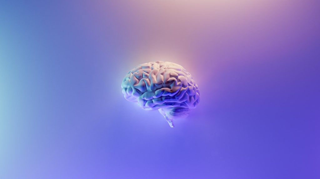
Depiction of a human brain
Image by Fakurian Design on Unsplash

A new study at Tel Aviv University showed significant improvements in social skills and the condition of the autistic brain through hyperbaric therapy. The study which is reported in the journal International Journal of Molecular Sciences, was conducted on lab models of autism.

Hyperbaric medicine, where patients sit in special high-pressure chambers while breathing pure oxygen, is considered safe and, besides treating decompression sickness in divers, is already in use for other conditions. The use of hyperbaric medicine to treat autism is contentious, with many holding that it is based on pseudoscience. In recent years, scientific evidence has been accumulating that unique protocols of hyperbaric treatments improve the supply of blood and oxygen to the brain, thereby improving brain function.

Changes observed in the brain included a reduction in neuroinflammation, which is known to be associated with autism. A significant improvement was also found in the social functioning of the animal models treated in the pressure chamber. The study’s success has many implications regarding the applicability and understanding of treating autism using pressure chamber therapy.

The breakthrough was led by doctoral student Inbar Fischer, from the laboratory of Dr Boaz Barak of Tel Aviv University.

Improved brain function

“The medical causes of autism are numerous and varied, and ultimately create the diverse autistic spectrum with which we are familiar,” explains Dr Barak. “About 20% of autistic cases today are explained by genetic causes, that is, those involving genetic defects, but not necessarily ones that are inherited from the parents. Despite the variety of sources of autism, the entire spectrum of behavioural problems associated with it are still included under the single broad heading of ‘autism,’ and the treatments and medications offered do not necessarily correspond directly to the reason why the autism developed.”

In the preliminary phase of the study, a girl carrying the mutation in the SHANK3 gene, which is known to lead to autism, received treatments in the pressure chamber, conducted by Prof Shai Efrati. After the treatments, it was evident that the girl’s social abilities and brain function had improved considerably.

In the next stage, and in order to comprehend the success of the treatment more deeply, the team of researchers at Dr Barak’s laboratory sought to understand what being in a pressurised chamber does to the brain. To this end, the researchers used lab models carrying the same genetic mutation in the SHANK3 gene as that carried by the girl who had been treated. The experiment comprised a protocol of 40 one-hour treatments in a pressure chamber over several weeks.

“We discovered that treatment in the oxygen-enriched pressure chamber reduces inflammation in the brain and leads to an increase in the expression of substances responsible for improving blood and oxygen supply to the brain, and therefore brain function,” explains Dr Barak. “In addition, we saw a decrease in the number of microglial cells, immune system cells that indicate inflammation, which is associated with autism.”

Increased social interest

“Beyond the neurological findings we discovered, what interested us more than anything was to see whether these improvements in the brain also led to an improvement in social behaviour, which is known to be impaired in autistic individuals,” adds Dr Barak. “To our surprise, the findings showed a significant improvement in the social behaviour of the animal models of autism that underwent treatment in the pressure chamber compared to those in the control group, who were exposed to air at normal pressure, and without oxygen enrichment. The animal models that underwent treatment displayed increased social interest, preferring to spend more time in the company of new animals to which they were exposed in comparison to the animal models from the control group.”

Inbar Fischer concludes, “the mutation in the animal models is identical to the mutation that exists in humans. Therefore, our research is likely to have clinical implications for improving the pathological condition of autism resulting from this genetic mutation, and likely also of autism stemming from other causes. Because the pressure chamber treatment is non-intrusive and has been found to be safe, our findings are encouraging and demonstrate that this treatment may improve these behavioral and neurological aspects in humans as well, in addition to offering a scientific explanation of how they occur in the brain.”

Source: Tel Aviv University

How Breathing Influences the Brain to Shape Mood and Behaviour

Depiction of a human brain
Image by Fakurian Design on Unsplash

A study published in the journal Psychological Review describes a possible mechanism by which breathing influences the brain and how breathing exercises influence mood. The researchers synthesised results from more than a dozen studies with rodent, monkey, and human brain imaging, and used it to propose a new computational model that explains how our breathing influences the brain’s expectations.

“What we found is that, across many different types of tasks and animals, brain rhythms are closely tied to the rhythm of our breath. We are more sensitive to the outside world when we are breathing in, whereas the brain tunes out more when we breathe out. This also aligns with how some extreme sports use breathing, for example professional marksmen are trained to pull the trigger at the end of exhalation,” explains Professor Micah Allen from the Department of Clinical Medicine at Aarhus University.

The study suggest that breathing is more than just something we do to stay alive, explains Prof Allen.

“It suggests that the brain and breathing are closely intertwined in a way that goes far beyond survival, to actually impact our emotions, our attention, and how we process the outside world. Our model suggests there is a common mechanism in the brain which links the rhythm of breathing to these events.”

Breathing can affect our mental health

Understanding how breathing shapes our brain, and by extension, our mood, thoughts, and behaviours, is an important goal in order to better prevent and treat mental illness.

“Difficulty breathing is associated with a very large increase in the risk for mood disorders such as anxiety and depression. We know that respiration, respiratory illness, and psychiatric disorders are closely linked. Our study raises the possibility that the next treatments for these disorders might be found in the development of new ways to realign the rhythms of the brain and body, rather than treating either in isolation,” explains Micah Allen.

The new study sheds light on how the brain its possible to stabilise the mind through breathing exercises. It suggests that there are three pathways in the brain that control this interaction between breathing and brain activity. It also suggests that our pattern of breathing makes the brain more “excitable,” meaning neurons are more likely to fire during certain times of breathing.

Prof Allen says that research is underway into investigating how different kinds of emotional and visual perception are influenced by breathing in the brain, as well as the impact of long COVID.

Source: Aarhus University

Hormonal Contraceptives’ Impacts on the Adolescent Brain

Photo by Reproductive Health Supplies Coalition on Unsplash

Hormonal contraceptives are safe and highly effective at preventing pregnancy, but their impact on the developing bodies of teenage girls, especially their brains, is not well understood.

New research in young rats links the synthetic hormones found in birth control pills, patches and injections with disordered signal transmission between cells in the prefrontal cortex, which is still developing during adolescence. Compared to control rats, the animals receiving hormonal contraceptives also produced higher levels of the stress hormone corticosterone, which is similar to cortisol in humans.

The Ohio State University scientists began investigating the prefrontal cortex, where mood is regulated, because some previous research has associated early adolescent use of hormonal contraceptives with adulthood depression risk. But the most important thing, the researchers said, is learning how birth control affects the developing brain so individuals can weigh the risks and benefits of their reproductive health choices.

“Birth control has had a major positive impact for women’s health and autonomy – so it’s not that we’re suggesting adolescents should not take hormonal contraceptives,” said senior study author Benedetta Leuner, associate professor of psychology at Ohio State.

“What we need is to be informed about what synthetic hormones are doing in the brain so we can make informed decisions – and if there are any risks, then that’s something that needs to be monitored. Then if you decide to use hormonal birth control, you would pay more attention to warning signs if you knew of any possible mood-related side effects.”

The research poster was presented to at Neuroscience 2022, the annual meeting of the Society for Neuroscience.

An estimated 2 in 5 teenage girls in the US have sexual intercourse between age 15 and 19, and the vast majority use a contraceptive, mostly condoms. Of those using birth control, almost 5% use hormonal contraceptives, also known as long-acting reversible contraceptives. These products are also prescribed to treat acne and heavy periods.

Despite their popularity, “there isn’t a lot known about how hormonal birth control influences the teen brain and behaviour,” said co-author Kathryn Lenz, associate professor of psychology at Ohio State. “Adolescence is a crucially under-investigated period of dramatic brain change and dramatic hormonal change that we really haven’t understood.”

The researchers gave a combination of synthetic estrogen and progesterone typically found in hormonal contraceptives to female rats for three weeks beginning about a month after they were born, an age equivalent to early adolescence in humans. Researchers confirmed the drugs disrupted the animals’ reproductive cycling — these birth control products work by stopping ovaries from producing hormones at levels necessary to generate eggs and making the uterine lining inhospitable for an egg to implant.

Blood samples showed the treated rats were producing more corticosterone than untreated animals, a sign that they were stressed. And after being subjected to and recovering from an experimental stressor, the treated rats’ corticosterone level remained high. Their adrenal glands were also larger, suggesting their stress hormone production was consistently higher than that of control animals.

An analysis of gene activation markers in the animals’ prefrontal cortex showed a decrease in excitatory synapses in that region of treated rats’ brains compared to controls, but no change to inhibitory synapses — a phenomenon that could set up an imbalance of normal signaling patterns and result in altered behavior. The loss of only excitatory synapses in the prefrontal cortex has been linked to exposure to chronic stress and depression in previous research.

“What this means for the function of particular circuits, we don’t know yet. But this gives us a clue of where to look next in terms of what the functional outcomes might be,” Lenz said.

The researchers are moving forward with additional studies targeting hormonal contraceptive effects on the brain between puberty and late adolescence – a tricky time to study the developing brain because it is undergoing constant change, Leuner said. The reasons behind the drugs’ effects are an open question, as well.

“These are synthetic hormones, so are they affecting the brain because of their synthetic properties, or are they affecting the brain because they’re blocking the naturally produced hormones?” she said. “It’s a difficult question to answer, but an important one.”

Source: Ohio State University

Exploring the Avoidance of Eye Contact in Autism

A reluctance to make eye contact is a hallmark of autism spectrum disorder (ASD). By simultaneously imaging the brains of people making eye contact, Yale University researchers found that eye contact between two individuals was associated with a specific area associated with social interaction, which synchronises when two people with normal neural development gaze at each other. The results, published in the journal PLOS ONE, showed that in people with ASD, there was less activity in this region when they attempted eye contact.

People with ASD have been showed to have reduced or altered neurological arousal from looking at images of faces or even robots. Although eye contact is a critically important part of social interactions, scientists have been limited in studying the neurological basis of live social interaction with eye-contact in ASD because of the inability to image the brains of two people simultaneously.

Now, using an innovative technology that enables imaging of two individuals during live and natural conditions, Yale researchers have identified specific brain areas in the dorsal parietal region of the brain associated with the social symptomatology of autism. The study finds that these neural responses to live face and eye-contact may provide a biomarker for the diagnosis of ASD as well as provide a test of the efficacy of treatments for autism.

“Our brains are hungry for information about other people, and we need to understand how these social mechanisms operate in the context of a real and interactive world in both typically developed individuals as well as individuals with ASD,” said co-corresponding author Joy Hirsch, Elizabeth Mears and House Jameson Professor of Psychiatry, Comparative Medicine, and of Neuroscience at Yale.

The Yale team, led by Hirsch and James McPartland, Harris Professor at the Yale Child Study Center, analysed brain activity during brief social interactions between pairs of adults – each including a typical participant and one with ASD – using functional near-infrared spectroscopy, a non-invasive optical neuroimaging method. Both participants were fitted with neuroimaging caps which measured brain activity during face gaze and eye-to-eye contact.

The investigators found that during eye contact, participants with ASD had significantly reduced activity in a brain region called the dorsal parietal cortex compared to those without ASD. Further, the more severe the overall social symptoms of ASD as measured by ADOS (Autism Diagnostic Observation Schedule, 2nd Edition) scores, the less activity was observed in this brain region. Neural activity in these regions was synchronous between typical participants during real eye-to-eye contact but not during gaze at a video face. This typical increase in neural coupling was not observed in ASD, and is consistent with the difficulties in social interactions.

“We now not only have a better understanding of the neurobiology of autism and social differences, but also of the underlying neural mechanisms that drive typical social connections,” Hirsch said.

Source: Yale University

Delayed COVID Recovery could be a Protective Mechanism against Hypoxia

Photo by Andrea Piacquadio on Unsplash

COVID patients placed on ventilators can take a long time to regain consciousness. New research published the Proceedings of the National Academy of Sciences now shows that these delays may serve a purpose: protecting the brain from oxygen deprivation.

The existence of such a brain-preserving state could explain why some patients wake up days or even weeks after they stop receiving ventilation, and it suggests that physicians should take these lengthy recovery times into account when determining a patient’s prognosis.

In their study, investigators connect the pattern seen among those who have survived severe COVID with similar delays known to occur in a small fraction of cardiac arrest patients.

“The delayed recoveries in COVID patients are very much like the rare cases we’ve documented in previous research. In this new paper, we describe a mechanism to explain what we’re seeing in both types of patients,” said study co-senior author Dr Nicholas D. Schiff, a neurology professor at Weill Cornell Medicine.

He suggests that this mechanism is the brain protecting itself, pointing to animals, most notably painted turtles, that can tolerate extended periods without oxygen.

More than a decade ago, Dr Schiff and his colleagues first observed these delays among comatose cardiac arrest patients who received cooling therapy to reduce brain damage caused by a loss of blood flow. In one such case, a 71-year-old patient took 37 days to awaken, before ultimately making a near-complete recovery.

During the pandemic, Dr Schiff performed neurology consultations for COVID patients, and he soon began seeing similar, delayed awakenings occurring when patients were taken off ventilators and stopped receiving movement-limiting sedatives.

In a separate analysis of a large cohort of COVID patients from Weill Cornell Medicine and two other major U.S. medical centres, Dr Schiff and his colleagues, including co-author of the current paper, Dr Emery N. Brown, professor of anaesthesia at Harvard Medical School, found that a quarter of patients who survived ventilation took 10 days or longer to recover consciousness. The more oxygen deprivation they suffered while on the ventilator, the longer that delay.

In the prior study of cardiac patients, the researchers recorded a distinctive pattern in brain activity, one also seen in patients under deep anaesthesia. (Recordings from COVID patients are extremely limited.) Dr Schiff read that a similar pattern had been seen in the brains of painted turtles, which can withstand up to five months without oxygen under ice in the winter. To do so, they activate the same inhibitory system within the brain targeted by anaesthetics given to human cardiac and COVID patients but in novel ways developed by evolutionary specialisations.

Drs Schiff and Brown propose that, by chance, the same protective response emerges in the patients.

“It is our theory that oxygen deprivation as well as practices in the ICU, including commonly used anaesthetics, expose elements of strategies that animals use to survive in extreme conditions,” Dr Schiff said.

“These observations may offer new insights into the mechanisms of how certain anaesthetics produce unconsciousness and new approaches for ICU sedation and for fostering recovery from disorders of consciousness,” Dr Brown added.

When patients fail to regain consciousness for an extended time, physicians may recommend withdrawing life-supporting care. This threshold is typically set at 14 days or less for cardiac patients, while no such guidelines exist for COVID.

In light of this new research, however, so long as they lack brain injuries, physicians should avoid making negative projections about these patients’ potential to recover, note the researchers.

Source: Weill Cornell Medicine

Brain Changes in Autism More Widely Spread than Previously Believed

In autism, brain changes are spread throughout the cerebral cortex instead of in areas thought affect social behaviour and language, according to a new study that significantly refines scientists’ understanding of how autism spectrum disorder (ASD) progresses at the molecular level.

The study, published in Nature, represents a comprehensive effort to characterise ASD at the molecular level. While neurological disorders like Alzheimer’s disease or Parkinson’s disease have well-defined pathologies, autism and other psychiatric disorders have had a lack of defining pathology, challenging efforts to develop more effective treatments.  

The new study finds brain-wide changes in virtually all of the 11 cortical regions analysed, regardless of whether they are higher critical association regions – those involved in functions such as reasoning, language, social cognition and mental flexibility – or primary sensory regions.  

“This work represents the culmination of more than a decade of work of many lab members, which was necessary to perform such a comprehensive analysis of the autism brain,” said study author Dr Daniel Geschwind, the Gordon and Virginia MacDonald Distinguished Professor of Human Genetics, Neurology and Psychiatry at UCLA. “We now finally are beginning to get a picture of the state of the brain, at the molecular level, of the brain in individuals who had a diagnosis of autism. This defines a molecular pathology, which similar to other brain disorders such as Parkinson’s, Alzheimer’s and stroke, provides a key starting point for understanding the disorder’s mechanisms, which will inform and accelerate development of disease-altering therapies.” 
 
Just over a decade ago, Geschwind led the first effort to identify autism’s molecular pathology by focusing on two brain regions, the temporal lobe and the frontal lobe. Those regions were chosen because they are higher order association regions involved in higher cognition – especially social cognition, which is disrupted in ASD.  
 
For the new study, researchers examined gene expression in 11 cortical regions by sequencing RNA from each of the four main cortical lobes. They compared brain tissue samples obtained after death from 49 people with ASD against 54 controls individuals.  
 
While each profiled cortical region showed changes, the largest changes in RNA levels were in the visual cortex and the parietal cortex, which processes information like touch, pain and temperature. The researchers said this may reflect the sensory hypersensitivity that is frequently reported in people with ASD. Researchers found strong evidence that the genetic risk for autism is enriched in a specific group of genes expressed in neurons that has lower expression across the brain, indicating that these correlated RNA changes in the brain are likely the cause of ASD rather than a result of the disorder. 

One of the next steps is to determine whether researchers can use computational approaches to develop therapies based on reversing gene expression changes the researchers found in ASD, Geschwind said, adding that researchers can use organoids to model the changes in order to better understand their mechanisms.  

Source: University of California, Los Angeles

Mapping the Neural Pathways for Vomiting after Eating Infected Food

Photo by Kyle Glenn on Unsplash

The urge to vomit after eating contaminated food is the body’s natural defensive response to get rid of bacterial toxins. However, exactly how the brain initiates the response has remained a mystery. Now, researchers have mapped out the detailed neural pathway of the defensive responses from the gut to the brain in mice. The study, published in the journal Cell, could help scientists develop better anti-nausea medications for cancer patients who undergo chemotherapy.

Many foodborne bacteria produce toxins in the host after ingestion. After sensing their presence, the brain will initiate a series of biological responses, including vomit and nausea, to expel the substances and develop an aversion toward foods that taste or look the same.

“But details on how the signals are transmitted from the gut to the brain were unclear, because scientists couldn’t study the process on mice,” says Peng Cao, the paper’s corresponding author at the National Institute of Biological Sciences in Beijing. Rodents cannot vomit, so scientists have been studying vomit in other animals like dogs and cats, but these animals are not comprehensively studied and thus failed to reveal the mechanism of nausea and vomiting. However, Cao and his team noticed that while mice don’t vomit, they retch – meaning they also experience the urge to vomit without throwing up.

The team found that after receiving Staphylococcal enterotoxin A (SEA), which is a common bacterial toxin produced by Staphylococcus aureus that also leads to foodborne illnesses in humans, mice developed episodes of unusual mouth opening. Mice that received SEA opened their mouths at angles wider than those observed in the control group, where mice received saline water. Moreover, during these episodes, the diaphragm and abdominal muscles of the SEA-treated mice contract simultaneously, a pattern seen in dogs when they are vomiting. During normal breathing, animals’ diaphragm and abdominal muscles contract alternatively.

“The neural mechanism of retching is similar to that of vomiting. In this experiment, we successfully build a paradigm for studying toxin-induced retching in mice, with which we can look into the defensive responses from the brain to toxins at the molecular and cellular levels,” Cao says.

In mice treated with SEA, the team found the toxin in the intestine activates the release of serotonin, a type of neurotransmitter, by the enterochromaffin cells on the lining of the intestinal lumen. The released serotonin binds to the receptors on the vagal sensory neurons located in the intestine, which transmits the signals along the vagus nerves from the gut to a specific type of neurons in the dorsal vagal complex – Tac1+DVC neurons – in the brainstem. When Cao and his team inactivated the Tac1+DVC neurons, SEA-treated mice retched less compared with mice with normal Tac1+DVC neuron activities.

In addition, the team investigated whether chemotherapy drugs, which also induce defensive responses like nausea and vomiting in recipients, activate the same neural pathway. They injected mice with doxorubicin, a common chemotherapy drug. The drug made mice retch, but when the team inactivated their Tac1+ DVC neurons or serotonin synthesis of their enterochromaffin cells, the animals’ retching behaviours were significantly reduced.

Cao says some of the current anti-nausea medications for chemotherapy recipients, such as Granisetron, work by blocking the serotonin receptors. The study helps explain why the drug works.

“With this study, we can now better understand the molecular and cellular mechanisms of nausea and vomiting, which will help us develop better medications,” Cao says.

Next, Cao and his colleagues want to explore how toxins act on enterochromaffin cells. Preliminary research shows that enterochromaffin cells don’t sense the presence of toxins directly. The process likely involves complex immune responses of damaged cells in the intestine.

“In addition to foodborne germs, humans encounter a lot of pathogens, and our body is equipped with similar mechanisms to expel these toxic substances. For example, coughing is our body’s attempt to remove the coronavirus. It’s a new and exciting field of research about how the brain senses the existence of pathogens and initiates responses to get rid of them.” Cao says, adding that future research may reveal new and better targets for drugs, including anti-nausea medicines.

Source: ScienceDaily

Body Self-perception is Based on The Brain’s Guesswork

Photo by Bruce Christianson on Unsplash

Researchers at Karolinska Institutet in Sweden have found that the perception of one’s own body is largely based on the brain making guesses that are based on probability theory, instead of direct sensory input. The researchers detailed their findings in a study recently published in the journal eLife.

The researchers posit that the way humans perceive their bodies is largely governed by probability assessments based on past experiences, combined with sensory information such as sight and touch, for example.

“The experience of one’s own body is a statistical estimate of reality based on sensory information, sensory uncertainty, and previous experiences that can be summarised in a mathematical model”, explains Henrik Ehrsson, professor at the Department of Neuroscience, Karolinska Institutet.

Why are these results important?

“The results clarify the computational functions that govern the perception of one’s own body. This perception thus arises, not only as a result of a “direct” interpretation of signals from sight, touch sense, and proprioception as the textbooks say, but rather is based on active “guesses” that the brain constantly makes based on probability theory and the information that can be extracted from the patterns of sensory signals”, says Henrik Ehrsson.

“When we varied the degree of time delay between the visual and tactile impressions in small steps, or blurred the image in the augmented reality glasses to increase uncertainty, the illusion changed in a way that can be described by equations and curves: increased delay gave a weaker feeling of the rubber hand as its own, while increased uncertainty (blurriness) made the illusion stronger”, says Marie Chancel, corresponding author of the study.

Based on the experiments, the researchers came up with a statistical explanatory model for the brain’s perceptual awareness of its own body.

Changes in body ownership

The next step is to try to understand how the statistical model that determines own-bodily awareness is implemented by neural networks in the brain. In a preliminary study, the researchers have shown that neural activity in posterior parietal cortex follows the Bayesian model well in experiments where they measure brain activity with functional magnetic resonance imaging. The researchers also want to investigate how their model can explain changes in bodily awareness in various psychiatric and neurological conditions, such as Schizophrenia and Anorexia.

Source: Karolinska Institutet

Do Women Have the Edge in Remembering Words?

Photo by Andrea Piacquadio on Pexels

Women are popularly believed at being better at finding and remembering words than men, but are the popular science textbooks which proclaim this actually correct? If so, this has relevance for tests such as measures of dementia. Researchers investigated this supposed difference, publishing their findings in Perspectives on Psychological Science.

Marco Hirnstein, professor at The University of Bergen, Norway, is unequivocal about the results. “Women are better. The female advantage is consistent across time and life span, but it is also relatively small.”

Prof Hirnstein is interested in how biological, psychological, and social factors contribute to sex/gender differences in cognitive abilities and what the underlying brain mechanisms are.

“So far, the focus has mostly been on abilities, in which men excel. However, in recent years the focus has shifted more towards women,” said Prof Hirnstein.

Textbooks and popular science books take it for granted that women are better at finding words. For example, when naming words that begin with the letter “F,” or words that belong to a certain category like animals or fruits. It has also been considered “fact” that women are better at remembering words.

Yet, the actual findings are much more inconsistent than textbooks imply: Some studies find a female advantage, some find a male advantage, some do not find any advantage.

“Most intellectual skills show no or negligible differences in average performance between men and women. However, women excel in some tasks, while men excel in others on average.”

Prof Hirnstein and his colleagues point out how their findings can be useful in diagnosis and in healthcare. The results help to clarify whether the female advantage is real but also have relevance for for interpreting the results of diagnostic assessments.

For example, to diagnose dementia, knowing that women are generally better in those tasks is critical to not under-diagnose women, due to their better average, baseline performance and not over-diagnose men. Currently, many but not all assessments take sex/gender into account.

The researchers conducted a meta-analysis of the available literatures, encompassing more than 500 measures from more than 350,000 participants. The researchers found that women are indeed better. The advantage is small but consistent across the last 50 years and across an individual’s lifespan.

Moreover, they found that the female advantage depends on the sex/gender of the leading scientist: Female scientists report a larger female advantage, male scientists report a smaller female advantage.

Source: University of Bergen

Adult Brains can Rewire to Recover from Inherited Blindness

Eye
Source: Daniil Kuzelev on Unsplash

A recent discovery has revealed that the adult brain has far greater potential to recover from inherited blindness than previously believed, with important implications for visually impaired people. The paper appears in Current Biology.

The research team was examining treatment for Leber congenital amaurosis (LCA), a group of inherited retinal diseases distinguished by severe visual impairment at birth. The condition, caused by mutations in any of over two dozen genes, results in degeneration or dysfunction in the retina’s photoreceptors.

Administering chemical compounds that target the retina, called synthetic retinoids, can restore a notable amount of vision in children with LCA. The UCI team wanted to find out if the treatment could make a difference for adults who have the condition.

“Frankly, we were blown away by how much the treatment rescued brain circuits involved in vision,” said corresponding author Sunil Gandhi, professor of neurobiology and behaviour. “Seeing involves more than intact and functioning retinae. It starts in the eye, which sends signals throughout the brain. It’s in the central circuits of the brain where visual perception actually arises.” Until now, scientists believed that the brain must receive those signals in childhood so that central circuits could wire themselves correctly.

The researchers were surprised by what they found in rodent models of LCA. “The central visual pathway signalling was significantly restored in adults, especially the circuits that deal with information coming from both eyes,” Prof Gandhi said. “Immediately after the treatment, the signals coming from the opposite-side eye, which is the dominant pathway in the mouse, activated two times more neurons in the brain. What was even more mind-blowing was that the signals coming from the same-side eye pathway activated five-fold more neurons in the brain after the treatment and this impressive effect was long-lasting. The restoration of visual function at the level of the brain was much greater than expected from the improvements we saw at the level of the retinae. The fact that this treatment works so well in the central visual pathway in adulthood supports a new concept, which is that there is latent potential for vision that is just waiting to be triggered.”

The finding opens exciting research possibilities. “Whenever you have a discovery that breaks with your expectations about the possibility for the brain to adapt and rewire, it teaches you a broader concept,” Prof Gandhi said. “This new paradigm could aid in the development of retinoid therapies to more completely rescue the central visual pathway of adults with this condition.”

Source: University of California – Irvine