Category: Medical Research & Technology

Device Uses Body to Charge Wearable Tech

Photo by Josh Riemer on Unsplash

A team from the National University of Singapore (NUS) has devised an innovative way to charge wearable devices such as medical monitors — by transmitting power through the body to other devices.

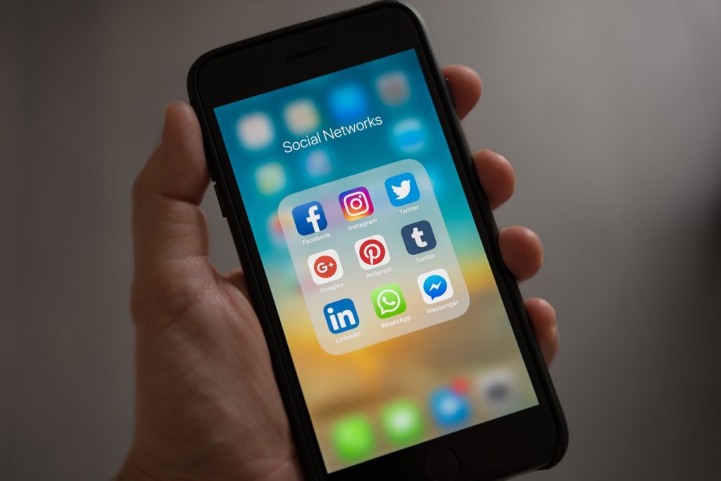
Advancements in wearable technology are reshaping the way we live, work and play, and also how healthcare is delivered and received. Wearable devices include wristbands, smartwatches, wearable mobile sensors, and other mobile hub medical devices that collect a large range of data from blood sugar and exercise routines to sleep and mood. 

Such devices can help patients and providers manage chronic conditions such as diabetes, heart conditions, and chronic pain. According to the Pew Research Center, 60% of US adults reported tracking their weight, diet, or exercise routine; 33% of US adults track health symptoms or indicators such as blood pressure, blood sugar, or sleep patterns; and 8% of adults specifically use medical devices, such as glucose meters.

One major obstacle of using wearables is keeping these devices properly and conveniently powered. The more wearable devices are worn, the more often there is the need to charge multiple batteries. Many users find it cumbersome to charge numerous devices every day, and inconvenient service disruptions occur when batteries run out.

A research team, led by Associate Professor Jerald Yoo from the Department of Electrical and Computer Engineering and the N.1 Institute for Health at NUS, has come up with an innovative solution to these problems. Their technology utilises the human body as a medium for power transmission, enabling a single device, such as a mobile phone placed in the pocket, to wirelessly power other wearable devices on a user’s body. The team’s novel system has an added advantage – it can harvest unused energy from electronics in a typical home or office environment to power the wearables.

Their achievement was first published in the journal Nature Electronics on 10 June 2021. It is the first of its kind to be established among existing literature on electronic wearables.

Power transmission through the body

To extend the battery life of wearable devices, power transmission and energy harvesting approaches are required. However, current approaches for powering up body area wearables are hampered by short distances, intervening obstacles and unstable power delivery. As such, none of the current methods are suitable for the sustainable provision of power to wearables placed around the entire human body.

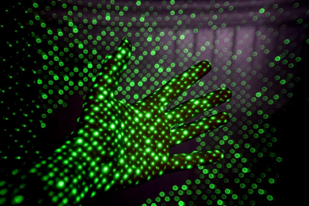
The NUS approach turned the obstacle of the human body into an advantage by designing a receiver and transmitter system that uses the human body as a medium for power transmission and energy harvesting. Each receiver and transmitter contains a chip that is used as a springboard to extend coverage over the entire body.

The power transmitter need only be on a single power source, such as a smart watch, while multiple power receivers can be placed anywhere on the person’s body. The system then harnesses energy from the source to power multiple wearables on the user’s body via a process termed as body-coupled power transmission. In this way, only one device needs to be charged, and the rest of the wearable devices can be powered from that source. The team’s experiments showed that a single, fully-charged power source to power up to 10 wearable devices on the body, for a duration of over 10 hours.

The researchers also found that typical office and home environments have parasitic electromagnetic (EM) waves that people are constantly exposed to from sources such as running computers. To tap this energy, their novel receiver scavenges the EM waves from the environment, and through a process referred to as body-coupled powering, the human body is able to harvest this energy to power the wearable devices.

Smaller wearables without batteries

On the benefits of his team’s method, Assoc Prof Yoo said, “Batteries are among the most expensive components in wearable devices, and they add bulk to the design. Our unique system has the potential to omit the need for batteries, thereby enabling manufacturers to miniaturise the gadgets while reducing production cost significantly. More excitingly, without the constraints of batteries, our development can enable the next generation wearable applications, such as ECG patches, gaming accessories, and remote diagnostics.”

The NUS team will continue to improve the efficiency of their transmitter/receiver system, so that hopefully any given power-transmitting device such as a smartphone can extend the battery life of other wearable technologies, some of which, like medical monitors, can be quite important.

Source: National University of Singapore

Journal reference: Li, J., et al. (2021) Body-coupled power transmission and energy harvesting. Nature Electronics. doi.org/10.1038/s41928-021-00592-y. 

A COVID Vaccine Without the Jab

Photo by Webstacks on Unsplash

University of Queensland scientists used a ‘patch’ to deliver a US-developed COVID vaccine without the jab, and successfully protected mice from the virus.

The vaccine candidate from University of Texas Hexapro was delivered via the high-density microarray patch (HD-MAP) and provided protection against COVID disease with a single, painless ‘click’ from a handheld applicator.

Dr David Muller, from UQ’s School of Chemistry and Molecular Biosciences, said the vaccine patch produced strong immune responses that were shown to be effective when the mice were exposed to SARS-CoV-2.

“When the Hexapro vaccine is delivered via HD-MAP applicator – rather than a needle – it produces better and faster immune responses,” Dr Muller said.

“It also neutralises multiple variants, including the UK and South Africa variants.

“And it’s much more user-friendly than a needle – you simply ‘click’ an applicator on the skin, and 5000 microscopic projections almost-imperceptibly deliver vaccine into the skin.

“The UQ team, together with Vaxxas, hope to take the technology to the world and are looking for funding opportunities to accelerate to clinical trials as soon as possible.”
Dr Muller said that Hexapro, delivered by the high-density microarray patch, could dramatically assist global vaccine rollout effort, particularly for billions of vulnerable people in low- and middle-income countries.

“We’ve shown this vaccine, when dry-coated on a patch, is stable for at least 30 days at 25 degrees Celsius and one week at 40 degrees, so it doesn’t have the cold chain requirements of some of the current options.”

High-density microarray patch (HD-MAP)

Vaxxas was founded in 2011 with the help of University of Queensland. The company’s president and CEO, David L Hoey, said he was extremely excited about the findings.

“These results are extremely clear – vaccination by HD-MAP produces much stronger and more protective immune responses against COVID-19 in model systems than via needle or syringe,” he said.

“We thank and recognise our incredible research collaborators at UQ for these important findings.

“The prospect of having a single-dose vaccine, that could be easily distributed and self-administered, would greatly improve global pandemic vaccination capabilities,” said Hoey

The research is currently undergoing peer review and has been published in BioRxiv (DOI: 10.1101/2021.05.30.446357).

Source: The University of Queensland

Precise Ultrasound Heating of Neurons Could Treat Neurological Disorders

Image source: Fakurian Design on Unsplash

A multidisciplinary team at Washington University in St. Louis has developed a new brain stimulation technique using focused ultrasound that is able to turn specific types of neurons in the brain on and off and precisely control motor activity without surgical device implantation.

Being able to turn neurons on and off can treat certain neurological disorders such as Parkinson’s disease and epilepsy. Used for over six decades, deep brain stimulation techniques have had some treatment success in neurological disorders, but those require surgical device implantation. 

The team, led by Hong Chen, assistant professor of biomedical engineering in the McKelvey School of Engineering and of radiation oncology at the School of Medicine, is the first to provide direct evidence showing noninvasive activation of specific neuron types in mammalian brains by combining an ultrasound-induced heating effect and genetics, which they have named sonothermogenetics. It is also the first work to show that the ultrasound- genetics combination can robustly control behaviour by stimulating a specific target deep in the brain.

The results of the three years of research were published online in Brain Stimulation

“Our work provided evidence that sonothermogenetics evokes behavioural responses in freely moving mice while targeting a deep brain site,” Chen said. “Sonothermogenetics has the potential to transform our approaches for neuroscience research and uncover new methods to understand and treat human brain disorders.”

Chen and colleagues delivered a viral construct containing TRPV1 ion channels to genetically-selected neurons in a mouse model. Then, they delivered small pulses of heat generated by low-intensity focused ultrasound to the selected neurons in the brain via a wearable device. The heat, only a few degrees warmer than body temperature, activated the TRPV1 ion channel, which then acted as a switch to turn the neurons on or off.

“We can move the ultrasound device worn on the head of free-moving mice around to target different locations in the whole brain,” said Yaoheng Yang, first author of the paper and a graduate student in biomedical engineering. “Because it is noninvasive, this technique has the potential to be scaled up to large animals and potentially humans in the future.”

Building on prior research from his lab, professor of biomedical engineering Jianmin Cui and his team found for the first time that ion channel activity can be influenced by ultrasound alone, possibly leading to new and noninvasive ways to control the activity of specific cells. They discovered that focused ultrasound modulated the currents flowing through the ion channels on average by up to 23%, depending on channel and stimulus intensity. Following this work, researchers found close to 10 ion channels with this capability, but all of them are mechanosensitive, not thermosensitive.

The work also builds on the concept of optogenetics, the combination of the targeted expression of light-sensitive ion channels and the precise delivery of light to stimulate neurons deep in the brain. While optogenetics has increased discovery of new neural circuits, it has limited penetration depth due to light scattering, requiring surgical implantation of optical fibres to reach deeper into the brain.

Sonothermogenetics has the promise to target any location in the mouse brain with millimetre-scale resolution without causing any damage to the brain, Chen said. She and her team are further refining the technique and validating their work.

Source: Sci Tech Daily

Journal information: Yaoheng Yang et al, Sonothermogenetics for noninvasive and cell-type specific deep brain neuromodulation, Brain Stimulation (2021). DOI: 10.1016/j.brs.2021.04.021

New Biomaterial Produced from Frog Skin and Fish Scales

Photo by Robert Zunikoff on Unsplash

Researchers at Nanyang Technological University, Singapore (NTU Singapore) have developed a new biomaterial made entirely from discarded bullfrog skin and fish scales that could help in bone repair.

The porous biomaterial, which contains the same compounds that are predominant in bones, acts as a scaffold for osteoblasts, or bone-forming cells, to adhere to and multiply, leading to new bone formation. Bone-forming cells successfully latched onto the biomaterial and started growing, and it was found to have a low inflammatory risk.

This kind of scaffold could help regenerate bone tissue lost to disease or injury, such as jaw defects from trauma or cancer surgery. It could also assist bone growth around surgical implants such as dental implants.

The current standard practice of using a patient’s own tissues means extra surgery is needed for bone extraction. The biomaterial used, frog skin and fish scales, are a significant waste stream produced by Singapore’s aquaculture industry and using them helps repurpose this waste.

‘Waste-to-resource’

“We took the ‘waste-to-resource’ approach in our study and turned discards into a high-value material with biomedical applications, closing the waste loop in the process,” said Dalton Tay, Assistant Professor, Nanyang Technological University. “Our lab studies showed that the biomaterial we have engineered could be a promising option that helps with bone repair. The potential for this biomaterial is very broad, ranging from repairing bone defects due to injury or ageing, to dental applications for aesthetics. Our research builds on NTU’s body of work in the area of sustainability and is in line with Singapore’s circular economy approach towards a zero-waste nation.”

To make the biomaterial, the team first extracted Type 1 tropocollagen (many molecules of which form collagen fibres) from the discarded skins of the American bullfrog and hydroxyapatite (a calcium-phosphate compound) from the scales of snakehead fish, commonly known as the Toman fish.

Collagen and hydroxyapatite (HA) are two predominant components found in bones, thus conferring on the biomaterial a structure, composition, and ability to promote cell attachment similar to bone, as well as toughness.

The scientists removed all impurities from the bullfrog skin, then blended it to form a thick collagenous paste that is diluted with water, from which collagen was extracted. “Using this approach, we were able to obtain the highest ever reported yield of collagen of approximately 70 per cent from frog skin, thus making this approach commercially viable,” said Asst Prof Tay, who is also from the NTU School of Biological Sciences (SBS).

HA was harvested from discarded fish scales through calcination – a purification process that requires high heat – to remove the organic matter, and then air-dried.

The biomaterial was synthesised by adding HA powder to the extracted collagen, then cast into a mould to make a 3D porous scaffold — a two-week process which the team believes can be shortened.

Testing the biomaterial

To assess the biological performance of the porous biomaterial scaffold for bone repair, the scientists seeded bone-forming cells onto the scaffold.

The cells proliferated, and after a week, the cells were uniformly distributed across the scaffold – an indicator that the scaffold could promote proper cellular activities and eventually lead to tissue formation. The scientists also found that the presence of HA in the biomaterial significantly enhanced bone formation.

The biomaterial was also tested for its tendency to cause an inflammatory response, which is common after a biomaterial is implanted in the body.

Using real-time polymerase chain reaction, the scientists found that the expression of pro-inflammatory genes in human immune cells exposed to the biomaterial stayed “relatively modest” compared to a control exposed to endotoxins, a compound known to stimulate immune response, said Asst Prof Tay.

For instance, the expression of the gene IL6 in the biomaterial group was negligible and at least 50 times lower than that of the endotoxins-exposed immune cells. This suggests that the risk of the NTU-developed biomaterial to trigger an excessive acute inflammatory response is low.

The team is now further evaluating the long-term safety and efficacy of the biomaterial as dental products. Further research would involve studying how the body responds to this biomaterial in the long term, as well its use in other applications such as skin wounds, along with further development of the waste-to-resource pipeline.

A preprint copy of the article is available as a PDF for download.

Source: Nanyang Technical University

New Early Warning System for Sudden Cardiac Death

Photo from Olivier Collett on Unsplash

Researchers at Tomsk Polytechnic University have developed a nanosensor-based system that can detect early abnormalities in the function of cardiac muscle cells, which otherwise can be recorded only with invasive procedures.

The nanosensor-based hardware and software complex can measure cardiac micropotential energies without filtering and averaging-out cardiac cycles in real time. The device allows registering early abnormalities in the function of cardiac muscle cells, which otherwise can be recorded only during open-heart surgery or by inserting an electrode in a cardiac cavity through a vein. Such changes can lead to sudden cardiac death (SCD). Nowadays, there are no alternatives to the Tomsk device for a number of key characteristics in Russia and the world. ).

The main method of detection of electrical pulses in the heart is electrocardiography (ECG). Nevertheless, ECG modern devices detect already critical changes in the function of the myocardium.

“Therefore, there is much concern about the creation of devices for early detection of these disorders, when it is still possible to restore cell function using medication and without surgical intervention. To implement this, it is required to record cardiac micropotential energies, electrical pulses emitted by separate cells. Here, there is a question of how to implement it noninvasive. Our research team have worked on this task for a long time, as a consequence, we jointly with the participation of our colleagues, doctors, have developed a hardware and software complex.

“The core principles of its operation are similar to ECG, however, we changed sensors: we made nanosensors instead of conventional sensors and managed to measure signals of nanovoltage and microvoltage layers without filtering and averaging-out in broadband. The use of nanosensors led to the necessity to apply original circuit solutions, write individual software.

“Ultimately, we gained a tremendous difference in sensitivity,” Diana Avdeeva, Head of the TPU Laboratory for Medical Engineering, a research supervisor of the project, said.

The system consists of a set of sensors, a tiny key device for recording incoming signals from sensors and data processing software. The sensors are fixed on a patient’s chest using a conducting gel, and the monitoring procedure takes about 20 minutes.

Conventional ECG machines operate on frequencies from 0,05 Hz to 150 Hz, while the device of the Tomsk scientists operates on much higher frequencies of up to 10 000 Hz.

“Silver chloride electrodes are usually used for recording ECG of high quality. Our sensors are also silver chloride electrodes, however, we used silver nanoparticles. There are up to 16 thin plates from porous ceramics in every our sensor, silver nanoparticles are placed in these pores. There are millions of particles in one sensor, where every particle is a silver chloride electrode capable to enhance an electric field of the heart. Silver and gold nanoparticles are capable to enhance an electromagnetic field: visible light by 10,000 folds and infrared radiation by 20 folds. We also refused to use filters for rejection network interference and noises, which are usually used in conventional ECG and significantly distort micropotentials,” Diana Avdeeva said.

The published study includes the monitoring data of one volunteer’s heart function, who took part in the research for four years and was monitored every 7-10 days.

“At the beginning of our research, we recorded clear violations of activity of cardiac muscle cells. His attending physician recommended surgery, he gained an inserted stent at the Cardiology Research Institute. Then, he continued to take part in the research and the device recorded the further gradual restoration of cardiac function,” the scientist noted.

“A task to create a sensitive, tiny and affordable complex was set up, in order in a long run, outpatient clinics and patients at home could use it. Moreover, the developed methods and devices can be used not only in cardiology.

“The fields of any electrophysiological research, such as electroencephalography, electromyography and so on are promising. Of course, before applying it to cardiology, we have to pass some essential stages. These are the collection of the required array of statistics, certification of the complex for medical use. All these stages require sponsorship, we are engaged in searching for partners and supporting programs,” said research team member Mikhail Yuzhakov, Engineer at the TPU Laboratory for Medical Engineering.

Source: Tomsk Polytechnic University

Researchers Test VR in Treatment of Chronic Pain

Photo by Bradley Hook from Pexels

A team of researchers at the University of East Anglia are starting a new project to see if virtual reality, combined with an innovative brain interface could help with the treatment of chronic pain.

Almost one in five South African adults are living with chronic pain, and the team will study whether new technology delivered via a VR headset could help ease their symptoms.

One very science-fiction-like aspect of the study the use of a ‘Brain-Computer Interface’ where participants will be taught to control elements of a VR game using only their thoughts. Previous studies have shown that VR therapy was effective in reducing chronic and acute pain in children and adults.  

Dr Jordan Tsigarides from UEA’s Norwich Medical School and a Rheumatology doctor at the Norfolk and Norwich University Hospital, said: “Virtual Reality is an emerging technology where the user gains a completely immersive, interactive and often transformative experience with the use of a head-mounted display.

“VR seems to flood the brain with a multitude of audiovisual signals, engaging the senses and diverting the brain’s attention from processing pain signals.”

“At present, there is evidence that VR can significantly decrease people’s acute pain. What we are looking to find out is whether this kind of technology can be used to help people suffering chronic pain as well.”

The team is working with industry partner, Orbital Innovations, and has developed an innovative VR program for patients with chronic pain conditions, such as fibromyalgia syndrome and chronic pain following a total knee replacement. It will be tested on patients recruited from the NNUH and Addenbrookes in partnership with rheumatology, orthopaedics and pain management departments.

Dr Tsigarides said: “The virtual worlds that we have created aim to transport patients to naturalistic, immersive environments whether its travelling down a cool snowy river or planting crops in a luscious warm forest. Through VR, we aim to engage patients through challenging and interactive games, with the objective of understanding more about how this technology can reboot the brain’s pain networks.

“We are working with neuroscientists at the UEA to look at the brain waves of people with chronic pain. This has the potential to allow us to see how this technology is working, as well as learn more about the brain function of people with these conditions.

“Moving things with your mind sounds like something straight from a sci-fi movie but with today’s technology, ‘Brain-Computer Interfaces’ are being used more and more in health research. We believe that this exciting new innovation when coupled with an immersive VR experience will ‘power up’ the pain relief seen with VR alone.

“Given that just under 28 million adults in the UK suffer from chronic pain, and that we are in the midst of an opioid epidemic, this treatment could be an important future intervention.”

Peter Brady, CEO of Orbital Innovations, said: “Orbital Innovations has been working towards the development of a virtual reality technology, which will offer a way of mitigating the effects of chronic pain. This innovative project collaboration with UEA, will be breaking new ground in fully exploring the exciting opportunities this technology will offer patients in the future.”

Source: University of East Anglia

Brain-computer Interface Lets Paralysed People Write Letters

Image by Gerd Altmann from Pixabay

Researchers have developed a new brain-computer interface (BCI) that can let paralysed people write by mentally writing letters by hand.

Working with a participant with paralysis who has sensors implanted in his brain, the team used an algorithm to identify letters in real time as he attempted to write them, putting the results on a screen.

This technology could be further developed to allow people with paralysis type rapidly without using their hands, said study coauthor Krishna Shenoy, a Howard Hughes Medical Institute Investigator at Stanford University who jointly supervised the work with Jaimie Henderson, a Stanford neurosurgeon.

By attempting handwriting, the study participant was able to ‘type’ 90 characters per minute — more than double the previous record for typing with such a brain-computer interface.

Thought-powered communication

Even if injury or disease the ability to move, the brain’s neural activity for being able to do so remains. By making use of this activity, researchers can help people with paralysis or amputations regain lost abilities.

In recent years, Shenoy’s team has decoded the neural activity associated with speech in the hopes of reproducing it. Patients with implanted sensors mentally pointed at and clicked on letters on a screen to type at about 40 characters per minute, the previous speed record for typing with a BCI.
Wanting to try something new and different, Frank Willett, a neuroscientist in Shenoy’s group, wondered if it might be possible to harness the brain signals evoked by writing by hand “We want to find new ways of letting people communicate faster,” he said. 

The team worked with a participant enrolled in a clinical trial involving BCIs. Henderson implanted two tiny sensors into the part of the brain that controls the hand and arm, making it possible for the person to, for example, move a robotic arm or a cursor on a screen by attempting to move their own paralysed arm.
The participant, who was 65 years old at the time of the research, had a spinal cord injury that left him paralysed from the neck down. A machine learning algorithm recognised the patterns his brain produced when he attempted to write each letter.

With this system, the man could copy sentences and answer questions at a rate similar to that of someone his age typing on a smartphone. The reason why this so-called “Brain-to-Text” BCI is so fast is because each letter elicits a highly distinctive activity pattern, making it relatively easy for the algorithm to distinguish one from another, Willett explained.

A new system

Shenoy’s team envisions using attempted handwriting for text entry as part of a more comprehensive system that also includes point-and-click navigation, much like that used on current smartphones, and even attempted speech decoding. “Having those two or three modes and switching between them is something we naturally do,” he said.
The team intends to next work with a participant who cannot speak, such as a person with amyotrophic lateral sclerosis, a degenerative neurological disorder leading to loss of movement and speech.

The new system could potentially help those suffering from paralysis caused by a number of conditions, Henderson added. Those include brain stem stroke, which afflicted Jean-Dominique Bauby, the author of the book The Diving Bell and the Butterfly. “He was able to write this moving and beautiful book by selecting characters painstakingly, one at a time, using eye movement,” Henderson said. “Imagine what he could have done with Frank’s handwriting interface!”

Source: Howard Hughes Medical Institute

Distraction a Big Problem in Teletherapy Sessions

Photo by Tracy le Blanc from Pexels

A small survey has found that although therapists appear to prefer virtual sessions over in-person meetings, a significant proportion admit to being distracted while delivering care.

A third of respondents admitted to providing lower-quality care to clients during online sessions. Overall, 39% admitted to checking emails and social media while providing virtual care.

These were the results of a survey of 600 therapists conducted and published by OnlineTherapy.com, a virtual directory for teletherapists and counselors. It’s also an affiliate of the controversial app BetterHelp, stating on their website that the company “may receive compensation from BetterHelp if you purchase products or services through the links provided.”

Online care is generally well received by therapists: nearly half said they prefer virtual sessions over in-person meetings. They appreciated many advantages of virtual therapy, such as working from home which allows therapists to keep a flexible schedule and increases their availability. Video sessions also provide therapists with a uniquely intimate look into their clients’ daily lives, making assessing their mental health easier.

However, teletherapy has introduced its own challenges. Besides the struggle to remain for therapists to stay focused themselves, 56% of those surveyed said their clients are more easily distracted during virtual sessions, and 48% reported technological issues as a major impediment to their practice.

Peter Yellowlees, MD, of UC Davis Health in Sacramento, California, noted with concern and confusion that 16% of therapists reported substance use before or during their sessions.

“There are all sorts of people in this world who call themselves therapists, most of whom have very reasonable training, but quite a number don’t,” Dr Yellowlees told MedPage Today, expressing serious doubts about whether these rates of social media distraction and substance use during virtual sessions would ring true for mental health clinicians with PhDs and MDs.

According to a spokesperson from OnlineTherapy.com, the survey only asked if respondents were currently practicing mental health professionals and did not ask for their credentials or certifications.

The results of the survey did, however, echo Dr Yellowlees’s own concerns of mental health problems on the rise of among therapists in general. The vast majority (90%) said that during the last year they suffered mental health issues, including anxiety disorders (50%) and depressive disorder (48%). This would likely impact the level of care that a therapist able to provide to their clients.

A further concern is that 17% of respondents reported seriously considered suicide since the start of the pandemic.

But  Dr Yellowlees also sees teletherapy as a way for therapists to start getting the help that they need, rather than simply treating themselves in fear of stigma or possible repercussions for their licences.

“It’s undoubtedly helped significantly,” Dr Yellowlees said. “We know that certain teletherapies are actually good for the mental health of providers, as well as the patients.”

Source: MedPage Today

Manganese Sharpens MRI Scans of Heart Attacks

Clinician prepares an MRI scanner. Image by Michal Jarmoluk from Pixabay

Manganese, a common trace mineral, could improve MRI scans of hearts after a heart attack and guide therapy, according to a new study.

By far the most widely used contrast agent for MRI is gadolinium, which improves the visibility of different organs and tissue types in MRI scans. However, it is taken up equally by cells regardless of their activity, and spreads out in damaged tissue. Furthermore, there are also extremely rare instances of serious kidney damage from its use. 

Manganese, besides being less toxic, has a useful property in that it competes with calcium uptake. Calcium handling is highly sensitive to altered heart muscle viability and changes rapidly after damage. Manganese ions enter heart muscle cells through calcium channels, and thus give a useful surrogate for heart tissue viability.

The contrast agent was tested first in vitro with heart muscle cells, and then in mice which had a myocardial infarction (heart attack) induced. The manganese contrast agent was administered with a calcium supplement or administered slowly to negate the effects of manganese interfering with the heart’s calcium channel. Findings were evaluated by examining the infarct size and blood supply at three key intervals: one hour, one day and 14 days after a myocardial infarction was induced. Overall, the manganese contrast agent was superior to gadolinium.

These findings could have major implications for heart attack treatment, if confirmed. They could also be greatly useful in preclinical evaluation of treatments for patients with cardiac ischaemia – where blood supply to the heart muscle is reduced, possibly leading to cardiac arrest.

Furthermore, if manganese-enhanced MRI is performed within the first few hours of a heart attack it could be used to determine the optimal treatment regime for individual patients – helping to regulate changes in the cardiac muscle and thereby further improving survival chances. 

“Magnetic resonance imaging (MRI) is increasingly used to diagnose and give information on heart conditions,” said lead researcher Dr Patrizia Camelliti, Senior Lecturer in Cardiovascular Science, University of Surrey. “This research using mice allows us to measure the health status of the heart muscle rapidly after a heart attack and could provide important information for optimizing treatments in patients.”

Source: News-Medical.Net

Journal reference: Jasmin, N.H., et al. (2021) Myocardial Viability Imaging using Manganese‐Enhanced MRI in the First Hours after Myocardial Infarction. Advanced Science. doi.org/10.1002/advs.202003987.

New Treatment may Regenerate Liver Scarring

Japanese researchers have come up with a new approach that could revolutionise the treatment and prevention of liver disease damage and possibly regenerate liver scarring.

This novel strategy involves small extracellular vesicles (sEVs), which are lipid-enclosed particles that are naturally released from a cell. The ones used in this study derived from interferon-γ (IFN-γ) pre-conditioned MSCs (γ-sEVs).

Cirrhosis (scarring of the liver) and other chronic liver diseases result in up to 2 million deaths reported annually around the world, these in turn account for approximately 3.5% of annual deaths globally. As the only treatment for clinically advanced cirrhosis liver transplantation, targeted therapies for modulating fibrosis and aiding tissue regeneration.  The ability to control fibrosis–the growth of fibrous tissue in response to damage– is often lost in livers under advanced cirrhosis.  The research builds upon this.

One of the most popular approaches is cell therapy, where mesenchymal stromal cells (MSCs) and macrophages have shown the potential to reduce liver fibrosis. MSCs are able to transform into a number of different cells. They are cost-effective, being available not only from bone marrow, but also from medical waste such as umbilical cord tissue, adipose (fatty) tissue, and dental pulp.

Apart from the ease of availability, MSCs can also be lab-grown. MSCs don’t replace tissue but instead have been shown to be medical signaling cells that indirectly produce cytokines, chemokines, growth factors, and exosomes that are crucial for repairing and regenerating damaged tissue.

Previous research showed that MSCs have anti-inflammatory, anti-fibrotic, and anti-oxidative effects through these humoural factors. MSCs also have lower potential for provoking an immune response and therefore rejection, enabling their use in both within the same individual and another.

In a series of experimental mice studies, researchers pre-conditioned fat extracellular vesicles with interferon gamma (IFN-γ), an important immune system signaller. They showed that this increases the number of anti-inflammatory macrophages, which are the key players in tissue repair, reducing fibrosis and promoting tissue regeneration.

They reported that both MSCs derived from fatty tissue (AD-MSC-sEVs) and AD-MSC-γ-sEVs can boost macrophage motility and phagocytic activity. In addition, they also show that AD-MSC-γ- sEVs can effectively control inflammation and fibrosis in mice with cirrhosis.

They found thatAD-MSC-derived sEVs can affect the shape and function of macrophages, effectively recruiting them into damaged areas to initiate tissue repair.

In an interview, researcher Dr Atsunori Tsuchiya at Niigata University, explained that, “Both mesenchymal stromal cells and macrophages are reported to have therapeutic effects for liver cirrhosis, however relationship of both cells and mechanisms of action was not clear. We challenged this problem.”

He continued, “We found the important fact that extracellular vesicles from interferon-γ can induce the tissue repair macrophages, which can regress fibrosis and promote liver regeneration effectively.” 

Dr Suguru Takeuchi, another of the researchers at Niigata University, concurred: “In our previous study, we reported that intravenous administration of mesenchymal stromal cells migrated to the lung, can work as ‘conducting cells’ and affect to macrophages ‘working cells’ in the liver.

“In this study we first elucidated that extracellular vesicles from mesenchymal stromal cells are key molecules to affect the macrophages.”

This study, which complements macrophage therapy, holds potential as a strategy for treating liver diseases using small extracellular vesicles pre-conditioned with IFN-γ. However, further development is needed, as well as uncovering the mechanisms by which they increase Treg cell count.

“Our results showed that modified extracellular vesicles can become a new therapeutic strategy for liver cirrhosis,” said Professor Shuji Terai, Niigata University.

Source: News-Medical.Net

Journal information: Takeuchi, S., et al. (2021) Small extracellular vesicles derived from interferon-γ pre-conditioned mesenchymal stromal cells effectively treat liver fibrosis. npj Regenerative Medicine. oi.org/10.1038/s41536-021-00132-4.