Category: Medical Research & Technology

SAMRC Honours Medical Scientists

Credit: South African Medical Research Council

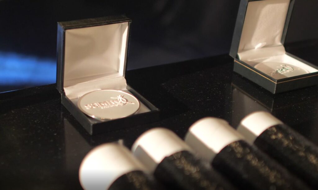
On Thursday, March the 10th, the South African Medical Research Council (SAMRC) honoured a selection of leading SA medical scientists and researchers at its 8th SAMRC Scientific Merit Awards at a hybrid event.

This year’s Presidential Award, which is awarded to scientists who have made exceptional lifelong contributions to medical research and public health, was bestowed upon Professor Koleka Mlisana, the country’s first black microbiologist. With over 40 years’ experience in health sciences, Prof Mlisana is the current executive manager of academic affairs, research, and quality assurance at the National Health Laboratory Service (NHLS) and Co-Chair of the COVID-19 Ministerial Advisory Committee (MAC). In the 1990s, she was one of the scientists investigating the unknowns of HIV. Her research focused on understanding the body’s response to acute HIV infection.

The Platinum Medal, for South Africans who have made seminal scientific contributions and who have also made an impact on health, especially for those living in developing countries, was awarded to Professor Andre Pascal Kengne. As a physician and an internationally renowned non-communicable diseases epidemiologist, his work focuses on cardiovascular disease, diabetes, and chronic kidney disease. He is the current Director of the SAMRC’s Non-Communicable Diseases Research Unit and holds conjoint appointments as Professor of Medicine at the University of Cape Town, as well as Extraordinary Professor of Global Health at Stellenbosch University.

In the Gold Medal category, which is for researchers who have made substantial and influential contributions that have impacted on health especially in the developing world, the awardees are Professors Tulio de Oliveira, Ntobeko Ntusi, Ambroise Wonkam and Grant Theron.

Silver Medals are conferred to emerging and upcoming scientists and those committed to capacity development. This year, the medal recipients are Professors Diane Gray, Marlo Moller, Rabia Johnson, and Dr Nasheeta Peer.

SAMRC President and CEO, Prof Glenda Gray said that scientific research remains fundamental for reducing the nation’s burden of disease and preventing mortality. “The knowledge produced by these exceptional scientists will carry our country’s legacy of science forward and continue to improve the lives of citizens as it is evident with COVID-19.” Their work shows the country’s ingenuity, she added, noting that “it was scientists in South Africa who first discovered and sounded the alarm on Omicron, which rapidly became the dominant variant of concern.”

Source: South African Medical Research Council (SAMRC)

Study Implicates High Leptin Levels in Androgen Deficiencies

Source: National Cancer Institute on Unsplash

Researchers have uncovered new clues about the cellular processes that can lead to androgen deficiencies, in which high leptin levels appear to play a role. The findings are published in the journal Cell Death & Disease.

Symptoms of testosterone deficiency include low sex drive, erectile dysfunction, depression, and fatigue. TD afflicts approximately 30% of men aged 40-79 years, with an increase in prevalence strongly associated with ageing and common medical conditions including obesity, diabetes, and hypertension.

“Although testosterone deficiency may be present in one in five men 40 years or older, the driving factors remain largely unknown,” said Himanshu Arora, PhD, assistant professor of urology.

Dr Arora’s lab examined the effect of different concentrations of leptin on the microenvironment of the testes. The research builds on prior studies of how Sertoli and peritubular myoid cells (PMC) in the testicular microenvironment help drive Leydig stem cell differentiation via the cellular desert hedgehog signalling pathway, which transmits information to embryonic cells that guides proper cell differentiation.

The researchers extracted cellular samples from men undergoing testes biopsies for sperm retrieval. When the testes microenvironment secreted leptin in low doses, they found that Leydig stem cells differentiated into adult Leydig cells producing normal levels of testosterone. Higher doses of leptin were observed to depress testosterone levels.

“Our findings identify leptin as a key factor within the testes microenvironment,” said Dr Arora, adding that the insight “holds important implications for androgen deficiency and could have further application in prostate cancer research.”

Noting that leptin is already used in treating patients for obesity, “Preclinical studies could indicate whether adjusting levels of this hormone would be helpful in patients with testosterone deficiency,” said Ranjith Ramasamy, MD, study co-author and associate professor and director of the Miller School’s Reproductive Urology Program.

Source: University of Miami Health System, Miller School of Medicine

X-Ray Images With Vastly Lower Radiation Doses

A new scintillation material developed by KAUST scientists can bring significant improvements to X-ray imaging in medicine, industry and security. Credit: KAUST

Scientists have successfully produced an exceptionally efficient, robust and flexible scintillation film to bring significant improvements in X-ray imaging, enabling much lower radiation doses to be used.

Scintillation materials release visible light, or “scintillate,” in response to absorbing  high-energy X-ray photons, enabling an image to be captured.

Researchers are continually exploring ways to make scintillation technology more sensitive, efficient and readily adaptable. The researchers, led by  Omar F Mohammed, Associate Professor of Chemical Sciences at King Abdullah University of Science and Technology (KAUST), sought to come up with an improved scintillation screen.

“Currently used materials suffer from several drawbacks, including complex and high-cost fabrication processes, radioluminescence afterglow and nontunable scintillation,” said Yang Zhou, a postdoc in Prof Mohammed’s lab.

Materials called lead halide perovskites have attracted considerable attention and shown significant promise. Novel perovskites are a category of materials that share the same crystal structure as the natural perovskite mineral calcium titanium oxide, but they include a variety of different atoms that replace all or some of those found in natural perovskite. 
To avoid toxicity problems and reduce cost, the researchers explored the use of elements besides lead. The newly developed screens are described in ACS Energy Letters.

The flexible scintillation screens the team developed can detect X-rays at ultralow levels, “approximately 113 times lower than a typical standard dose for X-ray medical imaging,” said Omar Mohammed, leader of the research group.

“Another vital advance is that the X-ray spatial resolution reported in this study is the highest achieved to date for powder-based screens,” said Dr Zhou.

“The physical flexibility of our films is also very important,” added Prof Mohammed. He explains that highly efficient flexible scintillation screens are urgently needed for using X-rays to better analyse awkward shapes.

The team plans to commercialise their advance, and to hope to refine their fabrication techniques.

Source: EurekAlert!

Sanofi’s Rare Disease Database Aids Healthcare Practitioners

Image source: CDC/Unsplash

Sanofi’s rare disease database that helps healthcare practitioners tackle their unique challenges – and knowing that treatments are available directly improves patients’ wellbeing. This comprehensive database has also aided rare disease research.

Johannesburg, 28 February 2022: Patients with rare diseases present unique challenges to healthcare practitioners (HCPs). Obstacles to caring for them include diagnostic delays and a lack of information, expertise, and treatment options for many rare diseases. HCPs play a vital role in enhancing the quality of life for patients and families living with a rare disease by making appropriate referrals to specialists, helping to coordinate care, and assisting patients in obtaining the proper support.1,2

A disease is defined as ‘rare’ when it affects fewer than 1 in 2000 people.3

Over 7000 rare diseases have been described to date, affecting over 350 million people worldwide.3,4 While most (70-80%) of rare diseases are genetic and inherited, some may be acquired, and 70% are exclusively paediatric in onset.5

Recent surveys showed that those living with rare diseases had a significantly higher prevalence of anxiety and depression compared to the general population.5,6 Levels of high stress can become even worse for carers when the person they are supporting has a diagnosis with no available treatment option.5,6

Monique Nel, Medical Advisor – Rare Diseases at Sanofi says: “Sanofi has been dedicated to researching and developing innovative treatments for rare diseases for 40 years. Currently, Sanofi has one of the largest rare diseases pipelines in the industry, across multiple diseases and modalities.7

“Our rare disease patient registries have grown to represent one of the largest collections of real-world data for rare diseases collected over the past 30 years. We have a presence in 68 countries worldwide, with more than 920 participating sites and more than 17 800 patients enrolled.”

These registries have helped researchers to publish studies describing the underlying biology of disease, identify risk factors impacting treatment outcomes, and share guidelines for monitoring and treatment.

A further useful resource for HCPs and patients is the list of rare diseases maintained by the Genetic and Rare Diseases Information Center (GARD) of the US National Institutes of Health.8          

Says Nel: “We understand the difficulty that healthcare professionals face when it comes to patient diagnosis of a rare disease, and that a coordinated approach to diagnosis and care for people living with rare diseases is needed. Rare diseases deserve the same amount of time, resources and dedication to finding effective treatments and therapies as any other conditions, which is a mission that Sanofi strives to promote every day, to help HCPs to improve diagnosis.”

References:

  1. Elliott E, Zurynski Y. Rare diseases are a ‘common’ problem for clinicians. Aust Fam Physician. 2015 Sep;44(9):630. http://www.ncbi.nlm.nih.gov/pubmed/26488039
  2. Dudding-Byth T. A powerful team: the family physician advocating for patients with a rare disease. Aust Fam Physician. 2015 Sep;44(9):634. http://www.ncbi.nlm.nih.gov/pubmed/264880401. NIH.
  3. Genetic and Rare Disease Information Center. FAQs About Rare Diseases. Available at: https://rarediseases.info.nih.gov/diseases/pages/31/faqs-about-rare-diseases
  4. Bogart KR, Irvin VL. Health-related quality of life among adults with diverse rare disorders. Orphanet J Rare Dis. 2017 Dec 7;12(1):177. doi: 10.1186/s13023-017-0730-1. PMID: 29212508; PMCID: PMC5719717.
  5. Nguengang Wakap S, Lambert DM, Olry A, et al. Estimating cumulative point prevalence of rare diseases: analysis of the Orphanet database. Eur J Hum Genet 2020;28:165–173. https://doi.org/10.1038/s41431-019-0508-0
  6. National Alliance for Caregiving. Rare Disease Caregiving in America. Available at: https://www.caregiving.org/wp-content/uploads/2020/05/NAC-RareDiseaseReport_February-2018_WEB.pdf
  7. Sanofi Your Health webpage. Rare Disease. https://www.sanofi.com/en/your-health/specialty-care/rare-diseases
  8. National Institutes of Health, Genetic and Rare Diseases Information Center. Caring for your patient with a rare disease.  Available at: https://rarediseases.info.nih.gov/guides/pages/122/caring-for-your-patient-with-a-rare-disease

Sound Waves Used to Regrow Bone

Image by Pawel Czerwinski on Unsplash

In a significant advance for the field of tissue engineering, researchers have used sound waves to turn stem cells into bone cells, a technology which may help regrow bone lost by cancer or disease.

Described in the journal Small, the innovative stem cell treatment from researchers at RMIT University offers a smart way forward for overcoming some of the field’s biggest challenges, through the precision power of high-frequency sound waves.

Tissue engineering is an emerging field that aims to rebuild bone and muscle by harnessing the human body’s natural ability to heal itself. A key challenge in regrowing bone is having sufficient amounts of bone cells that can thrive once implanted in the target area.

So far, turning stem cells into bone cells has needed complicated and expensive equipment, making widespread clinical use unrealistic.

The few clinical trials trying to regrow bone mostly used stem cells painfully extracted from a patient’s bone marrow.

In a new study published in the journal Small, the RMIT research team showed stem cells treated with high-frequency sound waves turned into bone cells quickly and efficiently.

Importantly, the treatment was effective on multiple types of cells including fat-derived stem cells, which are far less painful to extract from a patient.

Co-lead researcher Dr Amy Gelmi said the new approach was faster and simpler than other methods.

“The sound waves cut the treatment time usually required to get stem cells to begin to turn into bone cells by several days,” said Dr Gelmi. “This method also doesn’t require any special ‘bone-inducing’ drugs and it’s very easy to apply to the stem cells.

“Our study found this new approach has strong potential to be used for treating the stem cells, before we either coat them onto an implant or inject them directly into the body for tissue engineering.”

The high-frequency sound waves used in the stem cell treatment were generated on a low-cost microchip device developed by RMIT.

Co-lead researcher Distinguished Professor Leslie Yeo and his team have spent over a decade researching the interaction of sound waves at frequencies above 10MHz with different materials.

The sound wave-generating device they developed can be used to precisely manipulate cells, fluids or materials.

“We can use the sound waves to apply just the right amount of pressure in the right places to the stem cells, to trigger the change process,” Prof Yeo said.

“Our device is cheap and simple to use, so could easily be upscaled for treating large numbers of cells simultaneously – vital for effective tissue engineering.”

The next stage in the research is investigating methods to upscale the platform, working towards the development of practical bioreactors to drive efficient stem cell differentiation.

Source: RMIT

Do People’s Lives ‘Flash Before Their Eyes’ When They Die?

Source: Pixabay CC0

By chance, neuroscientists were able to record the activity of a dying human brain and discovered brain wave patterns similar to dreaming, memory recall, and meditation. An analysis of this case, reported in Frontiers in Aging Neuroscience suggests a possible explanation for near-death experiences.

Imagine reliving your entire life in the space of seconds. Like a flash of lightning, you are outside of your body, watching memorable moments you lived through. This process, known as ‘life recall’, can be similar to what it’s like to have a near-death experience. What happens inside your brain during these experiences and after death are questions that have puzzled neuroscientists for centuries. However, the present study suggests that your brain may remain active and coordinated during and even after the transition to death, and may in fact be programmed to orchestrate the whole ordeal.

When an 87-year-old patient developed epilepsy, Dr Raul Vicente of the University of Tartu, Estonia and colleagues used continuous electroencephalography (EEG) to detect the seizures and treat the patient. During these recordings, the patient had a heart attack and passed away. This unexpected event allowed the scientists to record the activity of a dying human brain for the first time ever.

“We measured 900 seconds of brain activity around the time of death and set a specific focus to investigate what happened in the 30 seconds before and after the heart stopped beating,” said Dr Ajmal Zemmar, a neurosurgeon at the University of Louisville, US, who organised the study.

“Just before and after the heart stopped working, we saw changes in a specific band of neural oscillations, so-called gamma oscillations, but also in others such as delta, theta, alpha, and beta oscillations.”

Brain oscillations (aka ‘brain waves’) are patterns of rhythmic brain activity normally present in living human brains. These different types of oscillations, including gamma, are involved in high-cognitive functions, such as concentrating, dreaming, meditation, memory retrieval, information processing, and conscious perception, just like those associated with memory flashbacks.

“Through generating oscillations involved in memory retrieval, the brain may be playing a last recall of important life events just before we die, similar to the ones reported in near-death experiences,” Dr Zemmar speculated. “These findings challenge our understanding of when exactly life ends and generate important subsequent questions, such as those related to the timing of organ donation.”

Though this is the first study to ever measure live brain activity during the process of dying in humans, similar changes in gamma oscillations have been previously recorded in rats kept in controlled environments. This raises the possibility that, during death, the brain organises and executes a biological response that could be conserved across species.

The interepretation of this however is complicated by the fact that these measurements are based on a single case and stem from the brain of a patient who had suffered injury, seizures and swelling. Nonetheless, Dr Zemmar plans to investigate more cases and sees these results as a source of hope.

“As a neurosurgeon, I deal with loss at times. It is indescribably difficult to deliver the news of death to distraught family members,” he said.

“Something we may learn from this research is: although our loved ones have their eyes closed and are ready to leave us to rest, their brains may be replaying some of the nicest moments they experienced in their lives.”

Source: Frontiers

Bacteria Uses Never-before-seen Method to Invade Tissue

Luallen Lab members pose in their lab at SDSU. Inset: Microscope image of Bordetella atropi (pink lines) infecting roundworm intestine (green). Credit: San Diego State University

Like something out of a horror movie, a new way that one type of bacteria invades tissue within a living organism has been identified by biologists from San Diego State University.

The study, published in Nature Communications, describes how a new species of bacteria, Bordetella atropi, invades its roundworm host. The name which comes from the Greek fate Atropos responsible for cutting the threads of life, is apt because the bacteria transforms into a long thread, growing up to 100 times the usual size of one bacterium in the span of 30 hours without dividing.

By altering the genes of B. atropi, the research team discovered that this invasive threading relies on the same genes and molecules that other bacteria use when they are in a nutrient-rich environment. However, these other bacteria only use this pathway to make subtly larger cells, whereas the B. atropi bacteria grows continuously.

Other bacteria often transform into threads, called filamentation, in response to dangerous environments or DNA damage. This lets them continue to grow in size, but delay cell division until they repair the damage inflicted by the stress.

Here, however, the researchers were the first to observe filamentation as a way of spreading from cell to cell in a living organism for a purpose other than the stress response. They believe that instead the new species is invading the host cells, detecting this rich environment and triggering filamentation in order to quickly infect more cells and access additional nutrients for their growth.

“We went from finding the worm in the ground, finding the bacteria, and carrying it all the way to the molecular mechanism of how the bacteria infects the worm,” explained Robert Luallen, biology professor and principal investigator of the study. “We’re seeing things that no one’s ever seen before.”

Though B. atropi does not infect humans, it is possible that human pathogens may also make use of its spreading mechanism. Separately, the nutrient-induced filamentation process might be used by other bacteria to form biofilms, which can coat the tubing of catheters and lead to complications for patients.

Source: EurekAlert!

Patients Could Control their Personal Health Information with NFTs

Image by Webstacks on Unsplash

In a new publication in the journal Science, researchers propose that NFTs, or nonfungible tokens, could help patients assert better control over their personal health information.

NFTs, or nonfungible tokens, created using blockchain technology, have been a big sensation in the art world as they serve as a platform to buy and sell digital art backed by a digital contract. Now, an international multidisciplinary team of scholars in ethics, law and informatics led by bioethicists have written one of the first commentaries on how this new emerging technology could be repurposed for the healthcare industry. NFT digital contracts could provide an opportunity for patients to specify who can access their personal health information and to track how it is shared.

“Our personal health information is completely outside of our control in terms of what happens to it once it is digitised into an electronic health record and how it gets commercialised and exchanged from there,” said Dr Kristin Kostick-Quenet, first author of the paper. “NFTs could be used to democratise health data and help individuals regain control and participate more in decisions about who can see and use their health information.”

“In the era of big data, health information is its own currency; it has become commodified and profitable,” said Dr Amy McGuire, senior author of the paper and Leon Jaworski Professor of Biomedical Ethics and director of the Center for Medical Ethics and Health Policy at Baylor. “Using NFTs for health data is the perfect storm between a huge market place that’s evolving and the popularity of cryptocurrency, but there are also many ethical, legal and social implications to consider.”

Presently, NFTs are still vulnerable to data security flaws, privacy issues, and disputes over intellectual property rights, the researchers noted. The complexity of NFTs may also prevent the average person from properly making use of them. The researchers believe it is important to consider potential benefits and challenges as NFTs emerge as a potential avenue to transform the world of health data.

Source: Baylor College of Medicine

A New Understanding of the Fundamental Order of the Abdomen

Source: Pixabay

In a research paper published in Communications Biology, researchers from the University of Limerick have detailed the development and structure of the mesentery. In doing this, they uncovered a new order by which all contents of the abdomen are organised or arranged – or the “fundamental order of the abdomen”, where organs are in one of two compartments.

Professor Calvin Coffey, Foundation Chair of Surgery at UL’s School of Medicine in Ireland, whose major discovery led to the reclassification of the mesentery as a new organ in 2016, has published new research on the makeup and structure of the abdomen.

The importance of these findings on the mesentery and the impact these have on our understanding of the abdomen have been further explained in a review article just published in the Lancet Gastroenterology and Hepatology.

Prof Coffey explained that his team have been looking at the development and structure of the mesentery since 2016.

“We showed how the mesentery is a single and continuous organ in and on which all abdominal digestive organs develop and then remain connected to throughout life,” he explained.

“These findings revealed a simplicity in the abdomen that was not apparent in conventional descriptions of anatomy.”

The international team of researchers used cutting edge techniques to clarify how the mesentery develops and the shape it has in adults.

Their work revealed that the organisation of the abdomen has a remarkably simple design.

“The abdomen is not the dauntingly complex collection of separate organs it was previously thought to be,” said Prof Coffey.

“Instead, all digestive organs are neatly packaged and arranged by the mesentery into a single digestive engine. That simplicity lay hidden until clarification of the nature of the mesentery.”

The model itself was described by the team in the most recent edition of Gray’s Anatomy. The supportive evidence was published in Communications Biology and the clinical importance was explained in the review in The Lancet Gastroenterology and Hepatology.

“The most important finding here was the discovery of the fundamental order of the abdomen. At the foundation level, all contents of the abdomen are simply organised into one of two compartments,” explained Prof Coffey.

“The fundamental order of any structure is of considerable importance, in particular when it comes to diagnosing patients with illness and treating their disease. The fundamental order is the foundation from which all science launches and clinical practice is based.

“The organisational simplicity of the abdomen now immediately explains the behaviours of viral and bacterial infections, cancer, inflammatory bowel disease, obesity, diabetes and many others,” he added.

Improvements in surgery have been made to surgery by a better understanding of the mesentery and its functions, and the new research builds on those advances. There are also exciting areas for future investigation, according to Prof Coffey.

“Patients are already benefiting from what we now call mesenteric-based approaches to the diagnosis and treatment of most abdominal conditions. The Mesenteric Model of Abdominal Anatomy – or the description of the order of the abdomen – is being incorporated into numerous reference curricula at this moment,” he said.

“Regarding the future, it is being argued that we are seeing a paradigmatic shift from old to new order. Already, intriguing questions are emerging that we can call ‘legitimate or admissible’ in the strictest scientific sense. Science can approach numerous questions in a new light.  Clinicians can design diagnostic and treatment approaches based on a new foundation,” Prof Coffey concluded.

Source: EurekAlert!

Robot Performs Surgery Without Human Assistance

Photo by Tara Winstead from Pexels

In a significant step toward fully automated surgery on humans, a robot has performed laparoscopic surgery on the soft tissue of a pig without human guidance. 

Designed by a team of Johns Hopkins University researchers, the Smart Tissue Autonomous Robot (STAR) is described in Science Robotics.

“Our findings show that we can automate one of the most intricate and delicate tasks in surgery: the reconnection of two ends of an intestine. The STAR performed the procedure in four animals and it produced significantly better results than humans performing the same procedure,” said senior author Axel Krieger, PhD, an assistant professor at John Hopkins University.

The robot excelled at intestinal anastomosis, which connects the two ends of an intestine. It is a procedure that requires a high level of repetitive motion and precision and is arguably the most challenging step in gastrointestinal surgery, requiring a surgeon to accurately and consistently suture. A slight hand tremor or misplaced stitch can result in a leak with potentially catastrophic complications for the patient.

The team developed a vision-guided system designed specifically to suture soft tissue. Their current iteration advances a 2016 model that repaired a pig’s intestines accurately, but required a large incision to access the intestine and more guidance from humans.

The team equipped the STAR with new features for enhanced autonomy and improved surgical precision, including specialised suturing tools and state-of-the art imaging systems that provide more accurate visualisations of the surgical field.

Soft-tissue surgery is especially hard for robots because of its unpredictability, forcing them to be able to adapt quickly to handle unexpected obstacles, Dr Krieger said. STAR features a novel control system that can adjust the surgical plan in real time, just as a human surgeon would.

As the medical field moves towards more laparoscopic approaches for surgeries, it will be important to have an automated robotic system designed for such procedures to assist, Dr Krieger said.

“Robotic anastomosis is one way to ensure that surgical tasks that require high precision and repeatability can be performed with more accuracy and precision in every patient independent of surgeon skill,” Dr Krieger said.

“We hypothesise that this will result in a democratised surgical approach to patient care with more predictable and consistent patient outcomes.”

Source: John Hopkins University