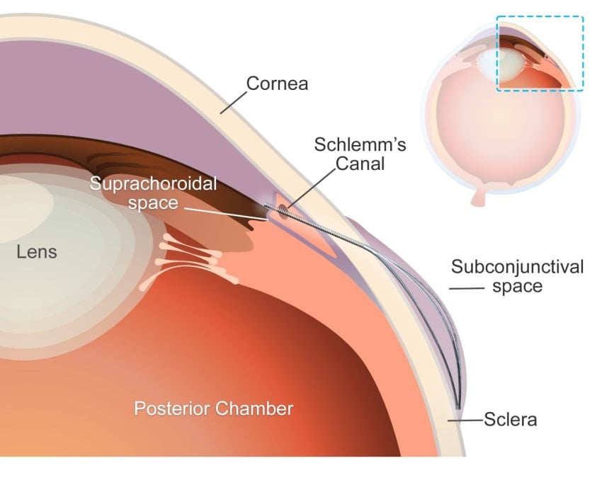
A schematic of the eye’s anterior segment, demonstrating the anatomical placement of the microstent. The stent diverts aqueous humour from the anterior chamber to the suprachoroidal space through the flexible tube, creating a subconjunctival bleb supported by the expanding element. Credit: Yunlan Zhang, Zhong You, Jared Ching.
A team of researchers at the University of Oxford have unveiled a pioneering ‘microstent’ which could revolutionise treatment for glaucoma, a common but debilitating condition. The study has been published in The Innovation, Cell Press.
Glaucoma is a leading cause of vision loss, second only to cataracts. Globally, 7.7 million people were blind or visually impaired due to glaucoma in 2020. The condition can cause irreversible damage to the optic nerve, due to increased pressure within the eyeball. Current treatment options – principally surgery to create openings in the eye or insert tubes to drain fluid – are highly invasive, carry risk of complications, and have limited durability.
‘Our deployable microstent represents a significant advancement in glaucoma treatment,’ said lead author Dr Yunlan Zhang (University of Oxford at the time of the study/University of Texas). ‘Current surgical implants for this type of glaucoma have been shown to have limited long-term effectiveness, being susceptible to failure due to fibrosis (scarring) in the eye.’
The new microstent features a unique structural shape that allows it to expand once in the eye. At 200µm, less than a quarter of a millimetre, the stent’s tiny diameter enables it to fit within the needle of a standard hypodermic syringe, for minimally-invasive insertion. Once in place and expanded, the microstent spans the fluid-filled space between the white of the eye and the membrane that covers it.
By supporting this space, the stent reduces the excessive fluid buildup and resulting intraocular pressure in the eye which is responsible for the most common type of glaucoma, primary open-angle glaucoma. Initial trials carried out in rabbits found that the microstents lowered eye pressure in less than a month with minimal inflammation and scarring. Furthermore, the microstent achieved a greater reduction of eye pressure than a standard tubular implant.
This development has the potential to transform the landscape of glaucoma therapy. By offering an enhanced solution in the minimally invasive glaucoma surgery field that combines mechanical innovation with biocompatibility, we hope to improve patient outcomes and quality of life.
Senior co-author Dr Jared Ching (Department of Engineering Science, University of Oxford).
Senior co-author, Professor Zhong You (Department of Engineering Science, University of Oxford) said: ‘Our microstent is made from a durable and super-flexible nickel-titanium alloy called nitinol, renowned for its proven long-term safety for ocular use. Its unique material and structural properties help prevent subsequent movement, improve durability, and ensure long-term efficacy.’
The research team used advanced modelling techniques to guide the microstent’s design and ensure compatibility with the anatomy of the eye. The device’s superelastic properties enable it to accommodate how the eye changes and stretches over time without permanent deformation, enhancing its durability and functionality.
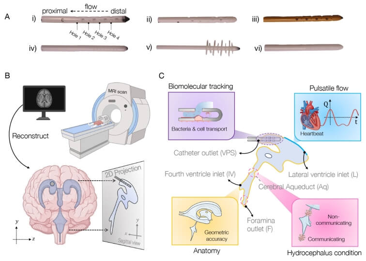
Schematic of approach to simulating brain shunt fluid dynamics. Credit: Harvard SEAS
Millions of people worldwide suffer from hydrocephalus, a condition which recently received greater attention when Billy Joel announced his diagnosis. Treatment usually involves surgical placement of shunts to divert cerebrospinal fluid away, but this procedure often leads to complications, infections, and multiple re-treatments.
Bioengineers in the Harvard John A. Paulson School of Engineering and Applied Sciences (SEAS) have now developed a new computational model to aid the creation of shunts tailored to individual patients’ anatomy and needs. The model combines brain anatomy, fluid flow, and biomolecular transport dynamics to simulate shunt performance with pinpoint accuracy.
The work was supported by federal funding from the National Science Foundation and published in Proceedings of the National Academy of Sciences. It was led by SEAS postdoctoral fellow Haritosh Patel, who works in the labs of Joanna Aizenberg, Professor of Materials Science at SEAS and Professor of Chemistry and Chemical Biology; and Venkatesh Murthy, Professor of Molecular and Cellular Biology and Director of the Center for Brain Science.
Repeat surgeries due to infection or obstruction
Tens of thousands of shunt procedures are performed annually in the U.S. — many of which are repeat surgeries due to the inserted devices becoming blocked or obstructed, or the patient suffering an infection.
“Some elderly patients told me they had had over 10 surgeries — one every two to three years,” Patel said. “We really wanted to understand why this was happening, and we realised that many of these obstructions and infections were tied to shunt designs that didn’t fully consider fluid dynamics as a fundamental part of their geometry. We noticed that the tubing geometry used in shunts closely resembles the kind of piping we rely on in household plumbing. While that simplicity has its advantages, we saw an opportunity to explore more creative, biomimetic solutions that better suit the complexity of the brain’s environment.”
Pursuing the problem from both a material and design perspective, the team quickly realized there was no universally accepted fluid flow model for the brain ventricle space to guide them. “Okay, well, we can’t test our devices in a model, so why don’t we first make a better model?” Patel said.
Computational tool simulates fluid flow in brain
The result is their computational tool, called BrainFlow, which combines detailed anatomical and physiological features of the brain to simulate the flow of cerebrospinal fluid flow in the presence of shunt implants.
The model incorporates patient-specific medical imaging data along with pulse-induced flow to mimic a patient’s cerebrospinal fluid dynamics, all to offer insight into optimal shunt design, placement, and even choice of materials.
“We believe that our model, combined with novel geometries and materials improvements such as anti-biofouling coatings developed in my lab, could lead to smoother integration of optimized, patient-specific medical devices into patients’ brains, with less likelihood of complications, and a better quality of life,” Aizenberg said.
The Harvard team is currently conducting studies that use the model to test different designs of shunts and calculate their efficacy.
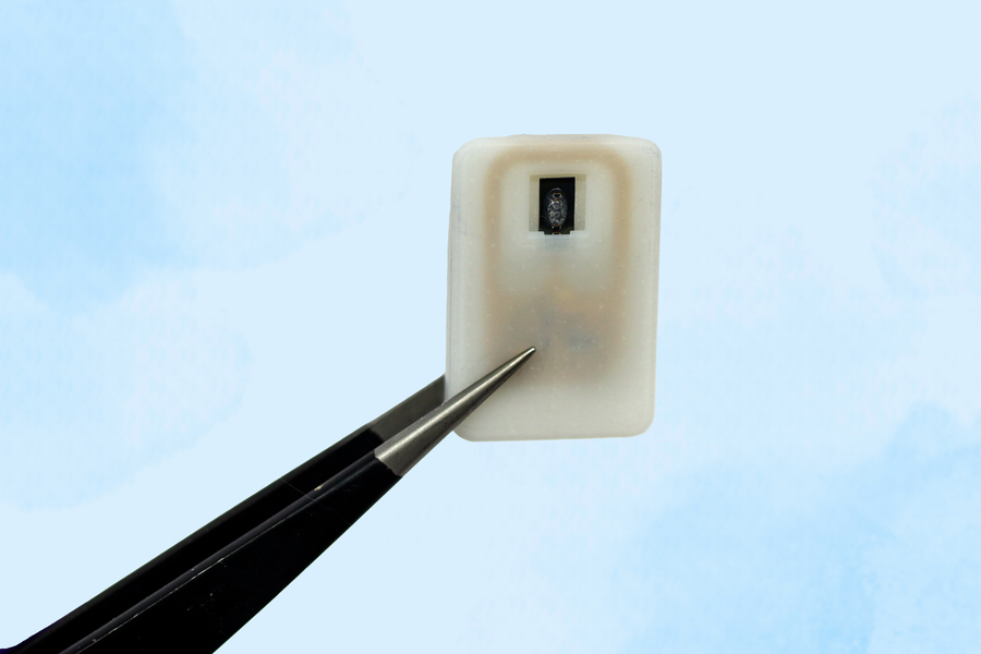
The new implant carries a reservoir of glucagon that can be stored under the skin and deployed during an emergency — with no injections needed.
Caption:A new implantable device carries a reservoir of glucagon that can be stored under the skin and could save diabetes patients from dangerously low blood sugar.
Image: Courtesy of the researchers
For people with Type 1 diabetes, developing hypoglycaemia, or low blood sugar, is an ever-present threat. When glucose levels become extremely low, it creates a life-threatening situation for which the standard treatment of care is injecting a hormone called glucagon.
As an emergency backup, for cases where patients may not realise that their blood sugar is dropping to dangerous levels, MIT engineers have designed an implantable reservoir that can remain under the skin and be triggered to release glucagon when blood sugar levels get too low.
This approach could also help in cases where hypoglycaemia occurs during sleep, or for diabetic children who are unable to administer injections on their own.
“This is a small, emergency-event device that can be placed under the skin, where it is ready to act if the patient’s blood sugar drops too low,” says Daniel Anderson, a professor in MIT’s Department of Chemical Engineering, a member of MIT’s Koch Institute for Integrative Cancer Research and Institute for Medical Engineering and Science (IMES), and the senior author of the study. “Our goal was to build a device that is always ready to protect patients from low blood sugar. We think this can also help relieve the fear of hypoglycaemia that many patients, and their parents, suffer from.”
The researchers showed that this device could also be used to deliver emergency doses of epinephrine, a drug that is used to treat heart attacks and can also prevent severe allergic reactions, including anaphylactic shock.
Siddharth Krishnan, a former MIT research scientist who is now an assistant professor of electrical engineering at Stanford University, is the lead author of the study, which appears today in Nature Biomedical Engineering.
Emergency response
Most patients with type 1 diabetes use daily insulin injections to help their body absorb sugar and prevent their blood sugar levels from getting too high. However, if their blood sugar levels get too low, they develop hypoglycaemia, which can lead to confusion and seizures, and may be fatal if it goes untreated.
To combat hypoglycaemia, some patients carry preloaded syringes of glucagon, a hormone that stimulates the liver to release glucose into the bloodstream. However, it isn’t always easy for people, especially children, to know when they are becoming hypoglycaemic.
“Some patients can sense when they’re getting low blood sugar, and go eat something or give themselves glucagon,” Anderson says. “But some are unaware that they’re hypoglycaemic, and they can just slip into confusion and coma. This is also a problem when patients sleep, as they are reliant on glucose sensor alarms to wake them when sugar drops dangerously low.”
To make it easier to counteract hypoglycaemia, the MIT team set out to design an emergency device that could be triggered either by the person using it, or automatically by a sensor.
The device, which is about the size of a quarter, contains a small drug reservoir made of a 3D-printed polymer. The reservoir is sealed with a special material known as a shape-memory alloy, which can be programmed to change its shape when heated. In this case, the researcher used a nickel-titanium alloy that is programmed to curl from a flat slab into a U-shape when heated to 40 degrees Celsius.
Like many other protein or peptide drugs, glucagon tends to break down quickly, so the liquid form can’t be stored long-term in the body. Instead, the MIT team created a powdered version of the drug, which remains stable for much longer and stays in the reservoir until released.
Each device can carry either one or four doses of glucagon, and it also includes an antenna tuned to respond to a specific frequency in the radiofrequency range. That allows it to be remotely triggered to turn on a small electrical current, which is used to heat the shape-memory alloy. When the temperature reaches the 40-degree threshold, the slab bends into a U shape, releasing the contents of the reservoir.
Because the device can receive wireless signals, it could also be designed so that drug release is triggered by a glucose monitor when the wearer’s blood sugar drops below a certain level.
“One of the key features of this type of digital drug delivery system is that you can have it talk to sensors,” Krishnan says. “In this case, the continuous glucose-monitoring technology that a lot of patients use is something that would be easy for these types of devices to interface with.”
Reversing hypoglycaemia
After implanting the device in diabetic mice, the researchers used it to trigger glucagon release as the animals’ blood sugar levels were dropping. Within less than 10 minutes of activating the drug release, blood sugar levels began to level off, allowing them to remain within the normal range and avert hypoglycaemia.
The researchers also tested the device with a powdered version of epinephrine. They found that within 10 minutes of drug release, epinephrine levels in the bloodstream became elevated and heart rate increased.
In this study, the researchers kept the devices implanted for up to four weeks, but they now plan to see if they can extend that time up to at least a year.
“The idea is you would have enough doses that can provide this therapeutic rescue event over a significant period of time. We don’t know exactly what that is — maybe a year, maybe a few years, and we’re currently working on establishing what the optimal lifetime is. But then after that, it would need to be replaced,” Krishnan says.
Typically, when a medical device is implanted in the body, scar tissue develops around the device, which can interfere with its function. However, in this study, the researchers showed that even after fibrotic tissue formed around the implant, they were able to successfully trigger the drug release.
The researchers are now planning for additional animal studies and hope to begin testing the device in clinical trials within the next three years.
“It’s really exciting to see our team accomplish this, which I hope will someday help diabetic patients and could more broadly provide a new paradigm for delivering any emergency medicine,” says Robert Langer, the David H. Koch Institute Professor at MIT and an author of the paper.
Other authors of the paper include Laura O’Keeffe, Arnab Rudra, Derin Gumustop, Nima Khatib, Claudia Liu, Jiawei Yang, Athena Wang, Matthew Bochenek, Yen-Chun Lu, Suman Bose, and Kaelan Reed.
The research was funded by the Leona M. and Harry B. Helmsley Charitable Trust, the National Institutes of Health, a JDRF postdoctoral fellowship, and the National Institute of Biomedical Imaging and Bioengineering.
Hearing loss doesn’t just affect how people hear the world — it can also change how they connect with it. New research from the University of Southern California, published in JAMA Otolaryngology – Head & Neck Surgery, is the first to link hearing aids and cochlear implants, surgically implanted devices that help those with profound hearing loss perceive sound, to improved social lives among adults with hearing loss.
“We found that adults with hearing loss who used hearing aids or cochlear implants were more socially engaged and felt less isolated compared to those who didn’t use them,” said lead researcher Janet Choi, MD, MPH, an otolaryngologist with Keck Medicine at USC. “This suggests that hearing devices may help prevent the social disconnection and broader health consequences that can follow untreated hearing loss.”
Hearing loss affects an estimated 40 million American adults, yet many go untreated. When left unaddressed, hearing loss can make communication difficult, leading people to withdraw from conversations and social activities, according to Choi.
Previous research has shown that over time, social withdrawal can reduce mental stimulation and increase the risk of loneliness, anxiety, depression, cognitive decline and dementia. It has also linked chronic social isolation to biological and neurological changes, including increased brain inflammation and alterations in brain structure.
“Understanding the link between hearing loss, hearing device use and social isolation is crucial,” said Choi. “Until this study, it has been unclear whether hearing devices could help reverse the isolation.”
Choi and her fellow researchers conducted a comprehensive, systematic review and meta-analysis of 65 previously published studies, encompassing over five thousand participants, on how hearing aids and cochlear implants affect three key measures: social quality of life, perceived social handicap, which refers to the limitations and frustrations hearing loss can create in social situations, and loneliness.
The researchers found that adults using hearing devices feel more socially connected and less limited in social situations. They are better able to engage in group conversations and feel more at ease in noisy or challenging listening environments. Participants also reported feeling less socially handicapped by their hearing loss, with fewer barriers and frustrations during interactions and an improved ability to stay engaged without feeling excluded. This increased confidence can help users connect more easily with family, friends and colleagues, leading to stronger feelings of belonging and reduced social anxiety. The study also suggested hearing devices may reduce loneliness, although further research is needed in this area, according to Choi.
Those with cochlear implants reported the most improvement in their social quality of life. This is likely because cochlear implants offer greater hearing restoration than hearing aids, especially for individuals with more severe hearing loss. As a result, they may experience more noticeable improvements in social engagement once their hearing is restored.
While it was outside the scope of the study to measure how better social lives relate to improved cognitive outcomes, Choi believes there may be a connection, as previous research has found managing hearing loss may be key to reducing the risk of cognitive decline and dementia. “While our study didn’t directly measure cognitive outcomes, the improvements we saw in communication and social engagement suggest that by restoring clearer communication, hearing devices may help preserve cognitive health by keeping the brain more actively involved and people more connected,” Choi said.
This research follows a January 2024 study by Choi showing that adults with hearing loss who use hearing aids have an almost 25% lower risk of mortality, suggesting that treating hearing loss can improve lifespan as well as social quality of life.
“These new findings add to a growing body of research showing that hearing health is deeply connected to overall well-being,” said Choi. “We hope this encourages more people to seek treatment and helps clinicians start conversations with patients about how hearing devices can improve their quality of life.”
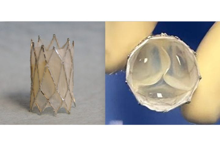
The Iris Valve, a transcatheter, growth-accommodating pulmonary valve designed for very young children, was developed at UC Irvine and is currently progressing toward FDA clinical approval. Arash Kheradvar
Researchers at the University of California, Irvine have successfully performed preclinical laboratory testing of a replacement heart valve intended for toddlers and young children with congenital cardiac defects, a key step toward obtaining approval for human use. The results of their study were published recently in the Journal of the American Heart Association.
The management of patients with congenital heart disease who require surgical pulmonary valve replacement typically occurs between the ages of 2 and 10. To be eligible for a minimally invasive transcatheter pulmonary valve procedure, patients currently must weigh at least 20.4kg. For children to receive minimally invasive treatment, they must be large enough so that their veins can accommodate the size of a crimped replacement valve. The Iris Valve designed and developed by the UC Irvine team can be implanted in children weighing as little as 7.7 to 10kg and gradually expanded to an adult diameter as they grow.
Research and development of the Iris Valve has been supported by the Eunice Kennedy Shriver National Institute of Child Health and Human Development; the National Heart, Lung, and Blood Institute; and the National Science Foundation.
This funding has enabled benchtop fracture testing, which demonstrated the valve’s ability to be crimped down to a 3mm diameter for transcatheter delivery and subsequently enlarged to 20mm without damage, as well as six-month animal studies that confirmed successful device integration within the pulmonary valve annulus, showing valve integrity and a favourable tissue response.
“We are pleased to see the Iris Valve performing as we expected in laboratory bench tests and as implants in Yucatan mini pigs, a crucial measure of the device’s feasibility,” said lead author Arash Kheradvar, UC Irvine professor of biomedical engineering. “This work represents the result of longstanding collaboration between our team at UC Irvine and Dr Michael Recto at Children’s Hospital of Orange County built over several years of joint research and development.”
Congenital heart defects affect about 1% of children born in the United States and Europe, with over 1 million cases in the US alone. These conditions often necessitate surgical interventions early in life, with additional procedures required to address a leaky pulmonary valve and prevent right ventricular failure as children grow.
The Iris Valve can be implanted via a minimally invasive catheter through the patient’s femoral vein. The Kheradvar group employed origami folding techniques to compress the device into a 12-French transcatheter system, reducing its diameter to no more than 3mm. Over time, the valve can be balloon-expanded up to its full 20mm diameter.
This implantation method, along with the ability to begin treatment earlier in very young patients, helps mitigate the risk of complications from delayed care and reduces the need for multiple surgeries in this vulnerable population.
“Once the Iris Valve comes to fruition, it will save hundreds of children at least one operation – if not two – throughout the course of their lives,” said Recto, an interventional paediatric cardiologist at CHOC who’s also a clinical professor of paediatrics at UC Irvine. “It will save them from having to undergo surgical pulmonary valve placement, as the Iris Valve is delivered via a small catheter in the vein and can be serially dilated to an adult diameter and also facilitate the future placement of larger transcatheter pulmonary valves – with sizes greater than 20 millimetres, like the Melody, Harmony and Sapien devices – if needed.”
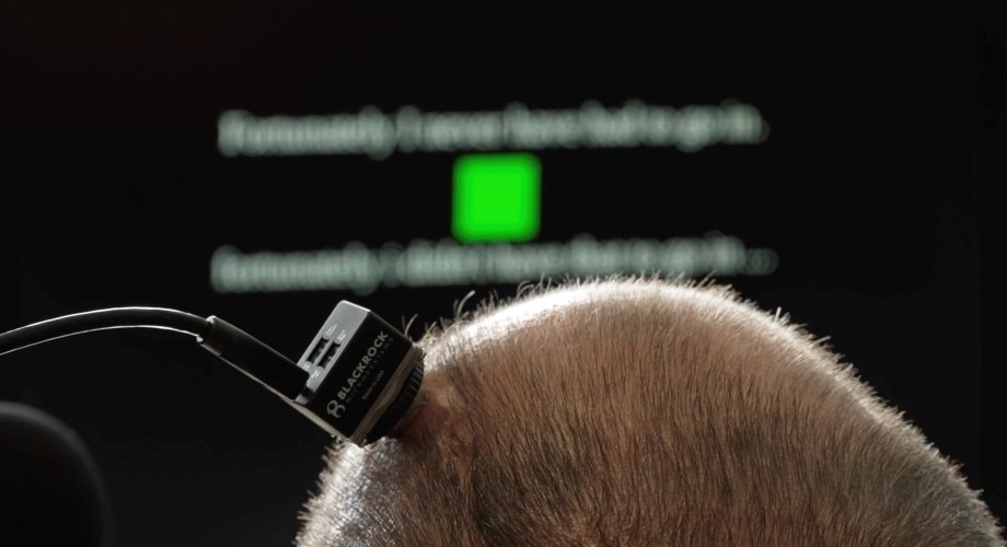
An investigational brain-computer interface (BCI) allows the study participant to communicate through a computer. Credit: UC Davis
Researchers at the University of California, Davis, have developed an investigational brain-computer interface that holds promise for restoring the ability to hold real-time conversations to people who have lost the ability to speak due to neurological conditions.
In a new study published in the scientific journal Nature, the researchers demonstrate how this new technology can instantaneously translate brain activity into voice as a person tries to speak – effectively creating a digital vocal tract with no detectable delay.
The system allowed the study participant, who has amyotrophic lateral sclerosis (ALS), to “speak” through a computer with his family in real time, change his intonation and “sing” simple melodies.
“Translating neural activity into text, which is how our previous speech brain-computer interface works, is akin to text messaging. It’s a big improvement compared to standard assistive technologies, but it still leads to delayed conversation. By comparison, this new real-time voice synthesis is more like a voice call,” said Sergey Stavisky, senior author of the paper and an assistant professor in the UC Davis Department of Neurological Surgery. Stavisky co-directs the UC Davis Neuroprosthetics Lab.
“With instantaneous voice synthesis, neuroprosthesis users will be able to be more included in a conversation. For example, they can interrupt, and people are less likely to interrupt them accidentally,” Stavisky said.
Decoding brain signals at heart of new technology
The man is enrolled in the BrainGate2 clinical trial at UC Davis Health. His ability to communicate through a computer has been made possible with an investigational brain-computer interface (BCI). It consists of four microelectrode arrays surgically implanted into the region of the brain responsible for producing speech.
These devices record the activity of neurons in the brain and send it to computers that interpret the signals to reconstruct voice.
“The main barrier to synthesising voice in real-time was not knowing exactly when and how the person with speech loss is trying to speak,” said Maitreyee Wairagkar, first author of the study and project scientist in the Neuroprosthetics Lab at UC Davis. “Our algorithms map neural activity to intended sounds at each moment of time. This makes it possible to synthesise nuances in speech and give the participant control over the cadence of his BCI-voice.”
Instantaneous, expressive speech with BCI shows promise
The brain-computer interface was able to translate the study participant’s neural signals into audible speech played through a speaker very quickly – one-fortieth of a second. This short delay is similar to the delay a person experiences when they speak and hear the sound of their own voice.
The technology also allowed the participant to say new words (words not already known to the system) and to make interjections. He was able to modulate the intonation of his generated computer voice to ask a question or emphasize specific words in a sentence.
The participant also took steps toward varying pitch by singing simple, short melodies.
His BCI-synthesized voice was often intelligible: Listeners could understand almost 60% of the synthesized words correctly (as opposed to 4% when he was not using the BCI).
Real-time speech helped by algorithms
The process of instantaneously translating brain activity into synthesized speech is helped by advanced artificial intelligence algorithms.
The algorithms for the new system were trained with data collected while the participant was asked to try to speak sentences shown to him on a computer screen. This gave the researchers information about what he was trying to say.
The electrodes measured the firing patterns of hundreds of neurons. The researchers aligned those patterns with the speech sounds the participant was trying to produce at that moment in time. This helped the algorithm learn to accurately reconstruct the participant’s voice from just his neural signals.
Clinical trial offers hope
“Our voice is part of what makes us who we are. Losing the ability to speak is devastating for people living with neurological conditions,” said David Brandman, co-director of the UC Davis Neuroprosthetics Lab and the neurosurgeon who performed the participant’s implant.
“The results of this research provide hope for people who want to talk but can’t. We showed how a paralyzed man was empowered to speak with a synthesized version of his voice. This kind of technology could be transformative for people living with paralysis.”
Brandman is an assistant professor in the Department of Neurological Surgery and is the site-responsible principal investigator of the BrainGate2 clinical trial.
Limitations
The researchers note that although the findings are promising, brain-to-voice neuroprostheses remain in an early phase. A key limitation is that the research was performed with a single participant with ALS. It will be crucial to replicate these results with more participants, including those who have speech loss from other causes, such as stroke.
People with Parkinson’s disease increasingly lose their mobility over time and are eventually unable to walk. Hope for these patients rests on deep brain stimulation. In a recent study, researchers at Ruhr University Bochum and Philipps-Universität Marburg, Germany, investigated whether and how stimulation of a certain region of the brain can have a positive impact on ambulatory ability and provide patients with a better quality of life. To do so, the researchers used a technique in which the nerve cells are activated and deactivated via light. Their report appeared in the journal Scientific Reports.
Improving ambulatory ability
If medication is no longer sufficient in alleviating restricted mobility in the advanced stage of Parkinson’s disease, one alternative is deep brain stimulation. An electrical pulse emitter is implanted within the brain, such as in the subthalamic nucleus, which is functionally part of the basal ganglia system.
The group under Dr Liana Melo-Thomas from Philipps-Universität Marburg was able to show in previous studies on rats that stimulation of the inferior colliculus, chiefly known for processing auditory input, can be used to overcome mobility restrictions. “There are indications that stimulation of this region of the brain leads to activation of the mesencephalic locomotor region, or MLR,” says Melo-Thomas.
Interestingly, the colliculus inferior – unlike the basal ganglia –is not affected by Parkinson’s disease. However, the research group under Melo-Thomas discovered that its stimulation activates alternative motor pathways and can improve patients’ mobility.
The current study aimed to further investigate this activating influence of the inferior colliculus on the MLR. “We suspected that this would have a positive effect on ambulatory ability,” says Melo-Thomas.
Optically influencing nerve cells
The Marburg group led by Professor Rainer Schwarting sought support by Dr Wolfgang Kruse from the Department of General Zoology and Neurobiology at Ruhr University Bochum. The team in Bochum led by Professor Stefan Herlitze played a significant role in co-developing the methods of optogenetics.
While doing so, the researchers ensure that the nerve cells of genetically modified test animals produce a light-sensitive protein in interesting regions of the brain. Light that reaches these nerve cells via small, implanted optical fibres allows the researchers to activate or inhibit them specifically. “This method is thus much more precise than electrical stimulation, which always affects the area around the cells as well,” says Kruse.
For the first time, the effect of the stimulation was directly documented with electrophysiological measurements of neuronal activity in the target structures. A multi-electrode system originally developed at Philipps-Universität Marburg was used for this purpose. By combining these methods, the researchers were able to directly understand the effect of the stimulation. Parallel measurement with up to four electrodes is also highly efficient, allowing minimisation of the number of animals used. Behavioural effects that can be triggered by the stimulation were monitored in conscious animals.
Stimulation of the inferior colliculus provides the desired effect
Optogenetic stimulation in the inferior colliculus predominantly triggered the expected increase in neuronal activity within it. “Simultaneous measurements in the deeper MLR region showed increased activity in the majority of cells, although nearly one quarter of the cells were inhibited by the additional activity in the inferior colliculus,” reports Kruse. The activation of individual nerve cells occurred with an average delay of 4.7 milliseconds, indicating a functional synaptic interconnection between the inferior colliculus and MLR.
Foundations for new types of therapy
Investigating circuits outside of the basal ganglia that are affected by Parkinson’s disease is a promising step in the search for a new therapeutic approach to alleviating motor deficits resulting from the disease. Such is the case with the connection between the inferior colliculus and the MLR that was investigated for this study.
“Even if the path toward new therapeutic approaches to alleviating the symptoms of Parkinson’s disease still appears long, such foundational research is immensely important,” emphasises Kruse. The exact mechanisms that lead to the observed relief of symptoms with deep brain stimulation in the basal ganglia are not fully understood. Further investigation of the underlying interconnections may provide new insight that could optimise therapy in the long term.
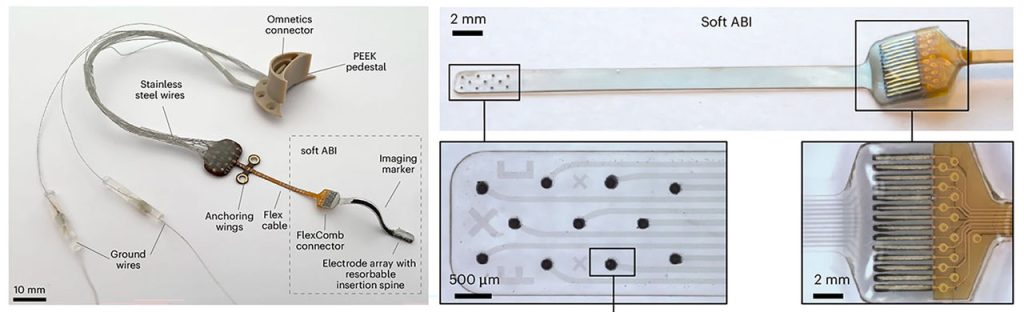
A new study co-led by Mass General Brigham researchers points to a promising new type of auditory brainstem implant (ABI) that could benefit people who are deaf due to Neurofibromatosis type 2 (NF2) and other severe inner ear abnormalities that prevent them from receiving cochlear implants. With further tests and trials, researchers hope it will provide a more effective treatment alternative than what is currently used.
In the new research, published in Nature Biomedical Engineering, scientists at Mass Eye and Ear, a member of the Mass General Brigham healthcare system, collaborated with scientists at the École Polytechnique Fédérale de Lausanne (EPFL) in Geneva, Switzerland, to report on a new class of soft, flexible ABIs that were designed to address the limitations of those currently used. These implants bypass damaged auditory structures and directly stimulate the brainstem’s sound-processing region to restore auditory function.
The new ABI was borne out of a decade-long collaboration between Mass Eye and Ear and EPFL scientists. It features an elastic, multilayer construct that includes ultra-thin platinum electrodes and silicone, a novel design that allows it to conform closely to the brainstem’s curved surface.
Conventional ABIs that are sometimes used in patients with NF2 rely on stiff electrodes that struggle to conform to the curved surface of the cochlear nucleus in the brainstem. That limits their effectiveness to modest benefits, typically providing only basic sound awareness to aid lip reading. The design can also cause side effects like discomfort that discourages long-term use.
The novel, soft electrode design was developed using advanced thin-film processing techniques, allowing for closer contact and more precise stimulation. In preclinical tests conducted in Switzerland, two macaques received the implants and underwent several months of behavioural testing. Results showed the animals could consistently distinguish between different patterns of stimulation – which indicated high-resolution auditory perception, a promising sign for eventual human use.
“While cochlear implants are life-changing for many, there remains a group of patients for whom current technology falls short,” said study co-senior author Daniel J. Lee, MD, FACS, Ansin Foundation Chair in Otolaryngology at Mass Eye and Ear. “Our research lays the groundwork for a future auditory brainstem implant that could improve hearing outcomes and reduce side effects in patients who are deaf and do not benefit from the cochlear implant.”
Hip replacement surgery, or total hip arthroplasty (THA), can lessen pain and improve function in individuals with hip osteoarthritis. Some patients, however, continue to experience long-term physical deficits, including muscle weakness, decreased functional mobility, and increased fall risk, after the procedure. New research published in the Journal of Orthopaedic Research reveals that a patient’s muscle quality before THA may predict their risk of such suboptimal recovery after surgery.
In the study, 10 people undergoing THA underwent imaging tests before surgery. Patients whose imaging results indicated poor muscle quality were more likely to perform poorly on movement tasks after surgery, compared with those with good muscle quality. The severity of patients’ osteoarthritis before surgery (as indicated by the imaging tests performed) was not linked to their functional abilities after surgery.
“The findings from this study indicate that hip muscle quality may be an important predictor of post-operative biomechanical recovery following hip replacement. Muscle quality is often overlooked, and magnetic resonance imaging is needed to visualise muscle composition, which is not routinely collected for hip replacement patients,” said corresponding author Jeannie F. Bailey, PhD, of the University of California, San Francisco. “Future studies will seek to understand possible implications for poor hip muscle quality on long-term functional outcomes.”
Titanium micro-particles in the oral mucosa around dental implants are common. This is shown in a new study from the University of Gothenburg, which also identified 14 genes that may be affected by these particles.
According to the researchers, there is no reason for concern, but more knowledge is needed.
“Titanium is a well-studied material that has been used for decades. It is biocompatible and safe, but our findings show that we need to better understand what happens to the micro-particles over time. Do they remain in the tissue or spread elsewhere in the body?” says Tord Berglundh, senior professor of periodontology at Sahlgrenska Academy, University of Gothenburg.
Found at all implants
Previous research has shown that titanium particles may occur in inflamed tissues around dental implants. The new study, published in Communications Medicine, showed that titanium micro-particles were consistently found at all examined implants—even those without signs of inflammation.
The researchers analysed tissue samples from 21 patients with multiple adjacent implants. Samples were taken both at healthy implants and at implants affected by peri-implantitis, an inflammatory disease in the tissue around the implant. Each patient thus served as their own control. The density of particles varied between patients, but not between sites with and without peri-implantitis within the same patient. The analyses were conducted in collaboration with Uppsala University, where researchers used an advanced method called µ-PIXE to map the distribution of titanium particles in the tissue samples.
Affected genes
Peri-implantitis is a microbial biofilm-associated inflammatory disease around dental implants, with features similar to those of periodontitis around teeth. The inflammatory process is complex and the resulting destruction of supporting bone in peri-implantitis may lead to loss of the implant.
“We observed that tissue samples with higher concentrations of titanium particles had an altered gene expression, especially genes related to inflammation and wound healing. We identified 14 such genes, but it is unclear whether the particles influence the local immune response or if the difference in gene expression reflects inter-individual variability in inflammatory conditions,” says Carlotta Dionigi, specialist in periodontology and researcher at the Department of Periodontology, Sahlgrenska Academy, University of Gothenburg.
The researchers suspect that titanium particles are released during the surgical installation procedure, when the screw-shaped implant is inserted into the prepared canal in the alveolar bone. In this context, the observation on differences in micro-particle densities between various implant systems deserves attention, since the surface structure of the implant may influence the deposition of micro-particles. This is now an important topic for continued research.