Category: Immune System

Regulatory T Cells Play a Surprising Role in Hair Growth

Photo by Engin Akyurt on Unsplash

In an unexpected finding in studying alopecia, scientists have uncovered an unexpected link between T cells and hair growth, which could potentially be used to treat the condition. The findings, published in Nature Immunology, describe how regulatory T cells interact with skin cells using a hormone as a messenger to generate new hair follicles and hair growth.

Alopecia is an autoimmune condition where the immune system attacks the hair follicles, resulting in hair loss.

“For the longest time, regulatory T cells have been studied for how they decrease excessive immune reactions in autoimmune diseases,” explained Ye Zheng, associate professor at the Salk Institute and the paper’s corresponding author. “Now we’ve identified the upstream hormonal signal and downstream growth factor that actually promote hair growth and regeneration completely separate from suppressing immune response.”

Initially, the researchers were investigating the roles of regulatory T (Treg) cells and glucocorticoid hormones in autoimmune diseases. (Glucocorticoid hormones are cholesterol-derived steroid hormones produced by the adrenal gland and other tissues.) They first investigated how these immune components functioned in multiple sclerosis, Crohn’s disease and asthma.

They found that glucocorticoids and Treg cells did not function together to play a significant role in any of these conditions. So, they thought they’d have more luck looking at environments where Treg cells expressed particularly high levels of glucocorticoid receptors (which respond to glucocorticoid hormones), such as in skin tissue. The scientists induced hair loss in normal mice and mice lacking glucocorticoid receptors in their Treg cells.

“After two weeks, we saw a noticeable difference between the mice — the normal mice grew back their hair, but the mice without glucocorticoid receptors barely could,” says first author Zhi Liu, a postdoctoral fellow in Associate Prof Zheng’s lab. “It was very striking, and it showed us the right direction for moving forward.”

The findings suggested that some sort of communication must be occurring between Treg cells and hair follicle stem cells to allow for hair regeneration.

The scientists then investigated how the regulatory T cells and glucocorticoid receptors behaved in skin tissue samples, and found that glucocorticoids instruct the Treg cells to activate hair follicle stem cells, leading to hair growth. This crosstalk between the T cells and the stem cells depends on a mechanism whereby glucocorticoid receptors induce production of the protein TGF-beta3, all within the regulatory T cells. TGF-beta3 then activates the hair follicle stem cells to differentiate into new hair follicles, promoting hair growth. Additional analysis confirmed that this pathway was completely independent of regulatory T cells’ ability to maintain immune balance.

However, Treg cells don’t normally produce TGF-beta3, as they did here. A database search revaled that this phenomenon occurs in injured muscle and heart tissue, similar to how hair removal simulated a skin tissue injury in this study.

“In acute cases of alopecia, immune cells attack the skin tissue, causing hair loss. The usual remedy is to use glucocorticoids to inhibit the immune reaction in the skin, so they don’t keep attacking the hair follicles,” said Associate Prof Zheng. “Applying glucocorticoids has the double benefit of triggering the regulatory T cells in the skin to produce TGF-beta3, stimulating the activation of the hair follicle stem cells.”

This study revealed that Treg cells and glucocorticoid hormones are not just immunosuppressants but also have a regenerative function. Next, the scientists will look at other injury models and isolate Treg cells from injured tissues to monitor increased levels of TGF-beta3 and other growth factors.

Source: Salk Institute

Autoimmune Clue in Some Schizophrenia Cases

Mirror symbolising schizophrenia
Source: Vince Fleming on Unsplash

Schizophrenia, which affects how people interact with reality, is difficult to treat because it has many possible causes. In a study published in Cell Reports Medicine, Japanese researchers have identified an autoantibody – an antibody which attacks the body’s own tissues – in some patients with schizophrenia.

Notably, they also found that this autoantibody caused schizophrenia-like behaviours and changes in the brain when they injected it into mice.

When considering possible autoantibodies that might cause schizophrenia, researchers from Tokyo Medical and Dental University (TMDU) had a specific protein in mind. Previous research has suggested that neural cell adhesion molecule (NCAM1), which helps facilitate synaptic connections, may have a role in the development of schizophrenia.

“We decided to look for autoantibodies against NCAM1 in around 200 healthy controls and 200 patients with schizophrenia,” explained lead author of the study Hiroki Shiwaku. “We only found these autoantibodies in 12 patients, suggesting that they may be associated with the disorder in just a small subset of schizophrenia cases.”

The research went on to find out whether these autoantibodies could cause any changes that commonly occur in schizophrenia, so they purified autoantibodies from some of the patients and injected them into the brains of mice.

“The results were impressive,” said the study’s senior author, Hidehiko Takahashi. “Even though the mice only had these autoantibodies in their brains for a short time, they had changes in their behaviour and synapses that were similar to what is seen in humans with schizophrenia.”

The mice given the patient autoantibodies had cognitive impairment and changes in their regulation of the startle reflex, which are both seen in other animal models of schizophrenia. They also had fewer synapses and dendritic spines, which are structures that are important for the connections between brain cells, and are also affected in schizophrenia.

The study findings hold promise for the diagnosis and treatment of schizophrenia can present very differently among patients and is often resistant to treatment. If schizophrenia is indeed caused by autoantibodies against NCAM1 in some patients, this will lead to important improvements in their diagnosis and treatment.

Source: Tokyo Medical and Dental University

T Cells Need a Break, Too

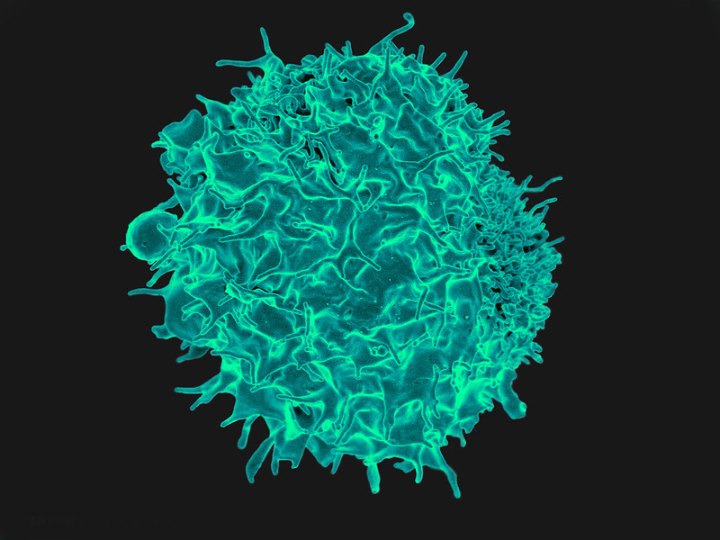
T cell
Scanning Electron Micrograph image of a human T cell. Credit: NIH/NIAID

While T cells are the body’s warriors against infection, without rest and maintenance T cells can die, leaving their hosts more susceptible to pathogens, researchers reported in the journal Science.

“We may have to change how we teach T cell biology,” said Professor Lieping Chen, who is the senior author of the study.

T cells remain in a quiescent state until pathogens are detected, but the molecular mechanisms of this state were previously unknown.

In the new study, researchers showed that a protein known as CD8a – which is found in a subset of T cells called CD8 cells – is crucial to keeping the cells in this dormant state. When scientists deleted this protein in mice, the protective CD8 cells were unable to enter a quiescent state and died, leaving the host vulnerable to infections.

Further, they identified another protein, PILRa, that provides a biochemical signal to CD8a. By disrupting this protein pair, both “memory” CD8 cells – previously been exposed to pathogens – and naïve cells died because they lacked the ability to stay in a quiescent state.

The researchers hope that understanding why this resting state is crucial to maintenance and survival of T cells can lead to improved immune system function.

Chen noted that as people age they tend to lose both naïve and memory T cells, making older individuals more susceptible to infections. It is possible that the inability of T cells to remain in a quiescent state could lead to people becoming more susceptible to infections and cancer, the authors suggest.

Source: Yale University

Hypoxia can Trigger Immune System Reaction

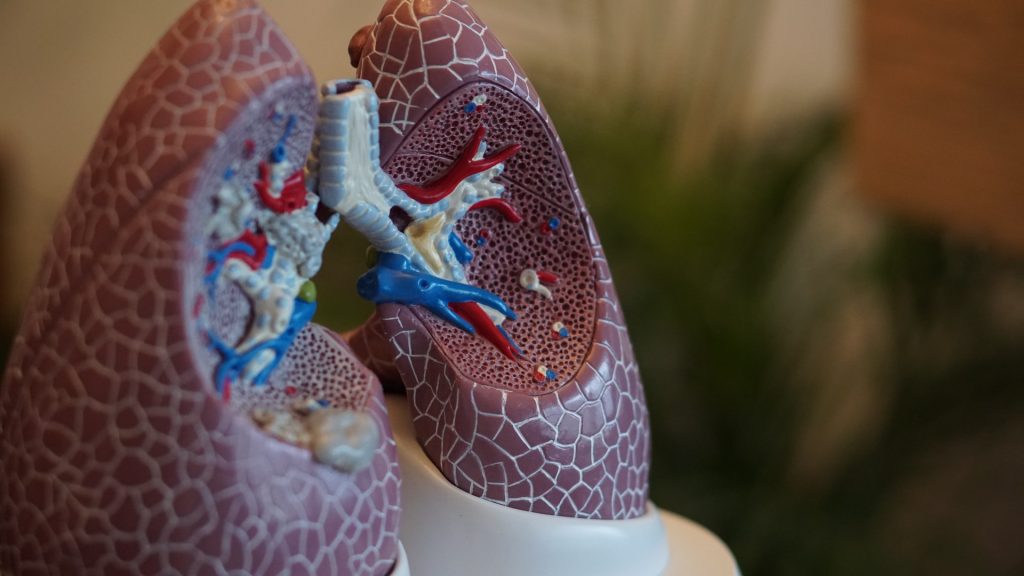
Anatomical model of lungs
Photo by Robina Weermeijer on Unsplash

New research from scientists at La Jolla Institute for Immunology (LJI), shows that hypoxia can activate the same group of immune cells that cause inflammation during asthma attacks. During hypoxia, these cells flood the airways with lung-damaging molecules.

Hypoxia is a known trigger for developing and worsening lung conditions such as severe asthma, chronic obstructive pulmonary disease (COPD), and fibrosis. To treat and prevent these diseases, researchers need to understand why a lack of oxygen would affect the immune system.

“We show how lack of oxygen can be part of a feedback loop that can contribute to even worse inflammation,” says LJI Professor and Chief Scientific Officer Mitchell Kronenberg, PhD, a member of the LJI Center for Autoimmunity and Inflammation. “This work gives us insight into the causes of fibrosis of the lung and severe asthma.”

Prof Kronenberg and colleagues worked with a genetically altered mouse model to mimic the signals of hypoxia in the airway’s epithelial cells, which line the paths to the lungs. They discovered that combining the hypoxia signals with inflammatory signals stimulated the “innate,” or rapidly responding immunity, and an immune cell type called an ILC2.

An ILC2’s job is to make signaling molecules – cytokines – that quickly alert other immune cells to react to a pathogen. Unfortunately, ILC2s sometimes over-react and respond to harmless environmental allergens. In these cases, ILC2s churn out cytokines that drive mucus production and inflammation in the lungs. All this swelling and mucus leads to hypoxia.

As they report in Journal of Experimental Medicine, ILC2s respond to hypoxia as well, adding to the lung damage already caused during an asthma attack.

“That hypoxia may then contribute further to inflammation,” said Prof Kronenberg.

The next step was to figure out exactly how epithelial cells activate ILC2 during hypoxia. LJI Postdoctoral Fellow Jihye Han, PhD, led the work to uncover an unexpected culprit: adrenomedullin (ADM). ADM is known for its role in helping blood vessels dilate, but until now it had no known role in immune function.

Prof Kronenberg was surprised to see ADM involved — but not shocked. “We’re finding that many molecules with no previously known role in the immune system can also be important for immune function,” said Prof Kronenberg. “We need to understand that more generally.”

The researchers showed that human lung epithelial cells exposed to hypoxia also produced ADM. This means ADM or its receptor could be targets for treating inflammatory and allergic lung diseases.

The challenge is to find a balance between dampening the harmful immune response without leaving the body vulnerable to infections. Prof Kronenberg points out that the epithelial cell-ADM-ILC2 connection protected mice from hookworm infections, which damage the lungs and gut.

“ADM is a new target for lung diseases and has been implicated in bacterial pneumonia as well,” said Prof Kronenberg. “But blocking it would have to be done carefully.”

Source: La Jolla Institute for Immunology

B Cells Play a Surprising Role in Bone Marrow Output

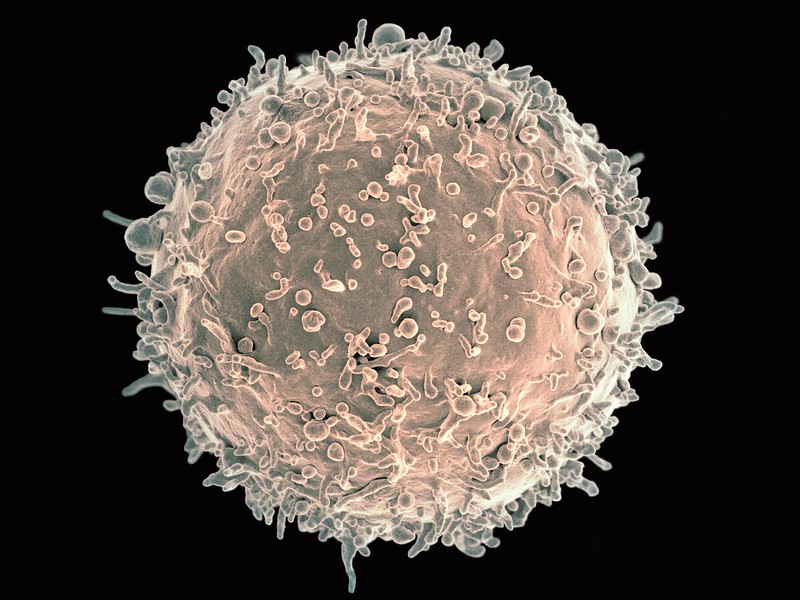
Scanning electron micrograph image of a human B cell. Credit: NIH/NIAID

New research in Nature Immunology has found that B cells play a surprising role in increasing or decreasing the bone marrow’s output of white blood cells. The findings may lead to new treatments for conditions that arise when white blood cell production goes out of balance.

Professor Matthias Nahrendorf, senior author of the study, explained that the nervous system plays a role in controlling blood cell production through neurotransmitters. “This is for instance important in people exposed to stress, where stress hormones — part of the ‘fight-or-flight’ response controlled by the sympathetic nervous system — may increase bone marrow activity and cardiovascular inflammation in response to the neurotransmitter noradrenaline,” he said. The parasympathetic nerves on the other hand, slow down responses and bring about a state of calm to the body, mainly through the neurotransmitter acetylcholine.

Because acetylcholine can have a protective effect against inflammation and heart disease, the researchers studied this neurotransmitter in the bone marrow. “When we looked into how acetylcholine acts on the production of blood cells, we found that it does the expected — it reduces white blood cells, as opposed to noradrenaline, which increases them,” said Prof Nahrendorf. “What was unexpected though was the source of the neurotransmitter acetylcholine.”

In the bone marrow, the typical nerve fibres that are known to release acetylcholine were not found. Instead, it was the antibody-producing B cells supplied the acetylcholine in the bone marrow. “Thus, B cells counter inflammation — even in the heart and the arteries — via dampening white blood cell production in the bone marrow. Surprisingly, they use a neurotransmitter to do so,” said Prof Nahrendorf.

Tapping into this process may help investigators develop strategies to block inflammation in cardiovascular conditions such as atherosclerosis. “Ultimately this may lead to new therapeutics that combat myocardial infarction, stroke, and heart failure,” said Prof Nahrendorf.

Source: Massachusetts General Hospital

Researchers Identify a Key Regulatory Mechanism in Inflammation

Firefighter
Source: Pixabay CC0

A newly published study has identified a key regulatory mechanism in inflammation that may lead to new targets for resolving that inflammation –and the inflammation of patients with sepsis, cancer and COVID.

In the journal PNAS, scientists reported their discovery of a regulatory pathway for immune response after infection or injury, such as burns. Dysregulation of this pathway could differentiate those who are at risk of fatal sepsis or help identify targets to resolve this unregulated inflammation.

“We are very excited about the findings in this paper and the far-reaching impacts it could have on understanding a key regulatory step in the immune response,” said co-lead author Cindy McReynolds, who holds a doctorate in pharmacology and toxicology.

In a rodent model, the research team found that the metabolites of linoleic acid formed by the enzyme, soluble epoxide hydrolase (sEH), drive damaging inflammation after injury. These metabolites, known as lipid mediators, regulate inflammation, blood pressure and pain. Drugs that inhibit the sEH enzyme and reduce inflammation could lead to better outcomes.

“Our previous work identified that these same lipid mediators were up-regulated in severe COVID infections, and we are now finding that these compounds play a role in modulating the immune response so that the body is unable to fight infection or respond properly to trauma without leading to a potentially fatal overreaction,” said Dr McReynolds.

“This dysregulation has fatal consequences in serious diseases such as COVID, cancer, sepsis, burn, where fatality rates can be as high as 40 percent in severe cases,” she said. “An understanding of these pathways can help identify patients at risk of developing serious disease or identify new therapeutic targets for treatment.”

“The immunological disbalance we see in many cases of severe burn injury, trauma and sepsis pose a huge clinical challenge as we lack the understanding of how to diagnose and treat it,” explained co-lead author Dr Christian Bergmann. “With this work, we reveal an important mechanism how immune cells are functionally disabled by sEH-derived metabolites of linoleic acid.”

“The natural compounds we are studying in this paper are metabolites of linoleic acid (LA), an essential fatty acid the body needs in very small amounts to survive and is only available through the diet,” Dr McReynolds elaborated. “At lower concentrations, these metabolites are necessary for regulating thermogenesis and heart health but promote inflammation at higher concentrations. LA is more stable and much cheaper than longer chain polyunsaturated fatty acids, so heavily processed foods have higher LA content to increase shelf-life. Additionally, agricultural practices, such as feeding animals corn-based diets, have increased LA in meats and dairy products.”

“As a result, we are consuming the highest amount of linoleic acid and have the highest recorded concentration of LA in our fatty tissue in human history,” McReynolds said. “As our bodies respond to stress or disease, we metabolise LA into the regulatory metabolites that were monitored in this paper. At higher concentrations, the immune system was unable to properly respond to infection, thereby promoting a sustained immune response. These observations are important in inflammatory-driven diseases, such as sepsis and COVID, but could also be important in understanding many of the increased chronic diseases we are seeing in our population.”

Source: UC Davis

Meat, Gut Bacteria and Multiple Sclerosis

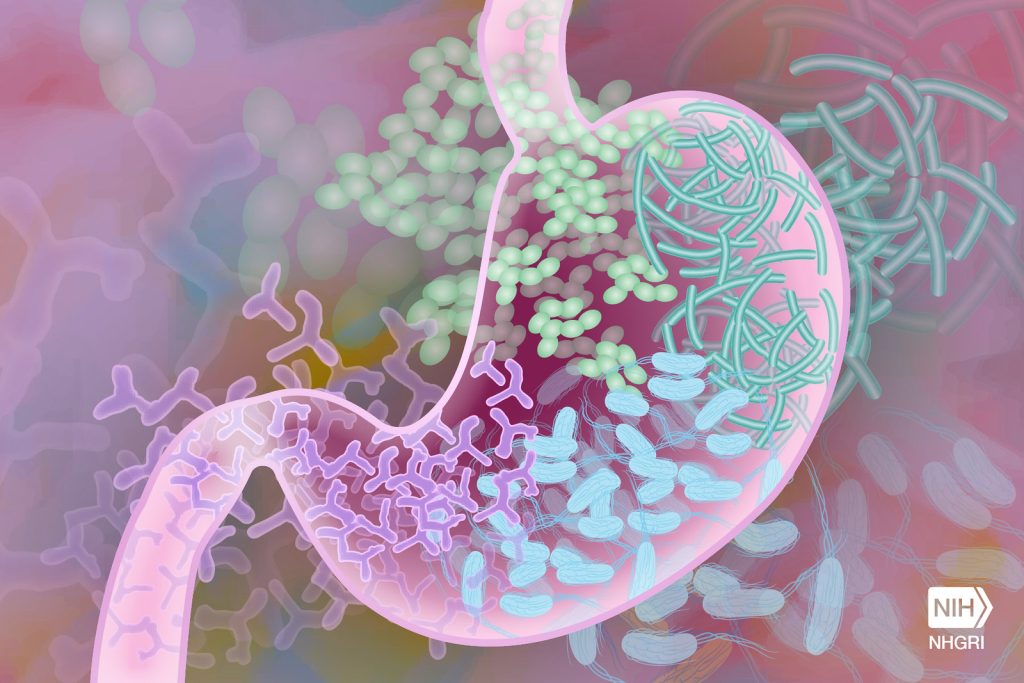
Gut microbiome. Credit: Darryl Leja, NIH

There appears to be a link between eating meat, gut bacteria and multiple sclerosis, according to new research published in EBioMedicine. The study teased out subtle connections that could lead to a better understanding of the causes of the disease.

The autoimmune disease multiple sclerosis (MS) is more prevalent in specific regions, particularly the northern mid-latitudes, suggesting that geography is somehow linked to the disease, perhaps involving diet. However, the exact relationships between diet, immune response, and MS has been a mystery. What exactly triggers the body to attack the myelin sheaths in MS in the first place is unknown.

Growing evidence suggests that bacteria might play a role. Gut bacteria affect the immune system, and diet affects the gut. Researchers studied the gut microbiome, immune systems, diet, and blood metabolites in 25 MS patients and 24 healthy controls to look for any subtle but important correlations.

“We found a number of gut bacteria associated with MS and severity of disability of MS patients,” said Dr Yanjiao Zhou. “We also found increased autoimmune markers and signature metabolites in MS. But what is really interesting is how these systems connect with each other, and how diet is involved in these connections. Using multi-OMICS approaches, we try to close the loop and show the associations between multiple systems.”

Meat eating was the strongest link in their analysis, where higher meat consumption saw a decrease in the population of Bacteroides thetaiotaomicron, a bacteria associated with digesting carbohydrates from vegetables.

Higher meat consumption, seen in the MS patients, was also linked to an increase in T-helper 17 cells in the immune system, and an increase in S-adenosyl-L-methionine (SAM) in their blood.

Meat eating was not a predictor of MS. But the evidence suggested that, in MS, something causes gut bacteria to disassociate with the immune system, leading to heightened T-helper 17 cells and autoimmune attacks on the nervous system. And it tends to be associated with eating meat.

Future research aims to recruit more volunteers, including those with more severe MS. Eventually they hope to understand more of the cause-and-effect between diet, bacterial ecosystems in the gut, and immune response, and potentially help prevent or mitigate MS symptoms in people suffering from the disease.

Source: University of Connecticut

Existing COVID Vaccines Trigger Lasting T Cell Response

Image from Pixabay

Scientists have found that four COVID vaccines (Pfizer-BioNTech, Moderna, J&J/Janssen, and Novavax) prompt the body to make effective, long-lasting T cells against SARS-CoV-2. These T cells can recognise SARS-CoV-2 Variants of Concern, including Delta and Omicron.

The new study, published in Cell, showed that the vast majority of T cell responses are also still effective against Omicron, reducing the odds of illness for up to six months, regardless of vaccine.

These data come from adults who were fully vaccinated, but not yet boosted. The researchers are now investigating T cell responses in boosted individuals and people who have experienced “breakthrough” COVID cases.

The study also shows that fully vaccinated people have fewer memory B cells and neutralising antibodies against the Omicron variant. This finding is in line with initial reports of waning immunity from laboratories around the world.

Without enough neutralising antibodies, Omicron is more likely to cause a breakthrough infection, and fewer memory B cells means a slower production of more neutralising antibodies.

Co-first author Camila Coelho, PhD, said: “Our study revealed that the 15 mutations present in Omicron RBD can considerably reduce the binding capacity of memory B cells.”

Neutralising antibodies and memory B cells are only two arms of the body’s adaptive immune response. , T cells do not prevent infection, rather they patrol the body and destroy cells that are already infected, which prevents a virus from multiplying and causing severe disease.

The team believes the “second line of defence” from T cells helps explain Omicron’s reduced severity in vaccinated people. The variant also appears to infect different tissues.

To know whether the vaccine-induced T cells they detected in their study were actually effective against variants such as Delta and Omicron, the scientists took a close look at how the T cells responded to different viral “epitopes.”

Every virus is made up of proteins that form a certain shape or architecture. A viral epitope is a specific landmark on this architecture that T cells have been trained to recognise. Current COVID vaccines were designed to teach the immune system to recognise specific epitopes on the initial variant of SARS-CoV-2, specifically targeting the Spike protein which the virus uses to access human cells. As the virus has mutated, its architecture has changed, and the concern is that immune cells will no longer recognise their targets.

The new study shows that while the architecture of Omicron is different enough to evade some neutralising antibodies and memory B cells, memory T cells still do a good job of recognising their targets, even on the highly mutated Omicron variant. Overall, at least 83 percent of the CD4+ (helper) T cell responses and 85 percent of the CD8+ T cell responses stayed the same, no matter the vaccine or the variant.

The memory B cells that do bind Omicron are likely to also contribute to protection against severe disease, forming multiple lines of defence. 

Researchers are now focusing on measuring T cells, B cells and antibody responses after COVID booster shots, and also characterising immune responses after a breakthrough infection.

Source: La Jolla Institute

How Immune Cells Fight Infection Using Body Fat

T lymphocyte. Credit: NIH/NIAID

A new study from the University of East Anglia and Quadram Institute sheds light on how our immune cells make use of body fat to fight infection. The research, published today in Nature Communications, could lead to new approaches to treating people with bacterial infections.

The work could one day help treat infections in vulnerable and older people, the researchers said. The team studied Salmonella bacteria and tracked fatty acid movement and consumption in live stem cells. They then examined the immune response to Salmonella bacterial infection, by analysing liver damage.

They uncovered how blood stem cells respond to infection, by acquiring high energy fatty acids from the body’s fat stores. In the bone marrow where blood stem cells are resident, infection signals drive adipocytes to release their fat stores as fatty acids into the blood.

And they identified that these high energy fatty acids are then taken up by blood stem cells, effectively feeding the stem cells and enabling them to make millions of Salmonella-fighting white blood cells. The researchers also identified the mechanism by which the fatty acids are transferred and discusses the potential impact this new knowledge could have on future treatment of infection.

Dr Stuart Rushworth, from UEA’s Norwich Medical School, said: “Our results provide insight into how the blood and immune system is able to respond to infection.

“Fighting infection takes a lot of energy and fat stores are huge energy deposits, which provide the fuel for the blood stem cells to power up the immune response.

“Working out the mechanism through which this ‘fuel boost’ works gives us new ideas on how to strengthen the body’s fight against infection in the future.”

Dr Naiara Beraza, from the Quadram institute, said: “Our results allow us to understand how our immune system uses fat to fuel the response to infection. Defining these mechanisms will enable us to develop new therapeutics to treat infections in the liver.”

Source: University of East Anglia

Differences in Influenza Responses According to Genetic Ancestries

Photo by Andrea Piacquadio on Unsplash

Researchers have uncovered differences in immune pathway activation to influenza infection between individuals of European and African genetic ancestry, according to a study published in Science. Many of the genes that were associated with these immune response differences to influenza are also enriched among genes associated with COVID disease severity. 

“The lab has been interested in understanding how individuals from diverse populations respond differently to infectious diseases,” said first author Haley Randolph, a graduate student at the University of Chicago. “In this study, we wanted to look at the differences in how various cell types respond to viral infection.”

The researchers examined gene expression patterns in peripheral mononuclear blood cells, a diverse set of specialised immune cells that play important roles in the body’s response to infection. These cells were gathered from men of European and African ancestry and then exposed the cells to flu in a laboratory setting. This let the team examine the gene signatures of a variety of immune cell types, and observe how the flu virus affected each cell type’s gene expression.

The results showed that individuals of European ancestry showed an increase in type I interferon pathway activity during early influenza infection.

“Interferons are proteins that are critical for fighting viral infections,” said senior author Luis Barreiro, PhD, Associate Professor of Medicine at UChicago. “In COVID-19, for example, the type I interferon response has been associated with differences in the severity of the disease.”

This increased pathway activation hindered the replication of the virus more and limited viral replication later on. “Inducing a strong type I interferon pathway response early upon infection stops the virus from replicating and may therefore have a direct impact on the body’s ability to control the virus,” said Barreiro. “Unexpectedly, this central pathway to our defense against viruses appears to be amongst the most divergent between individuals from African and European ancestry.”

The researchers saw a variety of differences in gene expression across different cell types, suggesting a constellation of cells that work together to fight disease.

Such a difference in immune pathway activation could explain influenza outcome disparities between different racial groups; Non-Hispanic Black Americans are more likely to be hospitalised due to the flu than any other racial group.

However, these results are not evidence for genetic differences in disease susceptibility, the researchers point out. Rather, possible differences in environmental and lifestyle between racial groups could be influencing gene expression, and affecting the immune response.

“There’s a strong relationship between the interferon response and the proportion of the genome that is of African ancestry, which might make you think it’s genetic, but it’s not that simple,” said Barreiro. “Genetic ancestry also correlates with environmental differences. A lot of what we’re capturing could be the result of other disparities in our society, such as systemic racism and healthcare inequities. Although some of the differences we show in the paper can be linked to specific genetic variation, showing that genetics do play some role, such genetic differences are not enough to fully explain the differences in the interferon response.”

These differences in viral susceptibility may not be confined to just influenza. Comparing a list of genes associated with differences in COVID severity, the researchers found that many of the same genes showed significant differences in their expression after flu infection between individuals of African and European ancestry.

“We didn’t study COVID patient samples as part of this study, but the overlap between these gene sets suggests that there may be some underlying biological differences, influenced by genetic ancestry and environmental effects, that might explain the disparities we see in COVID outcomes,” said Barreiro.

As they explore this further, the researchers hope to figure out which factors contribute to the differences in the interferon response, and immune responses more broadly, to better predict individual disease risk.

Source: EurekAlert!