Category: Diseases, Syndromes and Conditions

New Antimalarial Compound Traps Parasites in Cells

Photo by Егор Камелев on Unsplash

To combat the growing resistance of malaria to current treatments, researchers at the Francis Crick Institute and the Latvian Institute of Organic Synthesis have designed a new antimalarial compound which interrupts the malaria parasite life cycle by trapping them in their host cells.

While drugs and mosquito control have reduced levels of malaria over recent decades, with malaria being effectively wiped out in North America by the 1950s, the parasite still kills over 400 000 people every year, 90% of whom live in sub-Saharan Africa. It has now developed resistance to many existing antimalarial drugs, meaning new treatments that work in different ways are urgently needed.

If we can effectively trap malaria in the cell by blocking the parasite’s exit route, we could stop the disease in its tracks and halt its devastating cycle of invading cells.
Mike Blackman

The researchers developed an array of compounds designed to prevent the parasites bursting out of blood cells, a vital replication step. One compound in particular was found to be very effective in human cell tests.

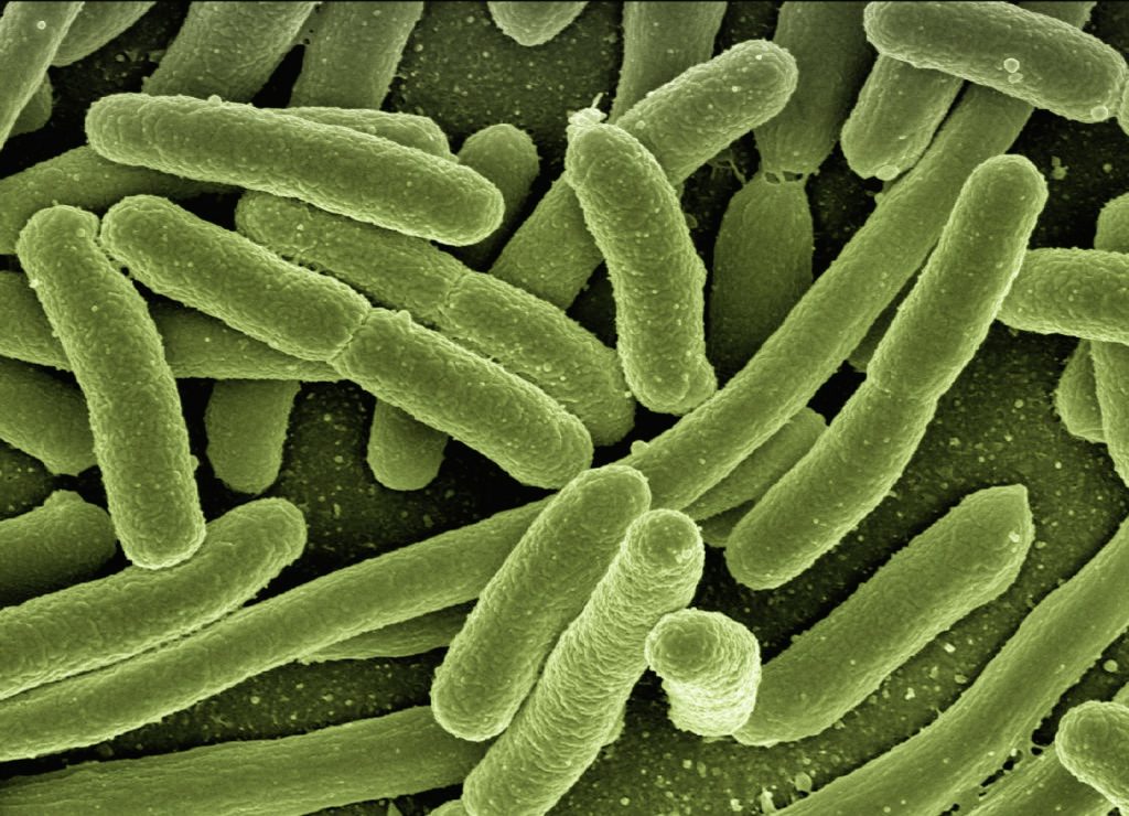
“Malaria parasites invade red blood cells where they replicate many times, before bursting out into the bloodstream to repeat the process. It’s this cycle and build-up of infected red blood cells which causes the symptoms and sometimes fatal effects of the disease,” says Mike Blackman, lead author and group leader of the Malaria Biochemistry Laboratory at the Crick.  

“If we can effectively trap malaria in the cell by blocking the parasite’s exit route, we could stop the disease in its tracks and halt its devastating cycle of invading cells.”

Blocking the parasite’s emergence

The compound works by blocking an enzyme called SUB1, needed for them to burst out of cells. Current antimalarials kill the parasite within the cell, so the researchers hope this alternative drug action will overcome the resistance the parasite has acquired.

The compound can penetrate both the cell wall and the compartment within where the parasites reside.

The researchers are further refining the compound making it smaller and more potent. Further tests are needed before it can be trialled in humans.

Study author Chrislaine Withers-Martinez and researcher in the Malaria Biochemistry Laboratory, said: “Many existing antimalarial drugs are plant derived and while they’re incredibly effective, we don’t know the precise mechanisms behind how they work. Our decades of research have helped us identify and understand pathways crucial to the malaria life cycle allowing us to rationally design new drug compounds based on the structure and mechanism of critical enzymes like SUB1.

“This approach, which has already been highly successful at finding new treatments for diseases including HIV and Hepatitis C, could be key to sustained and effective malaria control for many years to come.” 

Source: Francis Crick Institute

Sepsis Leaves a Dangerous Imprint in Immune System

E. Coli bacteria. Image by Gerd Altmann from Pixabay

New research suggests that sepsis can cause alterations in the functioning of defence cells that persist even after the patient is discharged from hospital.

This cellular reprogramming creates a disorder the authors term ‘post-sepsis syndrome’, symptoms of which include frequent reinfections, cardiovascular alterations, cognitive disabilities, declining physical functions, and poor quality of life.This explains why so many patients who survive sepsis die sooner after hospital discharge than patients with other diseases or suffer from post-sepsis syndrome, immunosuppression and chronic inflammation.

The article reviews studies done to investigate cases of septic patients who died up to five years after hospital discharge.

Sepsis is one of the main causes of death in intensive care units, sepsis is a life-threatening systemic organ dysfunction triggered by the body’s dysregulated response to a pathogen, usually a bacterium or fungus. While fighting the pathogen, the defence system injures the body’s own tissues and organs.

If not caught and treated in time, the condition can lead to septic shock and multiple organ failure. Patients with severe COVID and other infectious diseases have an increased risk of developing and dying from sepsis.

Worldwide, new sepsis cases are estimated to reach some 49 million per year. Hospital mortality from septic shock exceeds 40% globally, up to 55% in Brazil, according to the Sepsis Prevalence Assessment Database (SPREAD) study, conducted with support from FAPESP.

“The massive infection and the accompanying intense immune response with a cytokine outpouring during sepsis may promote irreversible cell metabolic reprogramming. Cell reprogramming is unlikely to occur in leukocytes or bone marrow only. This might happen in several tissues and cells that prompt systemic organ dysfunctions […] Bacteria can transfer genetic material to host cell DNA as eukaryotic cells develop tools to protect themselves against the microorganism invasion. The latter may induce cell biology and metabolic reprogramming that remains even after the infection’s elimination,” the investigators wrote in the article.

According to Raquel Bragante Gritte, joint first author with Talita Souza-Siqueira, one of the hypotheses was that metabolic reprogramming begins in the bone marrow, whose cells acquire a pro-inflammatory profile.

“Our analysis of blood samples from patients even three years after ICU discharge showed that monocytes [a type of defense cell] were activated and ready for battle. They should have been neutral. Monocytes are normally activated only when they are ‘recruited’ to the tissue,” Gritte told Agência FAPESP. Both Gritte and Souza-Siqueira are researchers at Cruzeiro do Sul University (UNICSUL) in the state of São Paulo, Brazil.

The researchers conducted a follow-up study of 62 patients for three years after discharge from the ICU at USP’s University Hospital, analysing alterations in monocytes, neutrophils and lymphocytes, as well as microRNAs, in order to identify prognostic markers and factors associated with post-sepsis syndrome.

“Our hypothesis is that white blood cells conserve a memory of sepsis, which helps explain why patients remain sick after they leave hospital,” said co-author Rui Curi, Professor at UNICSUL, and Director of Butantan Institute.

The investigators suggest that sepsis may create a specific macrophage phenotype that stays active even after hospital discharge. “Cell metabolism reprogramming is also involved in the functions and even generation of the different lymphocyte subsets. Several stimuli and conditions change lymphocyte metabolism, including microenvironment nutrient availability,” they wrote.

The next stage of research will be bone marrow studies to understand how cells are reprogrammed by sepsis. “We think the key to this alteration is in bone marrow,” she said. “However, another possibility is that activation occurs in the blood. We’ll need to do more in-depth research to find answers.”

Source:
News-Medical.Net

How Legionnaire’s Disease Digs In

A bunker from World War II, emulating how Legionella makes a protective shelter. Image by herb1979 from Pixabay

Scientists have discovered how the bacteria that causes Legionnaires’ disease digs in and makes a tiny shelter inside the cells of humans and other hosts. 

The findings, published in Science, could offer insights into how other bacteria are able to survive inside cells, knowledge that could lead to new treatments for a wide variety of infections.

Discovered in 1976, Legionella, an aerobic gram-negative bacillus is responsible for Legionnaires’ disease, a condition of severe pneumonia. Spread through aerosolised water particles, it is a common cause of hospital and community-acquired pneumonia.

“Many infectious bacteria, from listeria to chlamydia to salmonella, use systems that allow them to dwell within their host’s cells,” explained study leader Vincent Tagliabracci, Ph.D., assistant professor of molecular biology at UTSW and member of the Harold C Simmons Comprehensive Cancer Center. “Better understanding the tools they use to make this happen is teaching us some interesting biochemistry and could eventually lead to new targets for therapy.”

Dr Tagliabracci’s lab studies atypical kinases, unusual forms of enzymes that put phosphates onto proteins or lipids, changing their function. Legionella is a particularly rich source of these noncanonical kinases. According to the Centers for Disease Control and Prevention, nearly 10 000 cases of Legionnaires’ disease were reported in the US in 2018, though the true incidence is believed to be higher.      

After identifying a new Legionella atypical kinase named MavQ, Dr Tagliabracci and colleagues used a live-cell imaging technique in concert with a relatively new molecular tagging method to see where MavQ is found in infected human cells. However, rather than residing in a specific location, the researchers were surprised to see that the protein moved back and forth between the endoplasmic reticulum – a network of membranes important for protein and lipid synthesis – and bubble- or tube-shaped structures within the cell.

Further research suggests that MavQ, along with a partner molecule called SidP, remodels the endoplasmic reticulum so that Legionella can strip off sections of the membrane to help create and sustain the vacuole, a structure that the parasitic bacteria uses to shelter inside cells, protecting it from immune attack.

Dr Tagliabracci said that he suspects other bacterial pathogens may use similar mechanisms to co-opt existing host cell structures to create their own protective shelters. 

 Source: University of Texas

Journal information: Ting-Sung Hsieh, et al. Dynamic remodeling of host membranes by self-organizing bacterial effectors. Science, 2021; eaay8118 DOI: 10.1126/science.aay8118

Experimental Inhibitor Drug Shows Promise For The Deadly Marburg Virus

Photo by CDC on Unsplash

The lethal Marburg virus, a relative of the Ebola virus, causes a serious haemorrhagic fever with an extremely high fatality rate and has had no known treatment — until now. 

Marburg virus infects human and primates, the disease currently has no approved vaccine or antivirals for prevention or treatment. In two larger recent outbreaks in the DRC in 1998–2000, and in Angola in 2004–2005, Marburg had extremely high fatality rates of 83% and 90%.

A team of researchers is working to change that. In a new paper in the journal Antimicrobial Agents and Chemotherapy, investigators from Penn’s School of Veterinary Medicine, working together with scientists from the Fox Chase Chemical Diversity Center and the Texas Biomedical Research Institute, report encouraging results from tests of an experimental antiviral targeting Marburg virus.

The new compound prevents viruses from leaving infected cells, thus halting the spread of infection. In a first, this new class of inhibitors was shown to be effective against infection in an animal model.

Additionally, possible similarities in virus-host interactions between Marburg and SARS-CoV-2, prompted the team to conducted experiments on the coronavirus. Unpublished preliminary results appeared encouraging.

“It really is exciting. These viruses are quite different but may be interacting with the same host proteins to control efficient egress and spread, so our inhibitors may be able to block them both,” said co-corresponding author Ronald Harty, Professor, Penn’s School of Veterinary Medicine.

Prof Harty’s team have been developing an antiviral that instead of targeting the virus known as “host-oriented.” By blocking the proteins in host cells that viruses hijack during late stages of infection, preventing virus-host interactions.

This method helps prevent a virus evolving resistance, but it also makes it more likely that a drug could be used against multiple viruses, as many make use of the same machinery in the host cell to reproduce and spread.

The Marburg and Ebola viruses use protein known as VP40 to interact with a host protein called Nedd4 to allow the completed viruses to ‘bud off’ of the host cell, which is a key part of viral replication.

Previously, they had tested a variety of small molecule inhibitors of this process using laboratory tests that relied on non-infectious and more-benign viral models. Those assays led them to a promising candidate, FC-10696, for further study.

The researchers firstly tested the chosen inhibitor for safety and its useful duration within the body. Next, since the real Marburg virus is too dangerous to study safely in anything but a Biosafety Level 4 (BSL-4) laboratory, they used an assay to look at what are known as virus-like particles, or VLPs, which are non-infectious but can bud off of a host cell.

Using the Biosafety Level 2 laboratory at Penn, “it’s a very quick way we can test these inhibitors,” said Prof Harty.

The researchers saw a dose-dependent response to FC-10696 on VLP budding in cells tested the compound using the real Marburg virus. These studies were done in a BSL-4 lab at Texas Biomedical Research Institute and found the compound inhibited the budding and spread of live Marburg virus in two human cell types, including in macrophages, an immune cell type commonly infected by the virus.

As a final step, they tested the compound in mice infected with Marburg virus. That received the treatment took longer to display disease symptoms and had a reduced viral load.

“These are the first promising in vivo data for our compounds,” said Prof Harty. “Whereas the control group all became sick very quickly and died, with the treated animals there was one survivor and others showed delayed onset of clinical symptoms. It’s showing that our inhibitors are having an effect.”

Source: News-Medical.Net

Journal information: Han, Z., et al. (2021) Compound FC-10696 Inhibits Egress of Marburg Virus. Antimicrobial Agents and Chemotherapy. doi.org/10.1128/AAC.00086-21.

Night Shifts Increase Risk of Infection

Shift working and irregular working hours can affect our health and disrupt immune response, especially in men, according to new research from the University of Waterloo.

These health-related issues occur because the body’s circadian rhythm can be disrupted by inconsistent changes in the sleep-wake schedule and feeding patterns often caused by shift work. To study this, researchers at Waterloo developed a mathematical model to investigate how a disruption in the circadian clock affects the immune system in fighting off illness.

“Because our immune system is affected by the circadian clock, our ability to mount an immune response changes during the day,” said Anita Layton, professor of Applied Mathematics, Computer Science, Pharmacy and Biology at Waterloo. “How likely are you to fight off an infection that occurs in the morning than midday? The answer depends on whether you are a man or a woman, and whether you are among [the] quarter of the modern-day labor force that has an irregular work schedule.”

The researchers created new computational models, separately for men and women, which simulate the interplay between the circadian clock and the immune system. The model is composed of the core clock genes, their related proteins, and the regulatory mechanism of pro- and anti-inflammatory mediators. By adjusting the clock, the models can simulate male and female shift-workers.

The researchers’ simulation results demonstrate that the immune response varies with the time of infection. The model simulation indicates that the time just before people go to sleep is the “worst” time to get an infection. At this point during the day, the human body is least prepared to produce the pro- and anti-inflammatory mediators needed during an infection. An individual’s sex also impacts the effect significantly.

“Shift work likely affects men and women differently,” said Stéphanie Abo, a PhD candidate in Waterloo’s Department of Applied Mathematics. “Compared to females, the immune system in males is more prone to overactivation, which can increase their chances of sepsis following an ill-timed infection.”

Source: Medical Xpress

More information: Stéphanie M. C. Abo et al. Modeling the circadian regulation of the immune system: Sexually dimorphic effects of shift work, PLOS Computational Biology (2021). DOI: 10.1371/journal.pcbi.1008514

Predicting the Next Viral Pandemic

A group of experts has argued that trying to survey all of the viruses in the animal kingdom is a futile effort, and that we should rather focus on those most likely to cross over at the interface of humans and animals.

The observation that most of the viruses that cause human disease come from other animals has led some researchers to attempt “zoonotic risk prediction” to second-guess the next virus to cause a global pandemic. 
Zoonotic viruses, those that cross over from animal species into humans, have caused epidemics and pandemics in humans for centuries. This is exactly what is occurring today with the COVID pandemic: SARS-CoV-2—the coronavirus that causes the disease—emerged from an animal species, albeit which one is not yet known.

An essay published April 20th in the open access journal PLOS Biology, led by Dr Michelle Wille at the University of Sydney, Australia with co-authors Jemma Geoghegan and Edward Holmes outlines the great challenges in zoonotic risk prediction.

The authors argue that these zoonotic risk predictions are of limited value, and will not be able to predict which virus will cause the next great pandemic. Instead, they reason, the human-animal interface should be the target for intensive viral surveillance.

A key question is whether it is possible to predict which animal or which virus group will most likely cause the next pandemic. This has led to “zoonotic risk prediction,” in which researchers attempt to determine which virus families and host groups are most likely to carry potential zoonotic and/or pandemic viruses.

Dr Wille and her colleagues identified several key problems with zoonotic risk prediction attempts.

Firstly, they’re based on very small data sets. Despite decades of work, less than 0.001% of all viruses have likely been identified, even from the mammalian species from which the next pandemic virus is expected to emerge.

Second, these data are also already highly biased in favour of those the most infectious viruses  of humans or agricultural animals, or are already known to be zoonotic. Most animals have in fact not been surveyed for viruses, and that viruses evolve so quickly that any such surveys will soon be out of date and therefore be of limited value.

The authors instead argue that a new approach is needed, not trying to futilely survey all the viruses in the wild but instead undertaking extensive sampling at the animal-human interface. This would enable the detection of novel viruses as soon as they appear in humans. This kind of enhanced surveillance could help us forestall the next pandemic.

Source: Phys.Org

Journal information: Wille M, Geoghegan JL, Holmes EC (2021) How accurately can we assess zoonotic risk? PLoS Biol 19(4): e3001135. doi.org/10.1371/journal.pbio.3001135

Encapsulated Clusters of Noroviruses are Resistant to Disinfection

Encapsulated clusters of noroviruses which can cause stomach flu have been found to be resistant to detergent and ultraviolet disinfection.

Noroviruses are the leading cause of gastroenteritis around the world, with more than 21 million cases annually in the United States. The findings of this study show that there is a need to revise current disinfection, sanitation and hygiene practices which serve to protect against infection with noroviruses.

In 2018, the research team had found that noroviruses can be transmitted to humans in the form of membrane-enclosed packets that contain clusters of viruses. Previously, it was thought that viruses spread via exposure to individual virus particles, but the 2018 study, , showed how membrane-enclosed clusters arrive at a human cell and release a large number of viruses.

For the new study, Drs Danmeng Shuai, Nihal Altan-Bonnet and the study’s first author Mengyang Zhang, a doctoral student co-advised through a GW/NIH Graduate Partnerships Program, examined how such protected virus clusters behave in the environment. They found that the virus clusters could survive disinfection attempts with detergent solutions or even UV light. Water treatment plants use UV light to kill noroviruses and other pathogens, and is being widely used in the COVID pandemic.

“These membrane-cloaked viruses are tricky,” explained study co-author Danmeng Shuai, PhD, associate professor of civil and environmental engineering, George Washington University. “Past research shows they can evade the body’s immune system and that they are highly infectious. Our study shows these membrane enclosed viruses are also able to dodge efforts to kill them with standard disinfectants.”

“We have to consider these viral clusters cloaked in vesicle membranes as unique infectious agents in the public health arena,” added Nihal Altan-Bonnet, PhD, a senior investigator and the head of the Laboratory of Host-Pathogen Dynamics at the National Heart, Lung, and Blood Institute. “When it comes to virulence -; and now with this study, disinfection and sanitation -; the sum is much more than its parts. And these clusters are endowed with properties that are absent from other types of viral particles.”

Future studies are needed to determine whether certain kinds of cleaning solutions or more UV light exposure would degrade the protective membrane and/or kill the viruses inside. Such research would hopefully come up with improved disinfection methods that could be used for cleaning surfaces in the home, in restaurants and in places where norovirus can spread and cause outbreaks, like cruise ships.

“Our study’s findings represent a step towards recommendations for pathogen control in the environment, and public health protection,” Dr Altan-Bonnet said.

Source: News-Medical.Net

Journal information: Zhang, M., et al. (2021) Emerging Pathogenic Unit of Vesicle-Cloaked Murine Norovirus Clusters is Resistant to Environmental Stresses and UV254 Disinfection. Environmental Science and Technology. doi.org/10.1021/acs.est.1c01763.

Discovery Offers New Treatment for Sickle Cell Anaemia

In a promising step towards a new treatment for sickle cell anaemia, researchers have discovered a small molecule that boosts levels of foetal hemoglobin, a healthy form that adults normally do not make.

Current treatment options are few, including bone marrow transplants and gene therapy, and only address a subset of symptoms. Opioids are used for pain management, with their hazard for addiction and abuse.
The researchers presented their results at the spring meeting of the American Chemical Society (ACS).

“Using our proprietary small molecule probe and CRISPR guide RNA libraries, we screened a disease-relevant cell model that allowed us to pinpoint a treatment target,” said Ivan V Efremov, PhD, senior director, head of medicinal chemistry of Fulcrum Therapeutics.

Sickle cell disease occurs when genes for two of haemoglobin’s four proteins contains an error, resulting in a rigid, sickle-like shape. This has consequences in reduced oxygen transport, and painful blockages of the irregularly shaped cells called vaso-occlusive crises. The red blood cells die fast, leading to anaemia. These patients are also at high risk of developing stroke, heart disease, kidney failure and other potentially deadly conditions.

While in the womb, humans make “foetal” haemoglobin that carries oxygen normally but three or four months after birth, cells switch to an adult haemoglobin version. Although the adult haemoglobin expressed by sickle cell patients is defective, stem cells in their bone marrow still have the capacity to produce foetal haemoglobin.

Some individuals have a hereditary persistence of foetal hemoglobin, and so tap this resource automatically. “They have the sickle cell mutation, but additional mutations result in continued expression of fetal hemoglobin into adulthood,” said Christopher Moxham, PhD, chief scientific officer of Fulcrum Therapeutics. With foetal hemoglobin levels of around 25-30%, he said, enough red blood cells function well enough that patients may become asymptomatic.

The team developed a drug, called FTX-6058, that mimics the effect seen in patients with the hereditary persistence of foetal hemoglobin. It attaches to a protein inside bone marrow stem cells that will mature into red blood cells and reinstates their foetal haemoglobin expression. “What is really key is FTX-6058 upregulates fetal hemoglobin across all red blood cells, a pancellular distribution,” Dr Efremov said. “If some red blood cells did not express this, they could still sickle and cause disease symptoms.” Fulcrum began a phase 1 safety trial in healthy adult volunteers last year after preclinical experiments showed an increase in fetal hemoglobin levels to around 25-30%.

“What distinguishes FTX-6058 is that we are targeting the root cause of sickle cell disease,” Dr Moxham said. “Other drugs approved in this space, particularly since 2019, are treating the disease’s symptoms, either the anemia or the vaso-occlusive crises.”

Preclinical experiments showed that FTX-6058 outperformed another foetal heamoglobin booster, hydroxyurea, approved in the 1990s.

A phase 2 clinical trial is planned for people living with sickle cell disease which should begin by the end of 2021. The researchers are also further characterising the therapeutic molecule. Fulcrum is also considering exploring the use of FTX-6058 in people living with β-thalassemia, a blood disorder in which haemoglobin production is reduced.

Source: Medical Xpress

Mystery Brain Disease Baffles Canadian Doctors

Doctors in Canada are struggling to explain a spate of cases involving memory loss, hallucinations and muscle atrophy.

For more than a year public health officials in New Brunswick province have been tracking a “cluster” of 43 cases of suspected neurological disease with no known cause.

A leaked memo from the province’s public health agency asking physicians to be on the lookout for symptoms similar to Creutzfeldt-Jakob disease (CJD), a rare, fatal and largely sporadic disease caused by prion proteins. Symptoms such as memory loss, vision problems and abnormal jerking movements were similar enough to trigger an alert with Canada’s CJD surveillance network. However, it was confirmed that this disease was not CJD.

“We don’t have evidence to suggest it’s a prion disease,” said Dr Alier Marrero, the neurologist leading New Brunswick’s investigation.
Patients initially reported unexplained pains, spasms and behavioural changes, easily misdiagnosed as anxiety or depression.

However, over 18 to 36 months they began to develop cognitive decline, muscle wasting, drooling and teeth chattering. Some also began experiencing frightening hallucinations, including the sensation of crawling insects on their skin.  

Each time a possible case arises, a battery of tests is administered to determine if they match the cluster. Cases have risen from only one in 2015 to 24 in 2020, and so far five people are believed to have died from the illness.

“We have not seen over the last 20-plus years a cluster of diagnosis-resistant neurological disease like this one,” said Michael Coulthart, head of Canada’s CJD surveillance network.

The majority of cases are linked a sparsely populated region of the province, with the overall number of cases in the cluster remaining low. However, New Brunswick has a population of fewer than 800 000 people.

Dr Marrero and his team have consulted experts in neurology, environmental health, field epidemiology, zoonotics and toxicology to better understand the possible cause of the mysterious illness.

A growing team of researchers are trying to pin down a common cause or perhaps environmental effect.

“We don’t know what is causing it,” said Dr Marrero. “At this time we only have more patients appearing to have this syndrome.”

Valerie Sim, a researcher of neurodegenerative diseases at the University of Alberta cautioned against jumping to conclusions. “I don’t really know if we even have a defined syndrome. There just isn’t enough information yet,” she said.

She observed that key markers for degenerative neurological illnesses had not been identified, with the cluster’s wide range of symptoms being “atypical” for most brain diseases. Conversely, the scope of symptoms could be explained by certain cancers, dementia or even misdiagnoses.

Frustratingly, when the ailment is unclear a number of tools can be deployed, “and then the patient somehow recovers. You come away never knowing what they actually had,” said Sim.

“We see odd neurological syndromes from time to time. Sometimes we figure them out. Sometimes we don’t.”

Source: The Guardian

Premature Death Risk Doubled for Patients With Superbug on Their Skin

Adults middle-aged or older who carry methicillin-resistant Staphylococcus aureus (MRSA) ‘superbug’ on their skin are twice as likely to die within the next decade as people who do not, according to a study by the University of Florida (UF).

“Very few people who carry MRSA know they have it, yet we have found a distinct link between people with undetected MRSA and premature death,” said lead author Arch Mainous, PhD, a professor in the department of health services research, management and policy at UF.

The findings suggest that routine screening for undetected MRSA may be warranted in older people to prevent deaths from infection.

A third of Americans carry Staphylococcus aureus, or staph, on their skin or in nasal passages, and of these about 1% carry MRSA, the notorious antibiotic resistant staph strain.

MRSA carriers may not even be aware that they carry the bacteria unless they develop an infection or are tested for it. A quarter of people who carry MRSA without an active infection, known as colonised MRSA for at least a year, eventually develop a MRSA infection.

“MRSA can be part of normal body flora, but it can lead to infection when immune systems are compromised, especially in people who are hospitalized, have underlying disease, or after antibiotic use,” said Prof Mainous, also vice chair for research in the UF College of Medicine’s department of community health and family medicine.

According to a 2017 Centers for Disease Control and Prevention report, 119 000 Americans experienced a staph bloodstream infection and nearly 20 000 died. Hospitalised patients with colonised MRSA may be particularly vulnerable to  infection in hospital or after discharge. Among carriers, wounds, surgical incisions and use of medical devices, such as catheters, may also lead to MRSA infection.

In this study, researchers analysed data from the 2001-2004 National Health and Nutrition Examination Survey, a nationally representative study combining survey questions with laboratory testing, which includes nasal swabs for detecting MRSA.

Adjusting for risk factors including gender and ethnicity, the researchers linked data on participants ages 40-85 with data from the National Death Index to track deaths over an 11-year period.

The mortality rate among participants without MRSA was about 18% compared with 36% among those with colonised MRSA. There was no increased mortality risk for those with non-MRSA staph bacteria on their skin.

Although some states and hospital systems require MRSA testing for patients before hospital admission, policies for testing and treatment of colonised MRSA, which may include antibiotics use, vary widely betweens hospitals, Prof Mainous said.

“Without a uniform strategy, we are missing an opportunity to help prevent deaths caused by MRSA,” he said. “Maybe we should know who is carrying MRSA.”

Source: Medical-News.Net

Journal information: Mainous, A. G., et al. (2021) Methicillin-Resistant Staphylococcus Aureus Colonization and Mortality Risk Among Community Adults Aged 40-85. Journal of the American Board of Family Medicine.