Category: Diseases, Syndromes and Conditions

Inflammation Impedes the Development of Malaria Parasites

Photo by Ekamelev on Unsplash

Researchers have found that inflammation can slow down the development of malaria parasites in the bloodstream, which may lead to a new strategy for preventing or limiting severe disease.

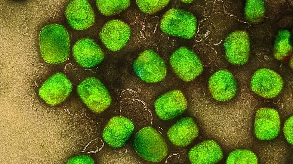
The malaria-causing Plasmodium parasites invade and multiply within red blood cells. Studies have shown that the parasites can rapidly sense and respond to conditions within the host by intimately syncing with their internal body clocks. While it is known that the body’s nutrient levels and daily circadian rhythms affect the parasites’ development, little was known about the impact of host inflammation on the parasites.

This animal-model study, led by the Peter Doherty Institute for Infection and Immunity (Doherty Institute) and the Kirby Institute and published in the journal mBio, reveals that when the body’s immune system responds to inflammation it alters the plasma’s chemical composition, directly impeding the maturation of the Plasmodium parasites in the bloodstream.

University of Melbourne’s Associate Professor Ashraful Haque, a senior author of the paper, said this work highlights the captivating dynamic of the host-parasite relationship.

“First, we discovered that inflammation in the body prevented the early stage of the parasites from maturing. We also noticed that inflammation triggered significant changes in the composition of the plasma – we were actually quite surprised by the magnitude of these changes,” said Associate Professor Haque.

“As we dug deeper, we found substances in the altered plasma that, we believe, are what may inhibit parasite growth in the body. This work reveals a new mechanism that slows down the malaria parasite’s development in the bloodstream. Our research was done using animal models, so it would be really interesting to study if such inhibitory mechanisms occur in humans too.”

Dr David Khoury, co-senior author of the paper, said the scientists found a remarkable response by the parasites to the changes in their environment.

“Parasites residing in red blood cells rapidly sense and respond to their new environment, showing fascinating adaptability. Using cutting-edge genome sequencing technology, we observed that even after just four hours in this changed plasma, the parasites adjusted their genetic and protein activity, resulting in slower maturation within red blood cells. It’s almost like the parasites actively sense an inhospitable host environment, and as a result trigger a coping mechanism,” said Dr Khoury.

“We believe this is the first study to show that inflammation can change how individual parasites behave genetically in the body.”

Professor Miles Davenport, co-senior author of the paper, said this work on the interaction between systemic host inflammation and malaria parasite maturation offers several potential benefits.

“This study, while based on animal models, broadens our understanding of malaria. It provides a foundation for further investigations into the specific mechanisms involved in the modulation of parasite maturation by inflammation, and opens avenues for future studies to explore the identified inhibitory factors, genetic changes and their implications for malaria development,” said Professor Davenport.

Source: The Peter Doherty Institute for Infection and Immunity

How the Hospital Pathogen Acinetobacter Baumannii can Adapt so Quickly

Photo by National Cancer Institute on Unsplash

Acinetobacter baumannii is a notorious hospital pathogen, and there is great pressure to devise novel therapeutic approaches to combat this growing threat. German researchers have now detected an unexpectedly wide diversity of certain cell appendages known as pili in A. baumannii that are associated with pathogenicity. This finding, published in PLOS Genetics, could lead to treatment strategies that are specifically tailored to a particular pathogen.

Each year, over 670 000 people in Europe fall ill because of antibiotic-resistant pathogens, and 33 000 die from the infections. Especially feared are pathogens with resistances against multiple, or even all, known antibiotics. One of these is the bacterium Acinetobacter baumannii, feared today above all as the “hospital superbug”: According to estimates, up to five percent of all hospital-acquired and one tenth of all bacterial infections resulting in death can be attributed to this pathogen alone. This puts A. baumannii right at the top of WHO’s list of pathogens for which there is an urgent need to develop new therapies.

Understanding which characteristics make A. baumannii a pathogen is one of the prerequisites for this. To this end, bioinformaticians led by Professor Ingo Ebersberger of Goethe University Frankfurt and the LOEWE Center for Translational Biodiversity Genomics (LOEWE-TBG) are comparing the genomes and the proteins encoded therein across a wide range of different Acinetobacter strains. Conclusions about which genes contribute to pathogenicity can be drawn above all from the differences between dangerous and harmless strains.

Due to a lack of suitable methods, corresponding studies have so far concentrated on whether a gene is present in a bacterial strain or not. However, this neglects the fact that bacteria can acquire new characteristics by modifying existing genes and thus also the proteins encoded by them. That is why Ebersberger’s team has developed a bioinformatics method to track the modification of proteins along an evolutionary lineage and has now applied this method for the first time to Acinetobacter in collaboration with microbiologists from the Institute for Molecular Biosciences and the Institute of Medical Microbiology and Infection Control at Goethe University Frankfurt.

In the process, the researchers concentrated on hair-like cell appendages, known as type IVa (T4A) pili, which are prevalent in bacteria and that they use to interact with their environment. The fact that they are present in harmless bacteria on the one hand and have even been identified as a key factor for the virulence of some pathogens on the other suggests that the T4A pili have repeatedly acquired new characteristics associated with pathogenicity during evolution.

The research team could show that the protein ComC, which sits on the tip of the T4A pili and is essential for their function, shows conspicuous changes within the group of pathogenic Acinetobacter strains. Even different strains of A. baumannii have different variants of this protein. This leads bioinformatician Ebersberger to compare the T4A pili to a multifunctional garden tool, where the handle is always the same, but the attachments are interchangeable. “In this way, drastic functional modifications can be achieved over short evolutionary time spans,” Ebersberger is convinced. “We assume that bacterial strains that differ in terms of their T4A pili also interact differently with their environment. This might determine, for example, in which corner of the human body the pathogen settles.”

The aim is to use this knowledge of the unexpectedly high diversity within the pathogen to improve the treatment of A. baumannii infections, as Ebersberger explains: “Building on our results, it might be possible to develop personalised therapies that are tailored to a specific strain of the pathogen.” However, the study by Ebersberger and his colleagues also reveals something else: Previous studies on the comparative genomics of A. baumannii have presumably only unveiled the tip of the iceberg. “Our approach has gone a long way towards resolving the search for possible components that characterize pathogens,” says Ebersberger.

Source: Goethe University Frankfurt

More Monkeypox Antibodies with Childhood Smallpox Vaccination

Mpox (monkeypox) virus. Source: NIH

In a study published in Cell Host & Microbe, scientists studied the sensitivity of MPXV, the virus that causes mpox (formerly monkeypox) to neutralising antibodies (NAbs) generated after infection with the virus and/or vaccination with IMVANEX. They found that those who had been born before 1980 had more antibodies in response to either IMVANEX vaccination or mpox infection, highlighting the lasting protection of smallpox vaccination.

The IMVANEX vaccine has been used as pre- and post-exposure prophylaxis in high-risk populations, but its effectiveness is not yet well characterised. To analyse the sensitivity of the virus, a team of scientists led by Pasteur Institut developed two cellular tests to quantify neutralising antibodies, using either the attenuated virus as a vaccine (MVA) or an MPXV strain isolated in a recently infected individual.

In 2022-2023, an unprecedented epidemic of 87 000 cases of mpox occurred in non-endemic areas, affecting people with no direct link to travel in Central or West Africa, where the virus has historically been present. MPXV is mainly transmitted to humans by rodents, with human-to-human transmission occurring via respiratory droplets or close contact. Symptoms are less severe than those of smallpox, and the case-fatality rate is lower. MPXV is still circulating at very low levels in non-endemic areas, which is why it is important to improve characterisation and analyse the immune response of people infected with the virus or vaccinated with IMVANEX, the third-generation vaccine currently available, initially developed for smallpox.

The large number of sera analysed provided good statistical power, meaning that the analysis could be narrowed to subgroups of patients based on various criteria such as age.

The study demonstrated the role of complement, already known for other poxviruses, and the neutralising activity of the antibodies generated by infection or vaccination. Robust levels of anti-MVA antibodies were detected after infection, vaccination with the historic smallpox vaccine, or administration of IMVANEX or another MVA-based vaccine candidate. MPXV was minimally sensitive to neutralisation in the absence of complement. The addition of complement from sera enhanced detection of individuals with antibodies and increased their level of anti-MPXV antibodies. Four weeks after infection, anti-MVA and -MPXV NAbs were observed in 94% and 82% of individuals, respectively. Two doses of IMVANEX generated anti-MVA and -MPXV NAbs that were detectable in 92% and 56% of vaccinees, respectively.

The highest level of antibodies was found in individuals born before 1980 (who had therefore been vaccinated for smallpox), whether after infection or after administration of IMVANEX, highlighting the impact of historic smallpox vaccination on immune responses to infection or administration of IMVANEX. This suggests that a sort of hybrid immunity was generated in infected individuals who were vaccinated in childhood.

The number of MPXV infections has been constantly on the rise since mass vaccination for smallpox was discontinued in the 1980s. “The neutralisation assays developed in connection with this research may help define correlates of protection against infection or disease severity. The assays can also be used to conduct epidemiological surveys, assess the duration of protection conferred by previous infection or by authorised and candidate vaccines, and analyse the use of immunotherapeutic intervention. The assays represent useful tools to understand the mechanisms of multiplication of MPXV and its effects on public health, and to optimsze patient treatment,” commented Olivier Schwartz, Head of the Institut Pasteur’s Virus and Immunity Unit and last author of the study.

Source: Institut Pasteur

Lactobacillus Combo Stops Bacteria that Cause Toxic Shock Syndrome

Methicillin resistant Staphylococcus aureus (MRSA) – Credit: CDC

A virulent strain of Staphylococcus aureus produces proteins that trigger toxic shock syndrome (TSS), a disease characterised by the quick onset of fever, a telltale rash, and, without treatment, multi organ failure. In the vagina, TSS is associated with a life-threatening reaction from the immune system. Research published in the journal Microbiology Spectrum shows that probiotics may help prevent the disease before the cytokine cascade ever begins.

Probiotics may help prevent the disease before the cytokine cascade ever begins. This study reports that strains of two bacteria, Lactobacillus acidophilus and Lacticaseibacillus rhamnosus, successfully inhibited the production of the superantigens that cause TSS, in lab experiments. L. acidophilus, in addition, inhibited the growth of the S. aureus strains that produce the problematic proteins.

A combination of the two could both prevent growth and inhibit the immune response. “It’s kind of a double whammy against S. aureus,” said microbiologist Patrick Schlievert, Ph.D., at the University of Iowa Carver College of Medicine, in Iowa City. “If any toxin is made, the probiotics still prevent inflammation.”

He noted that adding these probiotics to tampons or other menstrual products could reduce the risk of TSS associated with menstruation. Such a preventive measure has the potential to benefit millions of vulnerable people, Schlievert said. “We know that 20% of people over age 12 cannot make antibodies and never will make antibodies against toxic shock syndrome,” he said.

Schlievert has been studying TSS and its prevention for decades. In the early 1980s, he was the first researcher to identify the toxin that triggers an overreaction of the immune system, and to show how high-absorbency tampons facilitated production of that toxin if S. aureus was present.

The new work, he said, was motivated by observations made during an earlier study. A few years ago, he and his colleagues recruited 205 women to test whether a novel molecular mixture, when added to tampons, would inhibit pathogenic bacteria. That molecule proved effective against E. coli and other pathogens, but the researchers noticed an unexpected consequence.

“Some of the women in the treatment group had this tremendous growth of Lactobacilli,” Schlievert said.

Source: American Society for Microbiology

Scientists Unravel The Reason Why NSAIDs Exacerbate C. Diff Infections

Clostridioides difficile. Credit: CDC

Nonsteroidal anti-inflammatory drugs (NSAIDs) exacerbate gastrointestinal infections by Clostridioides difficile, the leading cause of antibiotic-associated diarrhoea worldwide – but the reason why has long eluded medical science. In a new paper published in Science Advances, researchers have begun to answer that question, showing that NSAIDs disrupt the mitochondria of cells lining the colon, sensitising them to damage by pathogenic toxins.

C. difficile is a bacterium that leads to a wide range of symptoms, from mild diarrhoea to complex infection and death. The factors that influence this wide spectrum of clinical outcomes remain largely unclear, but emerging evidence suggests that factors like diet and pharmaceutical drugs influence both susceptibility to infection and disease progression. However, little is known about how these factors impact the course of C. difficile infection.

Prior studies have shown that NSAIDs like indomethacin, aspirin, and naproxen negatively affect the gut, both in patients with C. difficile infection and other conditions like inflammatory bowel disease (IBD). Long-term NSAID use can lead to stomach ulcers and intestinal injuries. One hypothesis that this is due to the effects of NSAIDs on cyclooxygenase (COX) enzymes; a process that helps reduce inflammation and pain but also impairs mucosal function in the upper gastrointestinal tract. NSAIDs also have off-target effects and have been shown to affect cellular mitochondria by uncoupling cellular mitochondrial functions, but these had not been studied in C. difficile infections.

To define these effects, Children’s Hospital of Philadelphia (CHOP) researchers, led by graduate student Joshua Soto Ocaña, used in vitro and mouse models of C. difficile infection to test how permeable colonic epithelial cells are in the presence of the NSAID indomethacin. The researchers observed that both indomethacin and C. difficile toxins increased epithelial cell barrier permeability and inflammatory cell death. They also found that the effect was additive: the combined effect on cell permeability of both toxins and indomethacin was increased compared to each independently, suggesting a synergistic effect of NSAIDs and C. difficile in increasing this pathogen’s virulence.

Surprisingly, the researchers found that NSAIDs exacerbate C. difficile infection independent of COX inhibition and instead through off-target effects on mitochondria. They did so by treating colonic epithelial cells with a precursor molecule that is similar in structure to indomethacin but lacks the ability to inhibit the COX enzyme. Not only did they find that this NSAID-like molecule induced cell death, but they also found that adding selective COX inhibitors did not increase cell death, demonstrating that COX enzyme inhibition is not required to induce epithelial cell damage during C. difficile infection and that, instead, this damage occurs through off-target effects of NSAIDs.

To test the role of off-target effects during C. difficile infection, the researchers used mice pretreated either with indomethacin or the NSAID-precursor molecule. When exposed to C. difficile, both groups of mice showed equal enhancement in disease severity and mortality compared to untreated control mice infected with C. difficile only. The researchers also observed a similar effect in mice who were pretreated with the NSAID aspirin. To further define the specific mechanisms driving these off-target effects of NSAIDs, researchers looked at mitochondrial functions in colonic epithelial cells in vitro and in mice. They observed that the combination of NSAIDs and C. difficile toxins increased damage to colonic epithelial cell mitochondria and disrupted several important mitochondrial functions.

“Our work further demonstrates the clinical importance of NSAIDs in patients with C. diff infection and sheds light on why the combination of these two may be so detrimental,” said senior author Joseph P. Zackular, PhD, Investigator and Assistant Professor of Pathology and Laboratory Medicine at CHOP. “Our mechanistic findings are a starting point for further research that aims to understand the impact of mitochondrial functions during C. diff infection. These data could also inform how NSAID-mediated mitochondrial uncoupling affects other diseases, such as small intestinal injury, IBD, and colorectal cancer.”

Source: Children’s Hospital of Philadelphia

Peptides May Solve the Sticky Problem of Bacterial Biofilms

This image shows an intricate colony of Pseudomonas aeruginosa. The bacteria have self-organised into a sticky, mat-like colony called a biofilm, which allows them to cooperate with each other, adapt to changes in their environment, and ensure their survival.
Credit: Scott Chimileski and Roberto Kolter, Harvard Medical School, Boston

Researchers have developed peptides that can help combat bacteria growing in biofilms, which occur in up to 80% of human infections. Their results, published in Nature Chemical Biology, may offer a way to tackle antimicrobial-resistant infections. 

Treating infections becomes significantly more challenging when biofilms are present, as they not only reduce the effectiveness of antibiotics but also give rise to several medical complications. These complications include infections following joint replacements, prosthetic devices, as well as contamination in catheters and other medical equipment. The lack of specific treatments makes the management and treatment of biofilms exceptionally difficult.

The team of researchers, led by Dr Clarissa Melo Czekster and Dr Christopher Harding from the School of Biology at St Andrews, in collaboration with researchers at University of Dundee, developed antimicrobial peptides that can target the harmful bacteria growing in biofilms.

The team determined how a key enzyme (PaAP) in biofilms work and developed a revolutionary new strategy to inhibit the protein. Their inhibitor is potent and targets cells from the human pathogen Pseudomonas aeruginosa in biofilms. P. aeruginosa, a WHO pathogen of concern, causes chronic infections in patients with cystic fibrosis and other conditions, which means a biofilm inhibitor is urgently needed.

Dr Czekster and the team are currently working in collaboration with the University of St Andrews Technology Transfer Centre and industry partner Locate Bio, a biomedicine spinout of the University of Nottingham, to commercialise the technology. The Locate Bio team are trialling the peptides to see how they work with the company’s Programmed Drug Release technology to develop new orthobiologic solutions and products. The Technology Transfer Centre has filed a UK priority patent application.

Dr Czekster said: “Our research reveals how designed inhibitors can target a key enzyme in bacterial virulence, offering molecular insights applicable to aminopeptidases in diverse organisms.

“This remarkable new research presents an innovative strategy to target bacterial biofilms and pave the way for better treatment of bacterial infection.”

Source: University of St. Andrews

In Osteoarthritis, Molecular Changes Weaken Protective Films

Photo by Towfiqu barbhuiya: https://www.pexels.com/photo/person-feeling-pain-in-the-knee-11349880/

Although osteoarthritis has been extensively studied through a medical perspective, the molecular changes associated with osteoarthritis remain unclear. New research published in Biointerphases suggests that there may be an optimum concentration and size of molecules in the synovial fluid needed to form the protective film in joints.

Osteoarthritis is the most common degenerative joint disease, affecting 22% of adults over 40 globally. The cartilage in the joints, in concert with the synovial fluid, provides a smooth surface to support weight-bearing movements. The fluid contains several molecules, including hyaluronan (HA) and phospholipids. Since the cartilage environment cannot be quickly healed or repaired, researchers have tried to diagnose the early stages of joint disease by monitoring the molecular weight and concentration of HA.

“Although we know that in healthy joints there is very low friction, it is unclear which other molecules are involved and how they change during osteoarthritis,” said Rosa Espinosa-Marzal (EIRH), professor of environmental engineering & science, and materials science & engineering. “During the early stages of osteoarthritis, cartilage starts degrading, and previous research has shown that the molecular composition of the synovial fluid changes. We wanted to see if the two changes are related to each other.”

In a healthy joint, the molecular weight of HA varies between 2–20 MDa with a concentration ranging from 1–4 mg/mL. Studies have shown that in diseased joints, HA is broken down resulting in a lower molecular weight and its concentration is also reduced by 10x. Based on these observations, made by other researchers, the study looked at how the concentration and molecular weight of HA influences the structure of healthy and diseased joints.

To do so, the researchers combined vesicles with high and low molecular weight HA. Using neutron scattering and light scattering, they discovered that the molecular weight of HA can vastly change the structure of the vesicles. Lower molecular weight HA, which mimics osteoarthritis-diseased joints, results in larger vesicle size. Changes in HA’s molecular weight also changed the thickness of the phospholipid layers in the joints.

The researchers also studied how these differences can influence the formation of a protective film; in joints this film is responsible for the very low friction we need for unhindered motion. Once again, they used a combination of techniques, quartz crystal microbalance and atomic force microscopy, to examine how these molecules assemble on gold surfaces.

“The formation of a film is possible only when there is an optimal concentration of HA and phospholipids. Even though the gold surfaces have very little in common with cartilage, our studies indicate that there could also be an optimum concentration under biological conditions,” Espinosa-Marzal said. “This is an important observation because we can use the concentration changes as a diagnostic tool.”

The researchers are now testing this theory using cartilage. They are also interested in studying the other molecular components that are found in joints to build a more comprehensive model of the changes that are associated with osteoarthritis.

Source: Carl R. Woese Institute for Genomic Biology, University of Illinois at Urbana-Champaign

Pathogenic Bacteria Use A Sugar from Intestinal Mucus to Dig Themselves in

Source: CC0

A new study shows the sugar sialic acid, which makes up part of the protective intestinal mucus layer, fuels disease-causing bacteria in the gut. The findings, published in PNAS, suggest a potential treatment target for intestinal bacterial infections and a range of chronic diseases linked to gut bacteria, including inflammatory bowel disease (IBD), celiac disease, irritable bowel syndrome and short bowel syndrome.

The research by researchers at the University of British Columbia (UBC) and BC Children’s Hospital, used a mouse model of gut infections.

“Bacteria need to find a place in our intestines to take hold, establish and expand, and then they need to overcome all the different defences that normally protect our gut,” says Dr Bruce Vallance, a professor in the department of paediatrics at UBC and investigator at BC Children’s Hospital. “In the future, we can potentially target this sugar, or how pathogens sense it, to prevent clinically important disease.”

Inflammatory diseases such as IBD are on the rise in children, who are more susceptible to gut infections because of their immature immune systems. Dr Vallance and his team sought to understand what enables these bacterial pathogens to survive and expand inside our intestines.

For the study, the researchers examined Citrobacter rodentium, an intestinal bacterial pathogen of mice that’s used to model infections with human E. coli. The team discovered that the bacteria have genes involved in sialic acid consumption, and when these genes are removed, the bacteria’s growth is impaired.

Further investigation revealed that upon consuming the sugars, the bacteria produced two special virulence proteins that help the bacteria cross the colonic mucus layer and stick to the underlying epithelial cells. The findings reveal how the bacteria can change over time and actually worsen disease.

“You start off with IBD, your microbes change, they start digging their way into the cells lining your gut, causing more inflammation, and that may be one reason why IBD becomes chronic,” says Dr Vallance. “Specific nutrients such as sialic acid or other sugars might be the Achilles heels for them in terms of things you could target to remove dangerous bacteria from the intestine.”

Dr Vallance and his team are now examining the role other sugars in the gut may play in feeding pathogenic bacteria. They’re also looking for probiotics that could outcompete the dangerous bacteria, stealing the sugars away from them.

They also plan to explore potential interactions between resident and pathogenic bacteria. Pathogenic bacteria can’t access the sugars on their own and thus, some of the normally harmless resident bacteria must serve as accomplices.

“Basically, these accomplices cut the sugar off the mucus, and then either they hand it to the dangerous bacteria or the dangerous bacteria have come up with a way of stealing it from them,” he explains.

A better understanding of these interactions could provide new ways to block pathogenic bacteria, something Dr. Vallance says is urgently needed.

“In the past, our ancestors were constantly assaulted by dangerous bacteria,” says Dr. Vallance. “With the advent of more and more antibiotic resistance in bacteria, these bacterial infections are going to become a growing problem again. Without new antibiotics, we need to come up with novel ways to fight these bacteria, like starving them.”

Source: University of British Columbia

Ticks’ Trick: Using Static Electricity as a Grappling Hook

Photo by Pixabay

Ticks can be attracted across air gaps several times larger than themselves by the naturally accumulated static electricity of their hosts, researchers at the University of Bristol have discovered. This makes it much easier to finding hosts to parasitise because ticks cannot jumping, making this is the only mechanism by which they would be able to latch onto hosts that are beyond the reach of their tiny legs.

The findings, published today in Current Biology, are the first known example of static electricity being implicated in the attachment of an animal to another animal.

Lead author Sam England from Bristol’s School of Biological Sciences explained: “We knew that many animals, including humans, can accumulate quite significant electrostatic charges.

“We see this when we get a static shock after bouncing on a trampoline, or when rubbing a balloon on our hair, for example. But this electrostatic charging also happens to animals in nature when they rub against objects in their environment like grass, sand, or other animals. These charges are surprisingly high, and can be equivalent to hundreds if not thousands of volts — more than you get out of your plug sockets at home! Importantly, static charges exert forces on other static charges, either attractive or repulsive depending whether they are positive or negative.

“We wondered whether the static charges that mammals, birds, and reptiles naturally accumulate could be high enough that parasitic ticks could be lifted through the air by electrostatic attraction onto these animals, therefore improving their efficiency at finding hosts to feed on.”

The team initially tested the idea by bringing statically charged rabbit fur and other materials close to ticks and observing whether they were attracted to them.

They witnessed the ticks being readily pulled through the air across air gaps of several millimetres or centimetres (the equivalent of humans jumping up several flights of stairs) by these charged surfaces, and so investigated further.

Sam continued: “First, we used previous measurements of the typical charge carried by animals to mathematically predict the strength of the electric field that is generated between a charged animal and the grass that ticks like to sit on and wait for hosts to pass by.

“Then, we placed ticks underneath an electrode, with an air gap in between, and increased the charge on the electrode until the ticks were attracted onto the electrode. By doing this we were able to determine the minimum electric field strength at which the ticks could be attracted. This minimum electric field was within the order of magnitude predicted by the mathematical calculations of the electric field between a charged animal and grass, therefore it is likely that ticks in nature are attracted onto their hosts by static electricity.”

There are several wider implications and potential applications to these findings. Firstly, the phenomenon likely applies to many other parasitic species that want to make contact and attach to their hosts, such as mites, fleas, or lice, and so it could be a universal mechanism for animals to make contact with and attach onto each other.

Beyond the purely scientific implications, the discovery opens the door for new technologies to be developed to minimise tick bites in humans, pets, and farm animals, such as developing anti-static sprays.

Sam concluded: “We have now discovered that ticks can be lifted across air gaps several times larger than themselves by the static electricity that other animals naturally build up. This makes it easier for them to find and attach onto animals that they want to latch onto and feed from. Until now, we had no idea that an animal could benefit from static electricity in this way, and it really opens up one’s imagination as to how many invisible forces like this could be helping animals and plants live their lives.”

Now the team plan to investigate whether the ticks are capable of sensing the approaching electrostatic charge of their prospective hosts.

Source: University of Bristol

Researchers Link Lupus Flare-ups to Microbiota Blooms

A woman with Systemic Lupus Erythematosus. Source: Wikimedia CC0

Recurrent bouts of systemic lupus erythematosus, marked by the body’s immune system attack of its own tissues, closely tracked with upticks in growth in the gut of a certain species of bacteria. New research from NYU Grossman School of Medicine shows that bacterial blooms of the gut bacterium Ruminococcus blautia gnavus occurred at the same time as disease flare-ups in five of 16 women with lupus of diverse racial backgrounds studied over a four-year period.

Systemic lupus erythematosus involves damaging inflammation, especially in the kidneys, but also in joints, skin, and blood vessels. Four of these study patients with R. gnavus blooms had severe cases of the most common and kidney-specific form of the disease, lupus nephritis, while one had a severe example of lupus involving inflammation in multiple joints.

Published in the Annals of Rheumatic Diseases, the team’s analysis of these lupus patients’ gut bacterial blooms identified 34 genes that already had established links to the bacterium’s growth in people with inflammation. While the specific causes of lupus remain unknown, many experts suspect that bacterial imbalances trigger inherited genetic factors responsible for the disease.

This study also investigated how tightly these patients’ immune system antibodies bonded to structures in the bacterial wall, much like they would an invading virus. These antibodies showed a strong affinity to specific bacterial lipoglycan molecules that are known triggers of inflammation. These lipoglycans were found to be common in R. gnavus strains in lupus patients but not in healthy people. Antibodies are a major cause of the body damage in this disease, and this diagnostic antibody response, the researchers say, highlights the important role played by R. gnavus in the autoimmune disease.

“Our findings provide the strongest evidence to date that silent growths of Ruminococcus blautia gnavus are tied to active serious renal disease in lupus patients,” said study lead investigator Doua Azzouz, PhD.

“Interestingly, our study also established this common bacterial link among a racially diverse group of females with varying forms of lupus,” said Azzouz, a postdoctoral researcher. Lupus is more common in women than in men, and the disease affects more Blacks, Hispanics, and Asians than Whites.

“Our goal is to use our growing understanding of the biological pathways that underpin the disease to develop new treatments that prevent or treat flares for all forms of lupus,” said study senior investigator and immunologist Gregg Silverman, MD.

“Such future treatments for lupus, especially lupus nephritis, could potentially decrease the use of drugs designed to dampen the immune system and instead promote the use of less-toxic antibacterial agents, probiotics or dietary regimens that prevent imbalances such as Ruminococcal blooms in the local gut bacterial population, or microbiome,” said Silverman.

Previous research by Silverman’s team showed that R. gnavus blooms weaken the gut wall barrier, prompting bacterial leakages that in turn trigger inflammatory and overactive immune responses.

The team plans to extend the research to other medical centre and also plans further experiments in mouse models of lupus to see how R. gnavus colonisation triggers lupus. Using mouse models, they also want see whether if R. gnavus blooms speed up or otherwise affect the severity of flares and inflammation.

The researchers say they also want to conduct experiments on various lipoglycan molecules from different R. gnavus strains to see if any particular part of the molecular structure is key to triggering inflammation or if other lipoglycans also prompt an immune response tied to lupus or other diseases of the gut, including Crohn’s.

For the study, researchers used stool and blood samples from lupus patients being treated at NYU Langone. All study participants were being closely monitored for disease flare-ups. Test results were compared with those of 22 female volunteers of similar age and racial background who did not have lupus and were otherwise healthy.

As an autoimmune disease, systemic lupus erythematosus can lead to widespread inflammation and long-term tissue damage in affected organs. According to researchers, about half of patients develop lupus nephritis, of whom one-quarter are likely to experience end-stage renal disease that may require regular blood dialysis and even kidney transplantation.

Source: NYU Langone Health / NYU Grossman School of Medicine