Radiation is a powerful tool for treating cancer, but prolonged exposure can damage the skin. Radiation-induced skin injuries are painful and increase a person’s chances of infection and long-term inflammation. Now, researchers in ACS Biomaterials Science & Engineering report an aspirin-containing hydrogel that mimics the nutrient-rich fluid between cells and accelerates healing of skin damaged by radiation in animals. With further development, the new salve could provide effective and rapid wound healing for humans.
Most people undergoing radiotherapy for cancer will experience radiation-induced skin injury that can include redness, pain, ulcers, necrosis and infection. There are few treatments for these wounds, with the most common methods being debridement and hyperbaric oxygenation. Wound dressings made from hydrogels are gaining popularity because they are easy to apply and provide a wet environment for healing that is similar to the inside of the body. Glycopeptide-based hydrogels are especially promising: In laboratory and animal studies, the nanofibre structures have promoted cellular growth and regulated cell adhesion and migration. A research team led by Jiamin Zhang, Wei Wang, Yumin Zhang and Jianfeng Liu proposed loading aspirin, a common anti-inflammatory drug, into a glycopeptide-based hydrogel to create a multifunctional wound dressing for radiation-induced skin injuries.
In lab tests with cultured cells, the researchers found that the aspirin-contained hydrogel scavenged reactive oxygen species, repaired DNA double-strand breaks and inhibited inflammation caused by radiation exposure without affecting cellular growth. In mouse models of radiation-induced skin injury, the researchers found that dressing wounds for three weeks with the salve reduced acute injuries and accelerated healing – results that the team says point to its potential as an easy-to-administer, on-demand treatment option for reducing radiation damage and promoting healing in humans.
Skin cell (keratinocyte)
This normal human skin cell was treated with a growth factor that triggered the formation of specialised protein structures that enable the cell to move. We depend on cell movement for such basic functions as wound healing and launching an immune response. Credit: Torsten Wittmann, University of California, San Francisco
Fingerprints are one of the best-recognised examples of pattern formation by epithelial cells. The primary cells in the epithelium are the keratinocytes, and they are known to form patterns at the microscopic and macroscopic levels. While factors affecting this pattern formation have been reported, the exact mechanisms underlying the process are still not fully understood.
A team of researchers, led by Associate Professor Ken Natsuga at the Faculty of Medicine, Hokkaido University, have revealed that cell-cell adhesion governs pattern formation in keratinocytes. Their findings were published in the journal Life Science Alliance.
“In this study, we used an immortalised keratinocyte cell line, called HaCaT, which retains all the properties of normal keratinocytes,” Natsuga explained. “In order to ensure that our findings were accurate, we established single-cell cultures from this cell line.”
The team observed pattern formation in both the original heterogeneous cell line, as well as in single-cell-derived cultures. During culturing, the keratinocytes moved randomly and spontaneously formed high- and low-density regions, leading to pattern formation.
The pattern formation was markedly influenced by starvation. When the culture medium was renewed, patterns were obscured, but reappeared as the nutrients in the culture medium were consumed by the keratinocytes.
The team then examined the gene expression in the keratinocytes, which revealed that cell adhesion proteins and keratinocyte differentiation proteins were upregulated in high-density regions. “As cell adhesion is necessary for the development of high-cell-density regions, we specifically investigated the expression of adherens junction (AJ) molecules such as E-cadherin and actin,” Natsuga elaborated. “We found that these molecules were localised at the intercellular junctions of high-density regions.”
The authors then used a mathematical model to confirm that, under spatially uniform density and stress, strong cell adhesion leads to the formation of density patterns. They were also able to demonstrate that the keratinocyte patterns influenced cell proliferation and differentiation, and that serum starvation influences epidermal stratification (a type of differentiation) in skin cells from mice.
“Our study presents a novel and robust model of cell–cell adhesion-induced patterning (CAIP),” concludes Natsuga. “We have deepened our mechanistic insight into cellular organization and its consequences for cell fate decisions and epithelial stratification.” The team demonstrated that epithelial cell–cell adhesion is essential and sufficient for patterning. Future work will focus on adding more variables to the model to understand other processes that occur concurrently during development.
In a study in the Journal of Cosmetic Dermatology that included 60 individuals with mild to moderate acne, following the Mediterranean diet and taking omega-3 fatty acid supplements led to significant reductions in inflammatory and non-inflammatory skin lesions, as well as improved quality of life.
Notably, 98.3% of participants had omega-3 fatty acid deficits at the start of the study. Acne severity lessened significantly in those who reached target omega-3 fatty acid levels during the study.
“Lifestyle interventions, including dietary recommendations, should not be considered in opposition to prescription medications, but rather as a valuable adjunct to any modern acne treatment plan,” said corresponding author Anne Guertler, MD, of the Ludwig Maximilian University of Munich, in Germany. “Future studies should build on the foundation laid by our current findings in a randomised, placebo-controlled design to improve dietary recommendations for acne patients.”
Toxic chemicals used to flame-proof plastic materials can be absorbed into the body through skin, via contact with microplastics, new research shows. The study offers the first experimental evidence that chemicals present as additives in microplastics can leach into human sweat, and then be absorbed through the skin, into the bloodstream.
Many chemicals used as flame retardants and plasticisers have already been banned, due to evidence of adverse health effects including damage to the liver or nervous system, cancer, and risks to reproductive health. However, these chemicals are still present in the environment in older electronics, furniture, carpets, and building materials.
While the harm caused by microplastics is not fully understood, there is increasing concern over their role as conduits of human exposure to toxic chemicals.
The research team demonstrated in a study published last year, that chemicals were leached from microplastics into human sweat. The current study now shows that those chemicals can also be absorbed from sweat across the skin barrier into the body.
In their experiments, the team used innovative 3D human skin models as alternatives to laboratory animals and excised human tissues. The models were exposed over a 24-hour period to two common forms of microplastics containing polybrominated diphenyl ethers (PBDEs), a chemical group commonly used to flame retard plastics.
The results, published in Environment International, showed that as much as 8% of the chemical exposed could be taken up by the skin, with more hydrated – or ‘sweatier’ – skin absorbing higher levels of chemical. The study provides the first experimental evidence into how this process contributes to levels of toxic chemicals found in the body.
Dr Ovokeroye Abafe, now at Brunel University, carried out the research while at the University of Birmingham. He said: “Microplastics are everywhere in the environment and yet we still know relatively little about the health problems that they can cause. Our research shows that they play a role as ‘carriers’ of harmful chemicals, which can get into our bloodstream through the skin. These chemicals are persistent, so with continuous or regular exposure to them, there will be a gradual accumulation to the point where they start to cause harm.”
Dr Mohamed Abdallah, Associate Professor of Environmental Sciences at the University of Birmingham, and principal investigator for the project, said: “These findings provide important evidence for regulators and policymakers to improve legislation around microplastics and safeguard public health against harmful exposure.”
Professor Stuart Harrad, co-author of the paper, added “the study provides an important step forward in understanding the risks of exposure to microplastics on our health. Building on our results, more research is required to fully understand the different pathways of human exposure to microplastics and how to mitigate the risk from such exposure.”
In future research, the team plan to investigate other routes through which microplastics could be responsible for toxic chemicals entering the body, including inhalation and ingestion.
A certain biological pathway involving interleukin-17 drives the inflammation seen in the skin disease psoriasis, according to a new study published in the journal Immunity. The work could lead to improved therapies for all inflammatory skin diseases, including atopic and allergic dermatitis and a type of boil called hidradenitis suppurativa, say the study authors.
Led by researchers at NYU Langone Health, the new study found that the interleukin-17 (IL-17) pathway, whose activity is blocked by existing anti-inflammatory drugs, activates a protein called hypoxia inducible factor 1-alpha (HIF-1-alpha) in psoriasis. Researchers say that IL-17 has long been known to be active in inflammation, but the role of HIF-1-alpha has until now been unclear.
The research team also found that HIF-1-alpha let inflamed skin cells more actively break down sugar for energy, supporting their metabolism and leading to the production of a waste product called lactate. When consumed by inflammatory T cells, lactate triggered production of IL-17, fuelling even more inflammation.
The findings show that in human skin tissue samples from psoriatic patients, measures of gene activity around IL-17 and HIF-1-alpha were similar, suggesting that these factors are interconnected. Experiments in mice treated to develop psoriasis found that subsequent treatment with an experimental drug that blocks the action of HIF-1-alpha, called BAY-87-2243, resolved inflammatory skin lesions.
Further, skin samples from 10 patients successfully treated with anti-inflammatory drug etanercept showed diminished activity for both IL-17 and HIF-1-alpha, suggesting to researchers that when IL-17 is blocked, so is HIF-1-alpha.
“Our study results broadly show that activation of HIF-1-alpha is at the crux of metabolic dysfunction observed in psoriasis and that its action is triggered by IL-17, another key inflammatory-signaling molecule,” said corresponding study author Shruti Naik, PhD, associate professor at NYU Grossman School of Medicine.
Further experiments were performed on skin samples from five patients with psoriasis whose healthy and inflamed skin was separately treated with either BAY-87-2243 or an existing combination of topical drugs (calcipotriene and betamethasone dipropionate). Researchers then compared differences in inflammatory gene activity as a measure of impact and found that the HIF-1-alpha inhibitor had a greater effect than existing topical drugs. Specifically, skin samples that responded to HIF-1-alpha therapy had 2,698 genes that were expressed differently, while standard-of-care-treated samples had 147 differently expressed genes.
Genetic analysis of skin samples from another 24 psoriatic patients treated with the IL-17A-blocking drug secukinumab showed only decreased, not heightened, gene activity connected to HIF-1-alpha when compared to HIF-1-alpha gene activity in nine healthy patients with no psoriatic disease. Researchers say this indicates HIF-1-alpha’s blocked action was codependent on blockage of IL-17.
Additional experiments in mice showed that blocking glucose uptake in the skin slowed psoriatic disease growth by limiting glucose metabolism, or glycolysis. Both the number of immune T cells tied to inflammation and the cell levels of IL-17 also decreased. The researchers found further that levels of lactate, the main byproduct of glycolysis, in psoriatic skin cell cultures dropped once exposed to the glycolysis-inhibiting drug 2-DG.
Directly targeting lactate production in psoriatic mice using a topical skin cream containing lactate dehydrogenase, which breaks down lactate, also slowed disease progression in the skin, with reduced numbers of inflammatory gamma-delta T cells and reduced IL-17 activity. Gamma-delta T cells were shown to take up lactate and use it to produce IL-17.
“Evidence of HIF-1-alpha’s depressed action, or downregulation, could also serve as a biomarker, or molecular sign, that other anti-inflammatory therapies are working,” said study co-senior investigator Jose U. Scher, MD, professor at NYU Grossman School of Medicine.
Scher, who also serves as director of NYU Langone’s Psoriatic Arthritis Center and the Judith and Stewart Colton Center for Autoimmunity, says the team plans to develop experimental drugs that can block HIF-1-alpha and lactate action in the skin “to end the underlying vicious cycle of IL-17-driven inflammation in skin disease. Our research fundamentally expands the scope of feasible therapeutic options.”
Naik points out that while many available therapies for psoriasis, including steroids and immunosuppressive drugs, reduce inflammation and symptoms, they do not cure the disease. She said further experiments are needed to refine which experimental drug works best, with respect to HIA-1-alpha inhibition, before clinical trials could start.
In a novel study, a team of dermatologists evaluated the effect of ultraviolet (UV) exposure on appetite and weight regulation. They found that UV exposure raises norepinephrine levels, decreases leptin levels, and induces the browning of subcutaneous fat, thereby increasing energy expenditure. These results potentially pave the way for new approaches to prevent and treat obesity and metabolic disorders. Their findings appear in the Journal of Investigative Dermatology, published by Elsevier.
Co-first authors Qing-Ling Quan, MD, PhD, and Eun Ju Kim, PhD, Department of Dermatology, Seoul National University Hospital, explained, “Recent evidence has suggested that UV exposure limits body weight gain in mouse models of obesity. Subcutaneous fat is a critical organ in regulating energy homeostasis. Alongside previous studies on the effects of UV exposure on obesity and metabolic disorders, our team was inspired by our prior discovery that, although UV rays do not directly reach subcutaneous fat when exposed to the skin, they can regulate the metabolism of subcutaneous fat. This led us to hypothesise that skin exposure to UV rays could play a significant role in systemic energy homeostasis, prompting this research.”
Investigators discovered that when exposed to UV radiation consistently, mice fed a normal diet and those on a high-fat diet exhibited increased appetite due to a decrease in leptin, a key hormone in appetite regulation. But there was no weight increase – they found that UV radiation inhibits weight gain by enhancing secretion of the neurotransmitter norepinephrine, which not only decreases leptin but also increases energy expenditure through the “browning” of subcutaneous fat.
The increased energy intake, driven by heightened appetite, is converted to heat and burned before it can accumulate in subcutaneous fat, thus preventing weight gain.
This research provides new insights into the impact of UV exposure on appetite and weight regulation, opening possibilities for novel approaches in the prevention and treatment of obesity and metabolic disorders. Specifically, uncovering the mechanism by which UV radiation prevents weight gain could offer new approaches to dietary regulation and weight loss, providing innovative insights into health and obesity management that could positively impact human health.
Lead investigator Jin Ho Chung, MD, PhD, Department of Dermatology, Seoul National University Hospital, Seoul National University College of Medicine, explained, “This study elucidates the mechanism by which UV exposure can increase appetite while inhibiting weight gain. These findings contribute significantly to understanding the effects of UV radiation on energy metabolism and homeostasis and open new avenues for exploring prevention and treatment strategies for obesity and metabolic disorders. Notably, the fact that UV radiation lowers leptin levels and increases norepinephrine, thereby promoting the browning of subcutaneous fat and increasing energy expenditure, provides a groundbreaking clue for the development of obesity treatment strategies. This research demonstrates that UV exposure not only affects the skin but also plays a deep role in our body’s energy metabolism and homeostasis processes. However, further research is needed on the long-term effects and safety of UV exposure, and there should be significant interest in developing new therapeutic approaches that utilise the efficacy of UV radiation.”
However, as co-corresponding author Dong Hun Lee, MD, PhD, Institute of Human-Environment Interface Biology, Seoul National University, noted, “Because UV exposure can accelerate skin aging and promote skin cancer, it is advisable to minimise UV exposure and protect the skin with sunscreen. Thus, our research team plans to conduct follow-up studies to develop new strategies that could mimic the effects of UV radiation for obesity and metabolic regulation.”
Skin cell (keratinocyte). This normal human skin cell was treated with a growth factor that triggered the formation of specialised protein structures that enable the cell to move. We depend on cell movement for such basic functions as wound healing and launching an immune response. Credit: Torsten Wittmann, University of California, San Francisco
Millions of people worldwide suffer from unpredictable drug toxicities every year. In particular, drug eruptions which manifest through symptoms such as redness, blisters, and itching on the skin, are quite common. Severe drug eruptions can become life-threatening and can have long-lasting consequences.
Previous studies have identified specific variants of certain genes as potential causal agents of drug eruptions. Scientists believe that the genes encoding the human leukocyte antigen (HLA), a protein expressed on the surface of leucocytes known to play an important role in the immune system, are involved in the onset of drug eruption. But current theories cannot explain why HLA-related drug eruptions typically manifest on the skin rather than in multiple organs throughout the body.
To address this knowledge gap, a research team including Lecturer Shigeki Aoki, Kousei Ito, and Akira Kazaoka from the Graduate School of Medical and Pharmaceutical Sciences, Chiba University, conducted an in-depth study on the link between HLA and drug eruptions. Their findings were published in PNAS Nexus.
The researchers first conducted a series of experiments on mice keratinocytes. These keratinocytes, the most common type of skin cell, were engineered to express a specific variant of the HLA gene called HLA-B*57:01, which specifically bind to the antiviral drug abacavir. Then, they validated these findings in genetically modified mice expressing HLA-B*57:01, that were exposed to abacavir.
The researchers found that HLA-B*57:01-expressing keratinocytes that were exposed to abacavir exhibited endoplasmic reticulum (ER) stress responses, such as immediate release of calcium into the cytosol and elevated expression of heat shock protein 70 (HSP70). They also observed an increased production of cytokines and immune cell migration. Abacavir exposure triggered HLA misfolding in the ER, leading to ER stress. Moreover, the researchers observed that the ER stress could be reduced by using 4-phenylbutyrate (4-PB). By alleviating this stress, they managed to suppress the onset of severe drug eruption symptoms. This newfound knowledge could form the basis for innovative treatment options for management of drug eruptions.
HLAs – secondary players for the immune system
But how does this new information contrast with what was already known about HLA? “HLA molecules are an integral component of our immune system, that typically present foreign antigens to white blood cells, which judge these antigens as self or non-self. In this established role, HLAs are usually secondary players,” explains Dr Aoki. “However, our research highlights a novel function of the HLA molecule within skin cells. We revealed that a specific HLA genotype in keratinocytes can recognise certain drugs as foreign, triggering an endoplasmic reticulum stress response.”
Taken together, the findings of this study uncover a new role of HLA proteins in sensing and responding to potential threats in skin cells. Thus, their functions may extend well beyond mere antigen presentation for the immune system. Moreover, considering that the variant of HLA possessed by an individual can be determined through genetic testing, this study could help develop preventive measures and diagnostics against severe adverse drug reactions.
According to Dr Aoki, this is in line with current research directions and trends in medical science.
“In 10 years, we anticipate entering the ‘whole genome era,’ where personalised medicine based on individual genomes will become a standard practice,” he comments. He further adds, “Building on the findings of this study, we believe that a comprehensive understanding of the mechanism underlying HLA-dependent adverse drug reactions will enable the delivery of safe medical care, allowing patients to avoid unnecessary suffering due to side effects.”
Future research might minimise the occurrence of drug eruptions and save people from potentially fatal adverse drug reactions.
Melanoma is often detected later in people with darker skin complexions – and the consequences can be devastating, according to the results of a Mayo Clinic study published in the Journal of Surgical Oncology.
While melanoma may be found less frequently in people with darker complexions than fair ones, this aggressive form of skin cancer, accounting for 75% of all skin-cancer-related deaths, can strike anyone. The study, which consisted of 492 597 patients with melanoma, suggests that added vigilance in early screening is particularly needed for Black men, whose cancers are often found at later stages, leading to worse outcomes compared to white patients or Black women.
“We compared non-Hispanic Black patients to white patients and saw striking differences in how patients presented with the disease,” says surgical oncologist Tina Hieken, MD, senior author of the study and a researcher at Mayo Clinic Comprehensive Cancer Center. “We saw more extremity melanoma, and more later-stage disease.”
Extremity melanoma refers to skin cancer that can develop on the arms, legs, hands and feet. Various factors, including social risk factors and biological components, could be at play, but further research is needed to help determine why these differences exist.
Revealing differences in sex-based immune response
The research found that Black female patients with melanoma fared better than Black male patients. Men tended to be older at diagnosis and more likely to have cancer that had spread to their lymph nodes compared to women. This translated to worse survival rates: the five-year survival for Black men with stage 3 melanoma was only 42% chance, compared to 71% for Black women.
Most research on melanoma hasn’t focused on how race and sex affect outcomes and hasn’t looked at the influence of race and ethnicity across all groups. Dr Hieken says the study highlights the need to understand these differences better, noting that this is the first large study to confirm that sex-based differences in melanoma outcomes exist within the non-Hispanic Black population.
“When we talk about later-stage melanoma patients who are female versus male in that non-Hispanic Black patient cohort who ended up doing worse, some biological things may be going on here that are interesting,” says Dr Hieken.
One theory centres on variations in immune response.
“Several immune signals suggest that women may respond better to some immunotherapies than males,” says Dr Hieken.
Researchers note that more studies focused on melanoma in a broader range of people, including more Black participants in clinical trials, is key to bridging this knowledge gap and potentially identifying more effective treatments.
Healthcare professionals should screen carefully
Dr Hieken notes that this study is a wake-up call for everyone battling to diagnose and cure melanoma, regardless of the patient’s sex or skin tone.
She emphasises that healthcare professionals should carefully examine areas like palms, soles and under fingernails, where melanoma might be more challenging to spot on darker skin.
“We can incorporate screening for skin lesions or lesions under the nails into the visit for patients as part of their regular checkups,” says Dr Hieken. “What we want to do is elevate care for our patients.”
To improve the efficacy of treating skin blemishes with laser treatment while reducing complications, an Osaka Metropolitan University-led research group has developed an index of the threshold energy density, known as fluence, and the dependent wavelength for picosecond lasers. The use of this indicator, described in Lasers in Surgery and Medicine, is expected to help set irradiation conditions in clinical practice and reduce complications.
Picosecond lasers have in recent years been used to remove pigmented lesions. These lasers deliver energy beams in pulses that last for about a picosecond – a trillionth of a second. The lasers target melanosomes, which produce, store, and transport the melanin responsible for pigment.
Postdoctoral Fellow Yu Shimojo of OMU’s Graduate School of Medicine and Specially Appointed Professor Toshiyuki Ozawa and Professor Daisuke Tsuruta of the school’s Department of Dermatology were among the researchers who developed this first picosecond laser index for each of the wavelengths used in clinical practice in treating pigmented lesions.
Using a mathematical model based on the thresholds, the researchers quantitatively evaluated irradiation parameters for 532-, 730-, 755-, 785-, and 1064-nm picosecond laser treatments.
A suspension of melanosomes extracted from pig eyes was irradiated using picosecond lasers with varying fluence. The mean particle size of the irradiated melanosomes was measured by dynamic light scattering, and their disruption was observed by scanning electron microscopy to determine the disruption thresholds. A mathematical model was developed, combined with the threshold obtained and Monte Carlo light transport to calculate irradiation parameters required to disrupt melanosomes within the skin tissue.
The numerical results quantitatively revealed the relationship between irradiation wavelength, incident fluence, and spot size required to disrupt melanosomes distributed at different depths in the skin tissue. Comparing previously reported clinical studies, the researchers confirmed that clinical results showing low complication rates and high efficacy can be explained based on these wavelength-dependent indicators.
“The use of this indicator is expected to play an important part in setting irradiation conditions in clinical practice,” Postdoc Fellow Shimojo said. “In addition, the implementation of picosecond laser therapy based on scientific evidence, rather than relying solely on physicians’ experience, is expected to improve the safety and effectiveness of the treatment.”
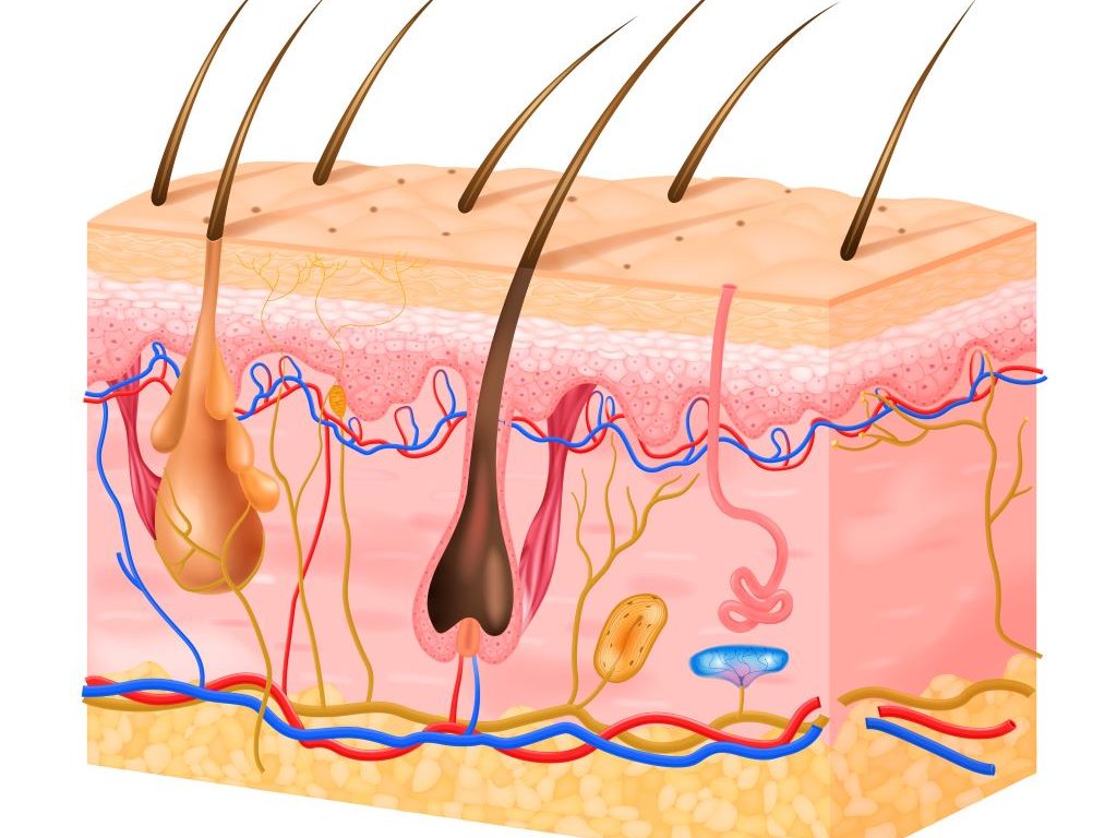
Chronic exposure of human skin to ultraviolet light causes premature aging, or photoaging. As the skin undergoes photoaging, type I collagen bundles, which are found in the dermis beneath the top layer of the skin and provide strength and support to skin, become fragmented. This leads to wrinkles, fragility and loss of support and elasticity.
“The best way to prevent damage to type I collagen by sunlight is to wear sunscreen consistently, daily if possible and particularly when spending time outdoors,” said Frank Wang, MD, the William B. Taylor Endowed Professor of Clinical Dermatology at U-M Medical School.
Experts observed in a new study that injection of the most popular type of dermal filler, cross-linked hyaluronic acid, into photoaged skin could reverse the dermal changes associated with photoaging.
These fillers are typically injected into the skin to reduce lines and wrinkles.
They are thought to provide clinical improvement by adding volume to the skin, but researchers have found that cross-linked hyaluronic acid also stimulates production of new type I collagen in the dermis.
The filler does so rapidly, stimulating collagen production within several weeks of injection, and is long-lasting, promoting the accumulation of more collagen over the course of a year.
These findings indicate how the filler improves the appearance of skin in the short-term – a combination of space-filling and collagen.
Additionally, since newly formed dermal collagen lasts many years, the findings also provide insight into how the filler can promote long-term clinical improvement, months or even a year after injection.
“A single injection of cross-linked hyaluronic acid dermal filler can lead to rapid and long-lasting improvement of skin by stimulating collagen deposition, and furthermore, repeat injections may add more collagen, eventually reducing the need for re-treatment,” Wang said.