Category: Cardiovascular Disease

Large Study Challenges Notion of Moderate Alcohol’s Cardiac Benefits

People clinking wine glasses
Photo by Jep Gambardella on Pexels

Though light alcohol consumption may provide heart-related health benefits has been suggested observational research, a large study published in JAMA Network Open showed a link between all levels of alcohol intake and higher risks of cardiovascular disease. The researchers found that the supposed benefits of alcohol consumption may in fact be attributable to other healthy lifestyle factors common among light to moderate drinkers.

The study included 371 463 adult participants from the UK Biobank, average age 57 and consuming an average of 9.2 drinks per week. In line with previous findings, researchers found that the lowest heart disease risk was in light to moderate drinkers, followed by people who abstained from drinking. People who drank heavily had the highest risk. However, light to moderate drinkers also tended to have healthier lifestyles than abstainers, such as more physical activity and vegetable intake, and less smoking. One a few lifestyle factors were taken into account, any benefit associated with alcohol consumption was significantly reduced.

The study also used new techniques in Mendelian randomisation, which uses genetic variants to determine whether an observed link between an exposure and an outcome is consistent with a causal effect. “Newer and more advanced techniques in ‘non-linear Mendelian randomisation’ now permit the use of human genetic data to evaluate the direction and magnitude of disease risk associated with different levels of an exposure,” said senior author Krishna G. Aragam, MD, MS, a cardiologist at MGH and an associate scientist at the Broad Institute. “We therefore leveraged these new techniques and expansive genetic and phenotypic data from biobank populations to better understand the association between habitual alcohol intake and cardiovascular disease.”

When such genetic analyses were performed on samples taken from participants, they found that individuals with genetic variants that predicted higher alcohol consumption were indeed more likely to consume greater amounts of alcohol, and more likely to have hypertension and coronary artery disease. The analyses also revealed significant differences in cardiovascular risk across the spectrum of alcohol consumption for both males and females, with minimal risk increase when going from zero to seven drinks per week, much higher risk increases when progressing from seven to 14 drinks per week, and greatly increased risk for 21 or more drinks per week. Notably, the findings suggest a rise in cardiovascular risk even at “low risk” levels (ie below two drinks per day for men and one per day for women).

This discovery of an exponential rather than liner relationship between alcohol intake and cardiovascular risk is was supported by an additional analysis of data on 30 716 participants in the Mass General Brigham Biobank. Therefore, cutting back on large consumption of alcohol may have even more clinical benefits than cutting back on moderate amounts.

“The findings affirm that alcohol intake should not be recommended to improve cardiovascular health; rather, that reducing alcohol intake will likely reduce cardiovascular risk in all individuals, albeit to different extents based on one’s current level of consumption,” said Dr Aragam.

Source: Massachusetts General Hospital

New ACC Expert Consensus Decision Pathway on Cardiovascular Sequelae of COVID

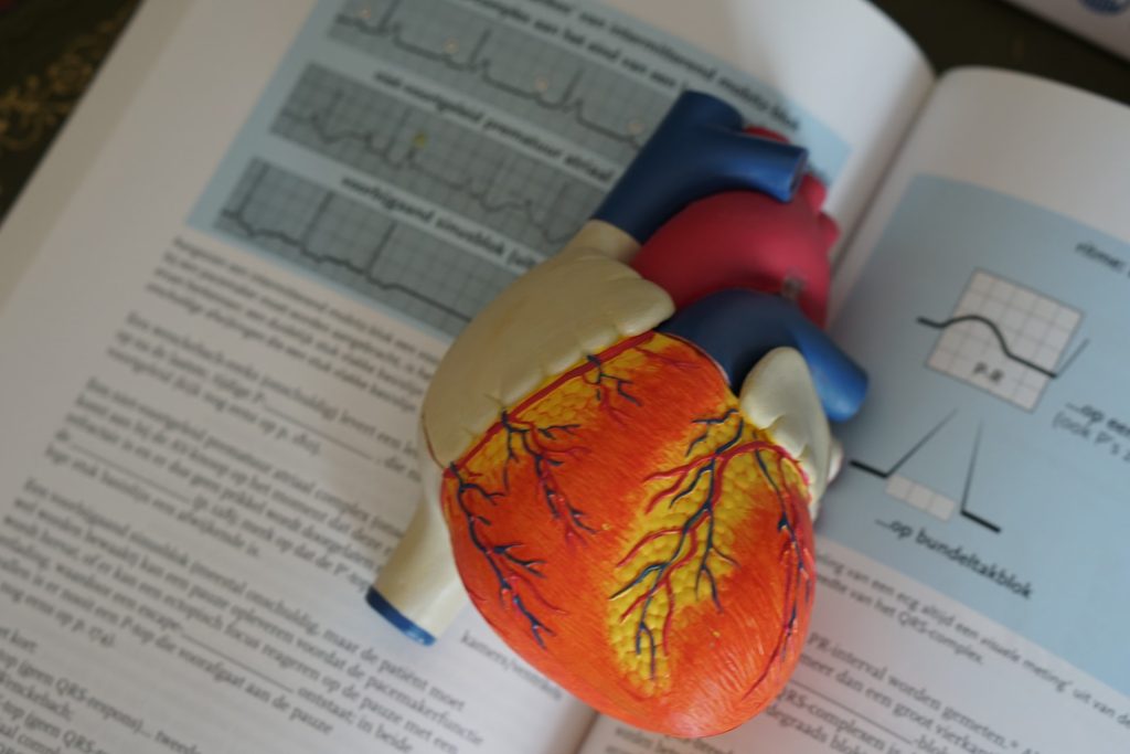
Anatomical model of a human heart
Photo by Robina Weermeijer on Unsplash

The American College of Cardiology has issued an expert consensus decision pathway for the evaluation and management of adults with key cardiovascular consequences of COVID. The document discusses myocarditis and other types of myocardial involvement, patient-centred approaches for long COVID and guidance on resumption of exercise following COVID. The clinical guidance was published today in the Journal of the American College of Cardiology.

“The best means to diagnose and treat myocarditis and long COVID following SARS-CoV-2 infection continues to evolve,” said Ty Gluckman, MD, MHA, co-chair of the expert consensus decision pathway. “This document attempts to provide key recommendations for how to evaluate and manage adults with these conditions, including guidance for safe return to play for both competitive and non-competitive athletes.”

Myocarditis

Myocarditis is a condition defined by the presence of cardiac symptoms such as chest pain, an elevated cardiac troponin, and abnormal ECG, cardiac imaging and/or cardiac biopsy findings.

Although rare, myocarditis with COVID is more commonly seen in men, and since it is associated with a higher risk of cardiac complications, a proactive management plan should be in place. For mild or moderate myocarditis, hospitalisation is recommended to closely monitor for worsening symptoms, while undergoing follow-up testing and treatment. Patients with severe myocarditis should ideally be hospitalised at appropriately equipped centres.

Myocarditis following COVID-19 mRNA vaccination is also rare and the benefits outweigh the risks. It is most commonly seen in younger males (40.6 cases per million for ages 12–29). Although most cases of myocarditis following COVID mRNA vaccination are mild, it should be diagnosed and treated similarly to myocarditis following COVID infection.

Long COVID

Post-acute sequelae of SARS-CoV-2 infection (PASC), or long COVID, is reported by up to 10-30% of infected individuals. It is defined by a constellation of new, returning or persistent health problems experienced by individuals four or more weeks after COVID infection. While individuals with this condition may experience wide-ranging symptoms, tachycardia, exercise intolerance, chest pain and shortness of breath represent some of the symptoms that draw increased attention to the cardiovascular system.

The writing committee has proposed two terms to better understand potential aetiologies for those with cardiovascular symptoms:

PASC-CVD, or PASC-Cardiovascular Disease, refers to a broad group of cardiovascular conditions (including myocarditis) that manifest at least four weeks after COVID infection.

PASC-CVS, or PASC-Cardiovascular Syndrome, includes a wide range of cardiovascular symptoms without objective evidence of cardiovascular disease following standard diagnostic testing.

Generally, patients with long COVID and cardiovascular symptoms should undergo evaluation with laboratory tests, ECG, echocardiogram, ambulatory rhythm monitor and/or additional pulmonary testing based on the clinical presentation. Cardiology consultation is recommended for abnormal test results, with additional evaluation based on the suspected clinical condition (eg, myocarditis).

Because multiple factors likely underlie PASC-CVS, evaluation and management may be best driven by the predominant cardiovascular symptom(s). For those with tachycardia and exercise intolerance, increased bedrest and/or a decline in physical activity may trigger cardiovascular deconditioning with progressive worsening of symptoms.

“There appears to be a ‘downward spiral’ for long COVID patients. Fatigue and decreased exercise capacity lead to diminished activity and bedrest, in turn leading to worsening symptoms and decreased quality of life,” said Nicole Bhave, MD, co-chair of the expert consensus decision pathway. “The writing committee recommends a basic cardiopulmonary evaluation performed upfront to determine if further specialty care and formalized medical therapy is needed for these patients.”

For PASC-CVS patients with tachycardia and exercise intolerance, upright exercise (walking or jogging) should be replaced with recumbent or semi-recumbent exercise (rowing, swimming or cycling) to avoid worsening fatigue. Exercise intensity and duration should be low initially, with gradual increases in exercise duration over time. Transition back to upright exercise can be done as  symptoms improve. Additional interventions (increased salt and fluid intake, elevation of the head during sleep, support stockings) and pharmacological treatments (beta-blockers) should be considered on a case-by-case basis.

Return to Play

Concerns arose about return to play for athletes after COVID due to observations of cardiac injury among some hospitalised COVID patients, along with uncertainty around cardiovascular sequelae after mild illness. However, data do not show a low prevalence of clinical myocarditis and no increase of cardiac events.

For athletes recovering from COVID with ongoing cardiopulmonary symptoms or those requiring hospitalisation with increased suspicion for cardiac involvement, further evaluation with triad testing (ECG, cardiac troponin and echocardiogram) should be performed. For those with abnormal test results, further evaluation with cardiac MRI should be considered. Individuals diagnosed with clinical myocarditis should abstain from exercise for three to six months.

Cardiac testing is not recommended for asymptomatic individuals following COVID infection. Individuals should abstain from training for three days to ensure that symptoms do not develop. For those with mild or moderate non-cardiopulmonary symptoms (fever, lethargy, muscle aches), training may resume after symptom resolution. For those with remote infection (≥ three months) without ongoing cardiopulmonary symptoms, a gradual increase in exercise is recommended without the need for cardiac testing.

Based on the low prevalence of myocarditis observed in competitive athletes with COVID-19, the authors note that these recommendations can be reasonably applied to high-school athletes (aged ≥ 14 years) along with adult recreational exercise enthusiasts. Future study is needed, however, to better understand how long cardiac abnormalities persist following COVID infection and the role of exercise training in long COVID.

Source: American College of Cardiology

Blood Pressure Rise on Standing up Linked to Cardiovascular Risk

Blood pressure cuff
BP cuff for home monitoring, Source: Pixabay

Among young and middle-aged adults with high blood pressure, a substantial rise in blood pressure upon standing may identify those with a higher risk of serious cardiovascular events, such as heart attack and stroke, according to new research published in the journal Hypertension.

“This finding may warrant starting blood-pressure-lowering treatment including medicines earlier in patients with exaggerated blood pressure response to standing,” said Professor Paolo Palatini, MD, lead author of the study.

Blood pressure usually falls slightly upon standing up. In this study, researchers assessed whether the opposite response – a significant rise in systolic blood pressure upon standing – is a risk factor for heart attack and other serious cardiovascular events.

Researchers recruited 1207 people aged 18-45 years old with untreated stage 1 hypertension, from the ongoing HARVEST study which started in 1990. Stage 1 hypertension was defined as systolic blood pressure of 140–159 mm Hg and/or diastolic BP 90–100 mm Hg. None had taken blood pressure-lowering medication prior to the study, and all were initially classed as low risk for major cardiovascular events based on lifestyle and medical history.

The researchers took six blood pressure measurements in various physical positions, including when lying down and after standing up. The 120 participants with the highest rise (top 10%) in blood pressure upon standing averaged an 11.4mmHg increase; all increases in this group were greater than 6.5mmHg. Remaining participants averaged a 3.8mmHg fall in systolic blood pressure upon standing.

The researchers compared heart disease risk factors, laboratory measures and the occurrence of major cardiovascular events (heart attack, heart-related chest pain, stroke, aneurysm of the aortic artery, clogged peripheral arteries) and chronic kidney disease among participants in the two groups. In some analyses, the development of atrial fibrillation, an arrhythmia that is a major risk factor for stroke, was also noted. Results were adjusted for age, gender, parental history of heart disease, and several lifestyle factors and measurements taken during study enrolment.

During an average 17-year follow-up, there were 105 major cardiovascular events among the participants. The most common were heart attack, heart-related chest pain and stroke.

People in the top 10% for rise in blood pressure:

  • had nearly twice the risk for a major cardiovascular event compared to the others;
  • did not generally have a higher risk profile for cardiovascular events during their initial evaluation (outside of the exaggerated blood pressure response to standing);
  • were more likely to be smokers (32.1% vs 19.9% in the non-rising group), yet physical activity levels were comparable, and they were not more likely to be overweight or obese, and no more likely to have a family history of cardiovascular events;
  • had more favourable cholesterol levels (lower total cholesterol and higher high-density-lipoprotein cholesterol);
  • had lower systolic blood pressure when lying down than the other group (140.5 mm Hg vs. 146.0 mm Hg, respectively), yet blood pressure measures were higher when taken over 24 hours.

After adjusting for average blood pressure taken over 24 hours, an exaggerated blood pressure response to standing remained an independent predictor of adverse heart events or stroke.

“The results of the study confirmed our initial hypothesis – a pronounced increase in blood pressure from lying to standing could be prognostically important in young people with high blood pressure. We were rather surprised that even a relatively small increase in standing blood pressure (6-7 mm Hg) was predictive of major cardiac events in the long run,” said Prof Palatini.

In a subset who had stress hormones measured from 24-hour urine samples, the epinephrine/creatinine ratio was higher in the people with rising BP compared to non-risers (118.4 nmol/mol vs 77.0 nmol/mol, respectively).

“Epinephrine levels are an estimate of the global effect of stressful stimuli over the 24 hours. This suggests that those with the highest blood pressure when standing may have an increased sympathetic response to stressors,” said Prof Palatini. “Overall, this causes an increase in average blood pressure.”

“The findings suggest that blood pressure upon standing should be measured in order to tailor treatment for patients with high blood pressure, and potentially, a more aggressive approach to lifestyle changes and blood-pressure-lowering therapy may be considered for people with an elevated blood pressure response to standing,” he said.

Source: American Heart Association

Cardiac CT Matches Coronary Angiography with Fewer Complications

Coronary artery showing atherosclerosis. Image source: Wikimidia CC0

A clinical trial found that cardiac computed tomography (CT) offers similar diagnostic accuracy to catheterisation – the current standard diagnostic test for intermediate-risk patients – in people with suspected coronary artery disease, as well as being associated with a lower risk of complications. The trial’s findings were published in the New England Journal of Medicine.

The current standard diagnostic test for coronary artery disease (CAD) is coronary angiography (often along with cardiac catheterisation). This minimally invasive procedure uses dye marker visible on X-ray imaging to detect arterial narrowing. Any narrowing detected in this manner can be treated during the procedure itself using stents, which prop open the newly widened blood vessels. More than 3.5 million of these procedures are carried out in European catheterisation laboratories every year, and more are carried out every year. Approximately two million of these do not involve immediate treatment in the cath lab. In these cases, the procedure is able to rule out narrowed or blocked coronary arteries.

The main question addressed by the DISCHARGE Trial Group was whether the low-risk, non-invasive coronary CT method can provide a safe alternative to catheterization in certain patients with suspected CAD. In order to test the effectiveness of both of these diagnostic imaging techniques in patients with stable chest pain, the project followed more than 3500 patients for a duration of four years. Patients were randomised to either computed tomography or cardiac catheterisation. If their initial evaluation ruled out obstructive coronary artery disease, participants were discharged back to their referring physician for further treatment – a step which gave the trial its name: DISCHARGE. Patients who were diagnosed as having the disease were managed in accordance with European guidelines at the time of the study.

Discussing the long-term results, trial leader Professor Dr Marc Dewey said: “The trial confirmed that a CT-based management is safe in patients with stable (ie, non-acute) chest pain and suspected coronary artery disease.”

Evaluation of safety was based on the incidence of major cardiovascular events over a period of up to four years. He added: “Among the patients referred for cardiac catheterisation and included in this trial, the risk of major adverse cardiovascular events was found to be similar in both the CT and catheterisation groups, occurring in 2.1% and 3.0% of patients, respectively. The incidence of major procedure-related complications was found to be four-times lower in patients managed with an initial CT strategy.”

Other outcome measures were included in the DISCHARGE trial, such as improvements in chest pain and quality of life over the course of the trial. This new strategy could help relieve pressure on health care systems by helping to reduce the volume of catheterisation procedures. Prof Dewey said: “Now that CT has been standardised and quality-tested as part of the DISCHARGE trial, this method could be made more widely available as part of the routine clinical care of people with intermediate CAD risk.”

As a next step, the trial’s method for estimating a person’s clinical risk of having coronary artery disease will need to be further evaluated to determine whether it can improve referral and indication for CT in routine clinical care. Health economics are an important component in making decisions about reimbursement in health care systems. As mentioned in the discussion of the publication, further methodologically very rigorous cost-effectiveness analyses of CT and cardiac catheterisation are necessary and will be conducted by the DISCHARGE Trial Group.

Source: University of Glasgow

Link Between LDL-C and CVD not as Strong as Thought

Source: Wikimedia CC0

New research has shown that the link between low density lipoprotein cholesterol (LDL-C) and cardiovascular disease may not be as strong as previously thought.

The study, published in JAMA Internal Medicine, provides evidence that calls into question the efficacy of statins when prescribed with the goal of lowering LDL-C and consequently cardiovascular disease (CVD) risk.

Numerous prior studies have suggested that using statins to lower LDL-C positively affects cardiovascular health outcomes, findings which are reflected in the various iterations of expert guidelines for the prevention of CVD. Several large clinical trials have indicated that for every 1-mmol/l reduction in LDL-C levels there is a 23% reduction in CVD risk.

The new findings contradict this theory, finding that this relationship was weaker than previously thought. Lowering LDL-C with statins in fact was found to have an inconsistent and inconclusive impact on CVD outcomes such as myocardial infarction (MI), stoke, and all-cause mortality.

Additionally, it indicates that the overall benefit of taking statins may be small and will vary depending on an individual’s personal risk factors.

Commenting on the findings, the paper’s lead author Dr Paula Byrne said: “The message has long been that lowering your cholesterol will reduce your risk of heart disease, and that statins help to achieve this. However, our research indicates that, in reality, the benefits of taking statins are varied and can be quite modest.”

The researchers go on to suggest that this updated information should be communicated to patients through informed clinical decision-making and updated clinical guidelines and policy.

Source: RCSI University

Two Biomarkers Predict CVD Risk in Psoriatic Disease

Photo by Hush Naidoo Jade Photography on Unsplash

In a new study published in Arthritis & Rheumatology, scientists have found that two biomarkers predict cardiovascular disease (CVD) risk in people with psoriatic disease. People with psoriatic disease, which includes psoriasis and psoriatic arthritis, are more likely to develop CVD than the general population.

The study, which included 1000 adults with psoriatic disease, found that elevated blood levels of two indicators of cardiovascular health, namely, cardiac high-sensitivity troponin I (cTnI) and N-terminal pro-brain-type natriuretic peptide (NT-proBNP), were associated with higher risks of experiencing cardiovascular problems independent of traditional risk factors such as hypertension and high cholesterol.

These findings pave the way for further studies exploring the clinical potential of measuring cTnI and NT-proBNP levels in helping assess the heart health of individual patients with psoriatic disease.

“Our study provides new insights regarding the pathophysiology of cardiovascular diseases in psoriasis and psoriatic arthritis. However, at this time, ordering tests of cardiac biomarkers is not recommended for risk stratification of asymptomatic patients with psoriatic disease,” said senior Lihi Eder, MD, PhD, associate professor of medicine at Women’s College Hospital and University of Toronto.

Source: Wiley

Comprehensive Bloodstream Lipid Level Test Can Predict CVD Decades Early

Source: Pixabay CC0

Lipidomics, measuring many different bloodstream lipid levels, can predict the risk of developing type 2 diabetes (T2D) and cardiovascular disease (CVD) years in the future, according to a new study in PLOS Biology. Such early prediction through lipidomic profiling may provide the basis for recommending diet and lifestyle interventions before disease develops.

At present, patient history and current risk behaviours are the main predictors for T2D and CVD, along with high- and low-density cholesterol ratios and levels. But there are over one hundred other types of lipids in the blood, which are thought to at least partially reflect aspects of metabolism and homeostasis throughout the body.

Nowadays, it is possible to measure thousands of individual lipids that make up the lipidome. Nuclear magnetic resonance spectrometry (NMR) metabolomics is also being increasingly used in large cohort studies to report on total levels of selected lipid classes, and relative levels of fatty acid saturation.

To find out if detailed lipid profiles could be better predictors, the authors drew on data and blood samples from a longitudinal health study of over 4000 middle-aged participants, first assessed from 1991 to 1994, with follow-up to 2015. Using baseline blood samples, the concentrations of 184 lipids were assessed. During the follow-up period, 13.8% of participants developed T2D, and 22% developed CVD.

The authors performed repeated training and testing on the data to create a risk model. Once the model was developed, individuals were clustered into one of six subgroups based on their lipidomics profile.

Compared to the group averages, the risk for T2D in the highest-risk group was 37%, an increase in risk of 168%. The risk for CVD in the highest-risk group was 40.5%, an increase in risk of 84%. Significant reductions in risk compared to the averages were also seen in the lowest-risk groups. The increased risk for either disease was independent of known genetic risk factors, and independent of the number of years until disease onset.

Rsk could be individually defined decades before disease onset, possibly in time to take steps to avert disease. Lipidomics could be combined with genetics and patient history to provide new insights into the beginnings of the disease. Additionally, new drug candidates could be identified from the lipids  contributing the greatest risk.

“The lipidomic risk, which is derived from only one single mass-spectrometric measurement that is cheap and fast, could extend traditional risk assessment based on clinical assay,” said lead researcher Chris Lauber of Lipotype. “In addition, individual lipids in blood may be the consequences of or contribute to a wide variety of metabolic processes, which may be individually significant as markers of those processes. If that is true, Lauber said, “the lipidome may provide insights much beyond diabetes and cardiovascular disease risk.”

Lauber added: “Strengthening disease prevention is a global joint effort with many facets. We show how lipidomics can expand our toolkit for early detection of individuals at high risk of developing diabetes and cardiovascular diseases.”

Source: EurekAlert!

Antiepileptics in Comatose Cardiac Arrest Survivors are Ineffective, Study Shows

Image by Falkurian Design on Unsplash

A large scale study of comatose intensive care (ICU) patients admitted after cardiac arrest and resuscitation has shown that antiepileptics to treat epilepsy-like brain activity has no effect, and may even prolong ICU stay.

Following a cardiac arrest and resuscitation, patients may need an ICU stay, and are in a coma. By that stage, the cardiac arrest may have damaged the brain to such an extent that half of the patients will not recover from coma. The other half will also have permanent damage, for example of memory functions. It is extremely difficult to predict if a patient will awaken and what their prognosis is, so clinicians make use of EEG (electroencephalography).

In 10–20% of the patients admitted to the ICU after cardiac arrest and resuscitation, there are signs of brain activity that appear similar to epilepsy: unlike an attack this activity is continuous. For a long time, it was unclear if anti-epileptic medication could help better recovery. As a result, some patients received this medication and some did not.

Now, a large-scale study done between 2014 and 2021 on 172 patients has proven that this medication is ineffective: it does not help recovery, even necessitating a longer ICU stay. The researchers, led by Professor Jeannette Hofmeijer of the University of Twente and Rijnstate Hospital in Arnhem, published their findings in the New England Journal of Medicine.

The conclusion from this study is that anti-epileptic medication does not lead to an improved recovery. The findings show that patients may need to stay longer at the ICU: for the patient an undesired situation, and it puts extra pressure on the health care system. 

Aside from patients who show continuous epileptic signals, a small group of patients show signs of a typical epileptic seizure: a short and heavy attack. In these situations, anti-epileptics could help, but this still needs further research.

“Although the outcome of the trial may be disappointing in terms of chances of recovery, it also takes away uncertainties from the family. The signals point at serious brain damage that would lead to a much longer stay at the ICU,” said Prof Hofmeijer.

Source: University of Twente

High CAC and Lipoprotein(a) Scores Greatly Worsen CVD Risk

Healthy red blood cells. Source: NIH

Having both a high lipoprotein(a) and high coronary artery calcium score (CAC) results in a 22% risk of heart attack or stroke over the following 10 years, nearly double the risk of having either condition alone. These are the findings are from a study published in the Journal of the American College of Cardiology (JACC).

Two decades ago, it was recognised that lipoprotein(a) (Lp(a)) concentrations were elevated in patients with cardiovascular disease (CVD). However Lp(a) was not yet proven to be important due to a lack of both Lp(a)-lowering therapy and evidence that reducing Lp(a) levels improves CVD risk. Recent research has added to the evidence 

“We are hopeful that by making the connection between Lp(a) and CAC as dual risk drivers, we can raise awareness in the medical community and improve earlier heart attack prevention for these patients,” said cardiologist Parag Joshi, MD, Associate Professor of Internal Medicine at UT Southwestern. “Our data may also expedite the development of treatments designed specifically for this high-risk population.”

About one sixth of people in the U.S. have high Lp(a), driven largely by genetics. Coronary artery calcium (CAC) is a marker of plaque deposits around the heart. 

Cardiology researchers confirmed the Lp(a) and CAC connection by comparing data from two landmark cardiovascular trials, the Dallas Heart Study, an ongoing comprehensive study of 6000 diverse and heart-healthy patients conducted from 2000 to present, and the Multi-Ethnic Study of Atherosclerosis (MESA) 6000-participant study investigating early-stage atherosclerosis.

The researchers found that participants with combined high Lp(a) and high CAC had a 22% 10-year risk of heart attack or stroke, compared with a 10-15% 10-year risk in patients who had either risk factor alone.

The team identified three distinct risk-related trends:

  • High Lp(a), high CAC: These individuals face the highest 10-year risk of heart attack or stroke.
  • High Lp(a), zero CAC: 10-year heart attack and stroke risk is low when there is no CAC, even if Lp(a) is high.
  • Low Lp(a), high CAC: 10-year heart attack or stroke risk is higher than average but lower than with high LP(a) and high CAC combined.

“Establishing the connection between Lp(a) and CAC means we can move to the important next phase of research, which will be defining and personalizing early screening protocols to identify patients at high risk of heart attack,” said Dr Joshi. “With further research, this could mean selectively scanning patients with high Lp(a) for their CAC score, and studying therapies specifically designed to reduce Lp(a) among patients with high CAC.”

Source: UT Southwestern Medical Center

CVD Risk Warning for Paracetamol That Contains Sodium

Graphical abstract from European Heart Journal editorial: sodium hidden in medication warrants warning labels by drug companies

Clinicians have recommended avoiding effervescent, soluble paracetamol that contains sodium, following findings from a large study that shows a link with a significantly increased risk of cardiovascular disease (CVD) and mortality in people who have hypertension and even in people with normal blood pressure.

The study of nearly 300 000 patients registered with UK GPs was published in the European Heart Journal.

Sodium is often used to help drugs such as paracetamol dissolve and disintegrate in water. However, effervescent and soluble formulations of 0.5g tablets of paracetamol can contain 0.44 and 0.39g of sodium respectively. If a person took the maximum daily dose of two 0.5g tablets every six hours, they would consume 3.5 and 3.1g of sodium respectively – a dose that exceeds the WHO-recommended total daily intake of 2g a day. In 2018, 170 people per 10 000 of the population in the UK were using sodium-containing medications, with a higher proportion among women. There are alternative formulations that contain little or no sodium.

Excessive salt in the diet remains a major public health problem and is associated with an increased risk of cardiovascular disease (CVD) and death among patients with hypertension. However, there is inconsistent evidence showing an increased risk in normotensive individuals.

Professor Chao Zeng led a team which analysed data from a medical database of UK GPs’ records. They looked at 4532 hypertensive patients who had been prescribed sodium-containing paracetamol and compared them with 146 866 hypertensive patients who had been prescribed sodium-free paracetamol. They also compared 5351 normotensive patients who were prescribed sodium-containing paracetamol with 141 948 normotensive patients prescribed sodium-free paracetamol. The patients were aged 60-90 years and followed up for one year.

The researchers found the risk of heart attack, stroke or heart failure after one year for patients with high blood pressure taking sodium-containing paracetamol was 5.6% (122 cases of CVD), while it was 4.6% (3051 CVD cases) among those taking sodium-free paracetamol. Mortality risk was also higher; the one-year risk was 7.6% (404 deaths) and 6.1% (5510 deaths), respectively.

A similar increased risk was seen among normotensive patients. Among those taking sodium-containing paracetamol, the one-year CVD risk was 4.4% (105 cases of CVD) and 3.7% (2079 cases of CVD) among those taking sodium-free containing paracetamol. The risk of dying was 7.3% (517 deaths) and 5.9% (5190 deaths), respectively.

Prof Zeng said: “We also found that the risk of cardiovascular disease and death increased as the duration of sodium-containing paracetamol intake increased. The risk of cardiovascular disease increased by a quarter for patients with high blood pressure who had one prescription of sodium-containing paracetamol, and it increased by nearly a half for patients who had five or more prescriptions of sodium-containing paracetamol. We saw similar increases in people without high blood pressure. The risk of death also increased with increasing doses of sodium-containing paracetamol in both patients with and without high blood pressure.”

Prof. Zeng said that clinicians and patients should be aware of the risks associated with sodium-containing paracetamol and avoid unnecessary consumption, especially when the medication is taken for a long period of time.

“Given that the pain relief effect of non-sodium-containing paracetamol is similar to that of sodium-containing paracetamol, clinicians may prescribe non-sodium-containing paracetamol to their patients to minimise the risk of cardiovascular disease and death. People should pay attention not only to salt intake in their food but also not overlook hidden salt intake from the medication in their cabinet,” he said.

“Although the US Food and Drug Administration requires that all over-the-counter medications should label the sodium content, no warning has been issued about the potentially detrimental effect of sodium-containing paracetamol on the risks of hypertension, cardiovascular disease and death. Our results suggest re-visiting the safety profile of effervescent and soluble paracetamol.”

Being an observational study it can only show only that there is an association between salt in paracetamol and CVD and deaths, rather than that salt causes these events. Other limitations include a lack of data on dietary intake of salt and excretion of salt from urinary samples. The use of over-the-counter paracetamol was not also recorded, however by restricting the study to those over 60 who qualify for free prescriptions in the UK, the risk of this is minimised.

Source: EurekAlert!