Category: Cardiovascular Disease

After Cardiac Event, Excessive Sedentary Time Led to Increased Risk of Another Event

People who sit or remain sedentary for more than 14 hours a day, on average, may have a higher risk of a cardiovascular event or death in the year after treatment at a hospital for symptoms of a heart attack such as chest pain, according to new research published today in the American Heart Association’s peer-reviewed scientific journal Circulation: Cardiovascular Quality and Outcomes.

Previous research from the study authors found that people who had experienced a heart attack were spending up to 12 to 13 hours each day being sedentary, defined as any awake activity that involved little-to-no physical movement. In this study, the researchers used a wrist accelerometer to track the amount of time each participant spent moving or being sedentary for a median of 30 days after discharge from a hospital’s emergency department. 

Wrist accelerometers measure the acceleration of motion in three directions: forwards and backwards, side-to-side, and up and down. These measurements allowed the researchers to infer the intensity of a participant’s physical activity, and they provide more accurate measurements of the participants’ time spent moving, rather than asking participants to remember. Some examples of moderate intensity physical activities are brisk walking, water aerobics, dancing, playing doubles tennis or gardening, and examples of vigorous-intensity activities are running, lap swimming, heavy yardwork such as continuous digging or hoeing, playing singles tennis or jumping rope.

”Current treatment guidelines after a cardiac event focus mainly on encouraging patients to exercise regularly,” said study lead author Keith Diaz, Ph.D., the Florence Irving Associate Professor of Behavioral Medicine at Columbia University Medical Center in New York City, a certified exercise physiologist and a volunteer member of the American Heart Association’s Physical Activity Science Committee. “In our study, we explored whether sedentary time itself may contribute to cardiovascular risk.”

Researchers followed more than 600 adults, ages 21 to 96, treated for a heart attack or chest pain in the emergency department at a single hospital system in New York City. Participants wore a wrist accelerometer for a median of 30 consecutive days after hospital discharge to measure the amount of time they spent sitting or being inactive each day. Additional cardiac events and deaths were evaluated one year after hospital discharge via phone surveys with patients, electronic health records and the Social Security Death Index. The study was focused on understanding the risk of sedentary behavior and identifying modifiable risk factors that may improve long-term outcomes in this high-risk group.

The analysis found:

  • Compared to participants in the group with the highest physical activity level, those in the group with the lowest activity level had a 2.58 times higher risk of having another heart problem or dying within the next year.
  • Replacing 30 minutes of sedentary time with 30 minutes of moderate to vigorous physical activity, daily, reduced the risk of adverse cardiovascular events or death by 61%; replacing the sedentary time with light-intensity physical activity reduced risk by 50%; and replacing the sedentary time with 30 minutes of sleep lowered risk by 14%.
  • According to accelerometer data, participants in the most physically active group had average daily physical activity measures of 143.8 minutes of light physical activity; 25 minutes of moderate-to-vigorous physical activity; 11.7 hours spent sedentary; and 8.4 hours of sleep.
  • Participants in the least physically active group had daily averages of 82.2 minutes each day of light physical activity; 2.7 minutes of moderate-to-vigorous physical activity; 15.6 hours spent sedentary; and 6.6 hours of sleep.
  • Participants in the group between most and least physically active had daily averages of 109.2 minutes of light intensity physical activity; 11.4 minutes of moderate-to-vigorous intensity physical activity; 13.5 hours spent sedentary and 7.8 hours of sleep.

“We were surprised that replacing sedentary time with sleep also lowered risk. Sleep is a restorative behavior that helps the body and mind recover, which is especially important after a serious health event like a heart attack,” Diaz said. “Our study indicates that one doesn’t have to start running marathons after a cardiovascular event to see benefits. Sitting less and moving or sleeping a little more can make a real difference. More physical activity and more sleep are healthier than sitting, so we hope these findings support health professionals to move toward a more holistic, flexible and individualized approach for physical activity in patients after a heart attack or chest pain.”

Physical activity and sleep are both key components of the American Heart Association’s Life’s Essential 8, a list of health behaviours and factors that support optimal cardiovascular health. Poor sleep is a known risk factor for cardiovascular disease, which claims more lives each year in the U.S. than all forms of cancer and chronic lower respiratory disease combined, according to the American Heart Association’s 2025 Statistical Update. In addition to sleep duration, a recent scientific statement from the Association highlighted the importance of sleep continuity, sleep timing, sleep satisfaction, sleep regularity, sleep-related daytime functioning and sleep architecture in cardiometabolic health.

The study had several limitations, including that the definition of sedentary behaviour was based only on the intensity level of physical movement, meaning that the study may have overestimated the time participants spent in sedentary behaviour. Additionally, there was no information about participants’ income and characteristics of the neighbourhoods where they live, which limits the study’s ability to account for social and environmental factors, including participants’ risk of one-year cardiac events and deaths. Also, hospital discharge information about whether patients were sent home, referred to rehabilitation or referred to other care centres such as skilled nursing facilities were not collected. This limited the study’s ability to fully assess whether the patients’ settings had an impact on their recovery.

Source: American Heart Association

Controlling 8 Risk Factors may Eliminate Early Death Risk from Hypertension

Pexels Photo by Thirdman

A new study led by researchers at Tulane University suggests that people with hypertension can significantly reduce, and possibly eliminate, their increased risk of premature death by controlling several key health risk factors at once.

The study, appearing in Precision Clinical Medicine, tracked more than 70 000 people with hypertension and over 224 000 without it, using data from the UK Biobank. Researchers followed participants for nearly 14 years to understand how managing these risk factors affected early mortality, defined as dying before age 80.

The eight health risk factors evaluated in the study include: blood pressure, body mass index, waist circumference, LDL “bad” cholesterol, blood sugar, kidney function, smoking status and physical activity. Notably, researchers found that hypertensive patients who had addressed at least four of these risk factors had no greater risk of an early death than those without hypertension. 

“Our study shows that controlling blood pressure is not the only way to treat hypertensive patients, because high blood pressure can affect these other factors,” said corresponding author Dr Lu Qi, professor of epidemiology at Tulane University. “By addressing the individual risk factors, we can help prevent early death for those with hypertension.” 

Hypertension, defined as a blood pressure of 130mmHg or higher, is the leading preventable risk factor for premature death worldwide. 

The study found that addressing each additional risk factor was associated with a 13% lower risk of early death, 12% lower risk of early death due to cancer and 21% lower risk of death due to cardiovascular disease, the leading cause of premature death globally.

“Optimal risk control” – having 7 or more of the risk factors addressed – was linked to 40% less risk of early death, 39% less risk of early death due to cancer and 53% less risk of early death due to cardiovascular disease. 

“To our knowledge, this is the first study to explore the association between controlling joint risk factors and premature mortality in patients with hypertension,” Qi said. “Importantly, we found that any hypertension-related excess risk of an early death could be entirely eliminated by addressing these risk factors.” 

Only 7% of hypertensive participants in the study had seven or more risk factors under control, highlighting a major opportunity for prevention. Researchers say the findings underscore the importance of personalised, multifaceted care – not just prescribing medication for blood pressure, but addressing a broader range of health behaviours and conditions.

Source: Tulane University

The Effect of Physical Fitness on Mortality is Overestimated

Photo by Ketut Subiyanto on Unsplash

Many observational studies have shown that people who exercise more and have good cardiorespiratory fitness early in life are at lower risk of premature death from causes such as cancer and cardiovascular disease. But a new study published in the European Journal of Preventive Cardiology suggests that the association between physical fitness and a reduced risk of mortality may be misleading.

“We found that people with high fitness levels in late adolescence had a lower risk of dying prematurely, for example from cardiovascular disease, compared to those with low fitness levels. But when we looked at their risk of dying in random accidents, we found an almost similarly strong association. This suggests that people with high and low fitness levels may differ in other important ways, which is something that previous studies have not fully taken into account,” says Marcel Ballin, associated researcher in epidemiology and lead author of the study.

Conscription data from over 1 million men

In the study, the researchers leveraged data from 1.1 million Swedish men who were conscripted for military service between the years 1972 and 1995. The men, who were on average 18 years old at the time of conscription, were divided into five groups based on their fitness level at the time. They were then followed until their 60s or until they died. With access to the National Cause of Death Register, the researchers were able to see their cause of death. They subsequently used different methods to study the association between fitness level in late adolescence and premature death.

The researchers started with a traditional analysis of mortality from cardiovascular disease, cancer and from all causes, as in previous observational studies. They adjusted their statistical models for factors such as BMI, age at conscription, year of conscription, and parents’ income and education level. The results showed that the group with the highest fitness level had a 58% lower risk of dying from cardiovascular disease, a 31% lower risk of dying from cancer, and a 53% lower risk of dying from all causes, compared with the group with the lowest fitness level.

Very similar risk of dying in random accidents

Next, the researchers examined how fitness was associated with the risk of dying in random accidents such as car accidents, drownings and homicides. They chose random accidents because they assumed that there ought to be no association between the men’s fitness in late adolescence and the risk of dying in random accidents. This method is called negative control outcome analysis and involves testing the validity of your results for a primary outcome by comparing them with an outcome where no association ought to be found. If, however, an association is found, it may indicate that the groups studied are not actually comparable, and that the study suffers from what is typically referred to as confounding. The researchers found that men with the highest fitness levels had a 53% lower risk of dying in random accidents. Yet, it is unlikely that the men’s fitness would have such a big effect on their risk of dying in random accidents.

These results were also confirmed when the researchers used the sibling comparison design. Using this method, the researchers compared the risk of premature death between siblings with different fitness levels to control for all the factors that the siblings share such as behaviours, environmental factors, and some genetic factors.

“It surprised us that the association with accidental mortality reflected the other associations, even after we controlled for all the factors that siblings share. This underlines how strong the assumptions are that you make in observational studies, since it appears to be very difficult to create comparable groups. The consequences may be that you overestimate the magnitudes of the effects you find,” says Marcel Ballin.

Picture confirmed in other studies

The study is one of the largest of its kind in which researchers used negative control outcomes to investigate whether the associations between fitness and mortality are in fact valid. The results in this study are also supported by other research.

“That the effects of good cardiorespiratory fitness may be overstated might sound controversial to some, but the fact is that if you look at the results from studies others than traditional observational studies, a more nuanced picture does emerge. A number of twin studies for example have found similar results. Some genetic studies also suggest that there are genes that affect both the propensity to be physically active or have a good fitness level, and the risk of developing diseases such as cardiovascular disease.”

Important to base interventions on correct estimates

Marcel Ballin also argues that there are many different reasons for promoting physical activity. However, large-scale interventions or policy changes intended to apply to the entire population must be based on reliable estimates – otherwise there is a risk of expecting effects that have in fact been overestimated.

“Our results should not be interpreted as if physical activity and exercise are ineffective or that you should not try to promote it. But to create a more nuanced understanding of how big the effects of fitness actually are on different outcomes, we need to use several different methods. If we just ask the question in the same way, we will always get the same answer. It’s only when we get the same answer to a question that we have asked in slightly different ways that we can be sure that the findings are accurate,” says Marcel Ballin.

Source: Uppsala University

Could a Transient Ischaemic Attack Leave Lasting Fatigue?

Photo by Karolina Grabowska on Pexels

A transient ischaemic attack (TIA) is typically defined as a temporary blockage of blood flow to the brain that causes symptoms that go away within a day, but a new study finds that people who have this type of stroke may also have prolonged fatigue lasting up to one year. The study is published in Neurology®, the medical journal of the American Academy of Neurology (AAN).

The study does not prove that TIAs, also known as mini-strokes, cause lasting fatigue; it only shows an association. “People with a transient ischaemic attack can have symptoms such as face drooping, arm weakness or slurred speech and these resolve within a day,” said study author Boris Modrau, MD, PhD, of Aalborg University Hospital in Denmark. “However, some have reported continued challenges including reduced quality of life, thinking problems, depression, anxiety and fatigue. Our study found that for some people, fatigue was a common symptom that lasted up to one year after the transient ischaemic attack.”

The study involved 354 people with an average age of 70 who had a mini-stroke. They were followed for a year.

Participants completed questionnaires about their level of fatigue within the first two weeks of the mini-stroke and again at three, six, and 12 months later. One questionnaire looked at five different types of fatigue, including overall tiredness, physical tiredness, reduced activity, reduced motivation and mental fatigue. Scores ranged from four to 20 with higher scores indicating more fatigue. Participants had an average score of 12.3 at the start of the study. At three months, the average score decreased slightly to 11.9, at six months to 11.4 and at twelve months to 11.1.

Researchers looked at how many participants experienced fatigue as defined as a score of 12 or higher. Of the participants, 61% experienced fatigue two weeks after the mini-stroke and 54% experienced fatigue at each of the three other testing time periods at three, six and 12 months.

Participants also had brain scans. Researchers found that the presence of a blot clot on a scan was equal between people with long term fatigue and those without it, so this did not explain the reason for the level of fatigue.

Researchers did find that previous anxiety or depression was twice as common in those participants who reported lasting fatigue.

“Long-term fatigue was common in our group of study participants, and we found if people experience fatigue within two weeks after leaving the hospital, it is likely they will continue to have fatigue for up to a year,” said Modrau. “For future studies, people diagnosed with a transient ischaemic attack should be followed in the weeks and months that follow to be assessed for lingering fatigue. This could help us better understand who might struggle with fatigue long-term and require further care.”

A limitation of the study was that while participants were asked to complete the questionnaires themselves, it is possible some responses may have been completed with assistance from relatives or caretakers and this may have influenced responses, including those around fatigue.

Source: American Academy of Neurology

Elevated Risk of Death or Complications from Broken Heart Syndrome

Credit: American Heart Association

Takotsubo cardiomyopathy, also known as broken heart syndrome, is associated with a high rate of death and complications, and those rates were unchanged between 2016 and 2020, according to new research published in the Journal of the American Heart Association, an open-access, peer-reviewed journal of the American Heart Association.

Takotsubo cardiomyopathy is a stress-related heart condition in which part of the heart temporarily enlarges and doesn’t pump well. It is thought to be a reaction to a surge of stress hormones that can be caused by an emotionally or physically stressful event, such as the death of a loved one or a divorce. It can lead to severe, short-term failure of the heart muscle and can be fatal. Takotsubo cardiomyopathy may be misdiagnosed as a heart attack because the symptoms and test results are similar.

This study is one of the largest to assess in-hospital death rates and complications of the condition, as well as differences by sex, age and race over five years.

“We were surprised to find that the death rate from Takotsubo cardiomyopathy was relatively high without significant changes over the five-year study, and the rate of in-hospital complications also was elevated,” said study author M. Reza Movahed, MD, PhD, an interventional cardiologist and clinical professor of medicine at the University of Arizona’s Sarver Heart Center in Tucson, Arizona. “The continued high death rate is alarming, suggesting that more research be done for better treatment and finding new therapeutic approaches to this condition.”

Researchers reviewed health records in the Nationwide Inpatient Sample database to identify people diagnosed with Takotsubo cardiomyopathy from 2016 to 2020.

The analysis found:

  • The death rate was considered high at 6.5%, with no improvement over period.
  • Deaths were more than double in men at 11.2% compared to the rate of 5.5% among women.
  • Major complications included congestive heart failure (35.9%), atrial fibrillation (20.7%), cardiogenic shock (6.6%), stroke (5.3%) and cardiac arrest (3.4%).
  • People older than age 61 had the highest incidence rates of Takotsubo cardiomyopathy. However, there was a 2.6 to 3.25 times higher incidence of this condition among adults ages 46-60 compared to those ages 31-45 during the study period.
  • White adults had the highest rate of Takotsubo cardiomyopathy (0.16%), followed by Native American adults (0.13%) and Black adults (0.07%).
  • In addition, socioeconomic factors, including median household income, hospital size and health insurance status, varied significantly.

“Takotsubo cardiomyopathy is a serious condition with a substantial risk of death and severe complications,” Movahed said. “The health care team needs to carefully review coronary angiograms that show no significant coronary disease with classic appearance of left ventricular motion, suggesting any subtypes of stress-induced cardiomyopathy. These patients should be monitored for serious complications and treated promptly. Some complications, such as embolic stroke, may be preventable with an early initiation of anti-clotting medications in patients with a substantially weakened heart muscle or with an irregular heart rhythm called atrial fibrillation that increases the risk of stroke.”

He also noted that age-related findings could serve as a useful diagnostic tool in discriminating between heart attack/chest pain and Takotsubo cardiomyopathy, which may prompt earlier diagnosis of the condition and could also remove assumptions that Takotsubo cardiomyopathy only occurs in the elderly.

Among the study’s limitations is that it relied on data from hospital codes, which could have errors or overcount patients hospitalized more than once or transferred to another hospital. In addition, there was no information on outpatient data, different types of Takotsubo cardiomyopathy or other conditions that may have contributed to patients’ deaths.

Movahed said further research is needed about the management of patients with Takotsubo cardiomyopathy and the reason behind differences in death rates between men and women.

Source: American Heart Association
Lack of Sleep can Increase the Risk of Cardiovascular Disease

Photo by Andrea Piacquadio

Even a few nights with insufficient sleep promote molecular mechanisms linked to a greater risk of heart problems. This has been shown in a new study in which the researchers investigated how sleep deprivation affects biomarkers (in this case, proteins) associated with cardiovascular disease. The Uppsala University-led study is published in the journal Biomarker Research.

“Unfortunately, nearly half of all Swedes regularly experience disturbed sleep, and this is particularly common among shift workers. That is why we wanted to try to identify mechanisms that affect how lack of sleep can increase the risk of cardiovascular disease. Ultimately, the purpose was to identify opportunities to address these problems,” says Jonathan Cedernaes, physician and docent at Uppsala University, who led the study.

A chronic lack of sleep is a growing public health problem and in large population studies it has been linked to an increased risk of heart attack, stroke and atrial fibrillation. Heart health is influenced by several lifestyle factors, including sleep, diet and exercise. In order to separate out the effects of sleep, a number of conditions were controlled in the laboratory environment such as diet and physical activity.

How the study was conducted

The authors studied 16 healthy young men of normal weight. They all had healthy sleep habits. The participants spent time in a sleep laboratory where their meals and activity levels were strictly controlled in two sessions. In one session, participants got a normal amount of sleep for three consecutive nights, while during the other session, they got only about four hours of sleep each night. During both sessions, morning and evening blood samples were taken, and following high-intensity exercise lasting 30 minutes.

Inflammatory proteins increased after sleep loss

The researchers measured the levels of around 90 proteins in the blood and were able to see that the levels of many of these that are associated with increased inflammation rose when the participants were sleep-deprived. Many of these proteins have already been linked to an increased risk of cardiovascular disease such as heart failure and coronary artery disease.

“Many of the larger studies that have been done on the link between sleep deprivation and the risk of cardiovascular diseases have generally focused on slightly older individuals who already have an increased risk of such diseases. That is why it was interesting that the levels of these proteins increased in the same way in younger and previously perfectly healthy individuals after only a few nights of sleep deprivation. This means that it’s important to emphasise the importance of sleep for cardiovascular health even early in life,” says Jonathan Cedernaes.

The effects of exercise can be affected by lack of sleep

Physical exercise generated a slightly different response after lack of sleep. However, a number of key proteins increased equally, whether the person was sleep-deprived or not. Thus, proteins that can be linked to the positive effects of exercise increased, even if the person had too little sleep. The researchers have previously shown that exercise in the presence of sleep deprivation can result in a slightly increased load on the heart’s muscle cells.

“With this study, we have improved our understanding of what role the amount of sleep we get plays in cardiovascular health. It’s important to point out that studies have also shown that physical exercise can offset at least some of the negative effects that poor sleep can cause. But it’s also important to note that exercise cannot replace the essential functions of sleep,” says Jonathan Cedernaes.

Hopefully help to develop better guidelines

“Further research is needed to investigate how these effects might differ in women, older individuals, patients with heart disease, or those with different sleep patterns. Our ongoing research will hopefully help to develop better guidelines on how sleep, exercise and other lifestyle factors can be harnessed to better prevent cardiovascular diseases,” says Jonathan Cedernaes.

Source: Uppsala University

Does Anaemia During Pregnancy Affect Newborns’ Risk of Heart Defects?

Source: Pixabay CC0

New research published in BJOG: An International Journal of Obstetrics & Gynaecology found that mothers who are anaemic in early pregnancy face a higher likelihood of giving birth to a child with a heart defect.

The study assessed the health records of 2776 women with a child diagnosed with congenital heart disease who were matched to 13 880 women whose children did not have this condition.

Investigators found that 4.4% of children with congenital heart disease and 2.8% of children with normal heart function had anaemia. After adjusting for potential influencing factors, the odds of giving birth to a child with congenital heart disease was 47% higher among anaemic mothers.

“We already know that the risk of congenital heart disease can be raised by a variety of factors, but these results develop our understanding of anaemia specifically and take it from lab studies to the clinic. Knowing that early maternal anaemia is so damaging could be a gamechanger worldwide,” said corresponding author Duncan B. Sparrow, PhD, of the University of Oxford. “Because iron deficiency is the root cause of many cases of anaemia, widespread iron supplementation for women—both when trying for a baby and when pregnant—could help prevent congenital heart disease in many newborns before it has developed.”

Source: Wiley

Shingles Vaccine Reduces Heart Disease Risk for up to Eight Years

Photo by Gustavo Fring on Pexels

People who are given a vaccine for shingles have a 23% lower risk of cardiovascular events, including stroke, heart failure, and coronary heart disease, according to a study of more than a million people published in the European Heart Journal.

The protective effect of the vaccine lasts for up to eight years and is particularly pronounced for men, people under the age of 60 and those with unhealthy lifestyles, such as smoking, drinking alcohol and being inactive.

The study was led by Professor Dong Keon Yon from the Kyung Hee University College of Medicine, Seoul, South Korea. He said: “Shingles causes a painful rash and can lead to serious complications, especially in older adults and those with weak immune systems. Previous research shows that, without vaccination, about 30% of people may develop shingles in their lifetime.

“In addition to the rash, shingles has been linked to a higher risk of heart problems, so we wanted to find out if getting vaccinated could lower this risk.”

The study included nearly 1.3 million people aged 50 or older living in South Korea. Researchers gathered data, from 2012 onwards, on whether people received a shingle vaccine and combined this with data on their cardiovascular health and data on other factors that can influence health, such as age, sex, wealth and lifestyle.

The vaccine was a live zoster vaccine, meaning it contained a weakened form of the varicella zoster virus that causes shingles. In many countries, this type of vaccine is now being replaced with a non-live, recombinant vaccine, meaning it contains a protein from the varicella zoster virus.

The study showed that among people who received the vaccine, there was a 23% lower risk of cardiovascular events overall, with a 26% lower risk of major cardiovascular events (a stroke, heart attack or death from heart disease), a 26% lower risk of heart failure and a 22% lower risk of coronary heart disease.

The protective effect was strongest in the two to three years after the shingles vaccine was given, but researchers found that the protection lasted for up to eight years.

Professor Yon said: “Our study suggests that the shingles vaccine may help lower the risk of heart disease, even in people without known risk factors. This means that vaccination could offer health benefits beyond preventing shingles.

“There are several reasons why the shingles vaccine may help reduce heart disease. A shingles infection can cause blood vessel damage, inflammation and clot formation that can lead to heart disease. By preventing shingles, vaccination may lower these risks. Our study found stronger benefits in younger people, probably due to a better immune response, and in men, possibly due to differences in vaccine effectiveness.

“This is one of the largest and most comprehensive studies following a healthy general population over a period of up to 12 years. For the first time, this has allowed us to examine the association between shingles vaccination and 18 different types of cardiovascular disease. We were able to account for various other health conditions, lifestyle factors and socioeconomic status, making our findings more robust.

“However, as this study is based on an Asian cohort, the results may not apply to all populations. Since the live zoster vaccine is not suitable for everyone, more research on the recombinant vaccine is needed. While we conducted rigorous analysis, this study does not establish a direct causal relationship, so potential bias from other underlying factors should be considered.”

Professor Yon and his colleagues also plan to study the impact of the recombinant vaccine to see if it has similar benefits for reducing heart disease.

Source: European Society of Cardiology

Two Heart Transplants, One Epic Ride

Four Cyclists Take on the 2025 KAP sani2c to Inspire South Africans with a Message of Hope

L-R_Steph Bester CEO of AllLife with Billy McLeod

From 8 to 10 May 2025, four cyclists will take on one of South Africa’s most iconic and challenging mountain bike stage races – the KAP sani2c. Covering 265 kilometres over three days from the Southern Drakensberg to Scottburgh on the KwaZulu-Natal South Coast, the event is known for its tough terrain, stunning scenery, and relentless pace. But this year, one team will be racing with more than just medals in mind.

Team Change of Heart is made up of two heart transplant recipients – Billy Macleod (56) and Pedro Basson (20) – and their two experienced cycling partners: Steph Bester, CEO of AllLife, and long-time cycling companion Guy Dudley. Together, they’re not just riding to finish – they’re riding to inspire.

Billy, once an accomplished cyclist and triathlete, was sidelined for six years by heart disease and given just two weeks to live before receiving his transplant.          

He made a remarkable comeback in 2024 when he became the first South African heart transplant recipient to complete the KAP sani2c. This year, Billy returns with a renewed purpose – not just to ride, but to support 20-year-old Pedro through his very first stage race. For Billy, this ride is about passing the baton of hope and helping another transplant recipient believe in the impossible.

Pedro, the youngest team member at just 20 years old, will be attempting his first-ever mountain bike stage race. Having received his transplant as a teenager, Pedro’s journey to recovery has been nothing short of remarkable. Taking on sani2c is a bold and symbolic step, one he hopes will encourage other young people with chronic health conditions to dream big and push boundaries.

“This is more than a race – it’s a celebration of life and second chances,” says Steph Bester, who was set to ride alongside Billy last year but had to withdraw at the last minute due to a sudden illness. “Missing out in 2024 was heartbreaking for me, especially because this cause and this team are so close to my heart. I can’t wait to finally be on the bike with them this year. Billy and Pedro are living proof that your story doesn’t end with a diagnosis. At AllLife, we believe in the power of resilience, and this team embodies that belief. It’s an honour to support them – not only on the bike but in sharing their powerful message with the country.”

The team will be riding on e-bikes, enabling them to manage pace, terrain, and energy levels more effectively while still tackling the full challenge of the race. For Pedro, who has never raced before, the support of his three teammates – all experienced sani2c finishers – is crucial.

“Knowing I have a team behind me who understands the journey and is there to support every climb and descent makes all the difference,” Pedro shared. “It’s not just about the bike – it’s about proving to myself that anything is possible.”

AllLife is once again using this race to raise awareness for Hero777, a non-profit foundation focused on increasing organ and tissue donor registrations in South Africa. The foundation is deeply personal to the AllLife family as they deal with so many people in need of transplants daily.

It takes a community that truly understands both the unique challenges individuals face and the deep love for cycling to make something like this possible. The team is incredibly grateful for the support from AllLife, the wider cycling community, and especially race founder Glen Haw (Farmer Glen). Pedro will ride an e-bike generously sponsored by Cannondale, allowing him to tackle this life-affirming challenge alongside his team.

Whether it’s climbing steep hills or sharing laughs around the campfire, Team Change of Heart is ready to show that with support, courage, and a whole lot of heart – anything is possible.

Follow their journey on Instagram and Facebook (https://www.facebook.com/AllLifeSA and https://www.instagram.com/alllife_sa), and visit www.odf.org.za to learn more about organ and tissue donation or register as a donor today.

Micronanoplastics Found in Artery-clogging Plaque in the Neck

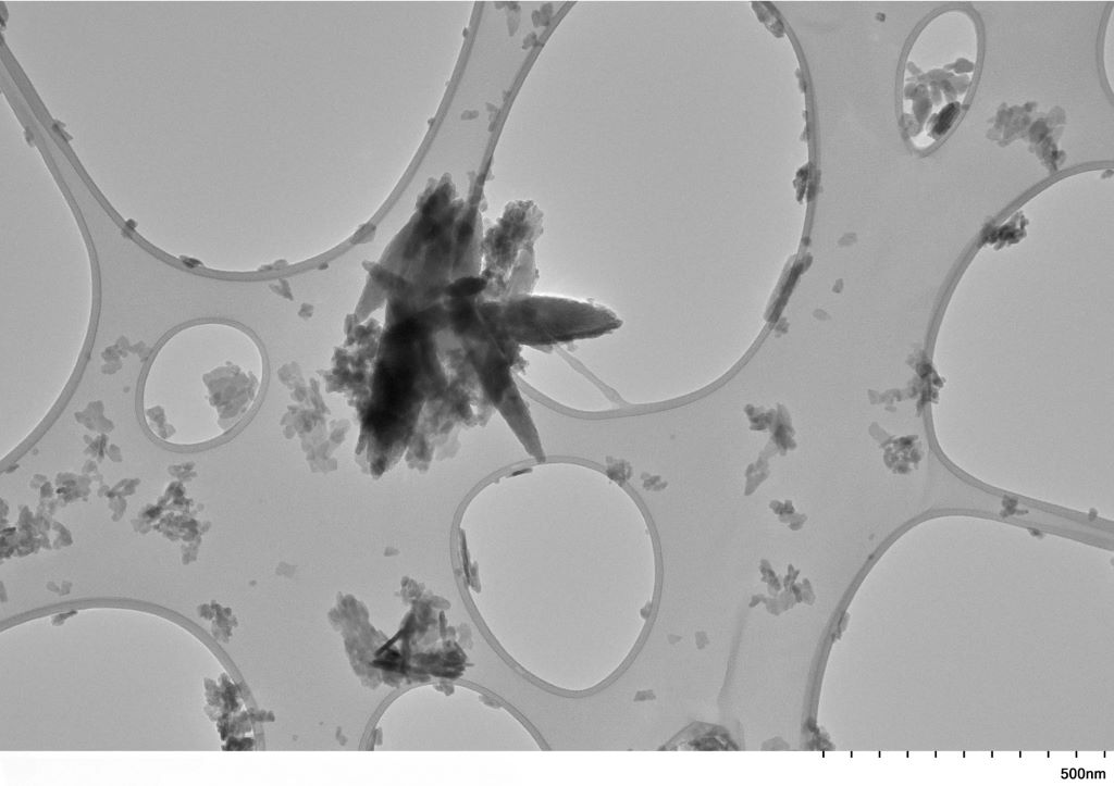
Transmission Electron Microscopy (TEM) picture of nanoplastic particles derived from carotid plaque. Copyright University of New Mexico

People with plaque in the blood vessels of their neck have a higher amount of tiny plastic particles in those vessels compared to people with healthy arteries. This increase was significantly higher in people who had experienced a stroke, mini-stroke or temporary loss of vision due to clogged blood vessels, according to preliminary research presented at the American Heart Association’s Vascular Discovery 2025 Scientific Sessions: From Genes to Medicine.

Micronanoplastics are tiny pieces of plastic created in industrial processes or from larger plastic objects as they degrade in the ocean or the soil. Micronanoplastics are not uniform in size and are a mixture of micro and nano plastic sizes. While microplastics are sometimes visible at less than 5 millimetres in size, nanoplastics are microscopic (invisible to the naked eye), less than 1000 nanometres across. This makes them more easily dispersed and able to penetrate cells and tissues in living organisms. Researchers suggest that terminology should gradually transition to nanoplastics because that is more precisely what is being studied.

“These types of plastics are commonly found in the environment, especially in ocean garbage patches. Over many years, these plastics break down, mix into the soil and water, and can build up in the food chain,” said lead study author Ross Clark, MD, MBA, RPVI, a vascular surgeon-scientist at the University of New Mexico in Albuquerque. “Many people think that micro and nanoplastics mainly come from using plastic utensils, cutting boards, packaging, water bottles and other plastic items. However, the main source is the food and water we eat and drink.”

In 2024, researchers in Italy reported finding micronanoplastics in plaque from some people without symptoms who underwent surgery to remove carotid artery plaque. Symptoms caused by carotid plaque buildup may include stroke, mini-stroke or temporary blindness. Followed for almost three years after surgery, people with micronanoplastics in their carotid plaque were significantly more likely to die or to have a non-fatal heart attack or stroke.

The current study, which included fewer than 50 participants, was built on the previous research conducted in Italy. Researchers compared the levels of micronanoplastics found in the carotid arteries of three groups: people with healthy arteries; those with plaque but no symptoms; and those experiencing symptoms due to plaque buildup. Researchers also compared plaques with low and high plastic levels to assess the effects of micronanoplastics on markers of inflammation, the gene activity of immune cells called macrophages and stem cells that help stabilise plaque.

The analysis found that the concentration of micronanoplastics in carotid arteries was:

  • 16 times higher (895 micrograms/gram vs. 57 micrograms/gram) in plaque among people without symptoms compared to the levels found in artery walls of deceased tissue donors of similar age with no plaque; and
  • 51 times higher (2888 micrograms/gram vs. 57 micrograms/gram) in plaque from people who had experienced stroke, mini-stroke or temporary loss of vision due to blockage of blood flow to the retina, in comparison to samples from age-matched, deceased tissue donors.

Comparing high-plastic and low-plastic plaque levels, the analysis found:

  • no link between the amount of micronanoplastics and signs of sudden inflammation; and
  • differences in gene activity in plaque-stabilizing cells and less activity in anti-inflammatory genes of plaque macrophage immune cells.

“These findings indicate that the biological effects of micronanoplastics on fatty deposits are more complex and nuanced than simply causing sudden inflammation,” Clark said. In their next phase of work, they will focus on better understanding the immunological effects of micronanoplastics in clogged arteries.

“It’s very important to study what these materials do to our bodies. However, we should be cautious about the early results of this study. We won’t fully understand the biological effects for many years to come,” Clark said.

The study has several limitations. It cannot prove that micronanoplastics in plaque were the cause of symptoms of carotid artery disease; micronanoplastics might be a sign of another health issue that caused these symptoms. Researchers did not have access to data detailing the sex or race/ethnicity of the tissue donors. Additionally, pyrolysis gas chromatography-mass spectrometry, used to measure plastic in biological samples may have limitations. This technique allows measurements to include nanoplastics and larger microplastic particles and uses high temperatures to break down plastics into smaller organic molecules. However, parts of the biological samples may also break down into similar molecules. For instance, fatty acids found in artery-clogging plaque could break down into compounds appearing similar to polyethylene.

“We are constantly improving our method to reduce the amounts of lipids in the samples to lessen their impact on the results. Lipids have a very similar spectral signature on gas chromatography as some plastic polymers (in particular polyethylene). It can be challenging to distinguish between the lipids and the polyethylene in the results. That’s why removing the lipids is so important. We believe our methods are currently the best way to address this specific criticism. However, new discoveries might change how we understand this data in the future,” Clark said.

“This is a very interesting and troubling study. To date, we have not considered exposure to plastic micronanoparticles a modifiable risk factor for stroke. Although it is important to understand the mechanism at play in the pathophysiology of symptomatic carotid atherosclerosis, this association presents a novel potential target for stroke prevention,” said Karen L. Furie, MD, MPH, FAHA, volunteer vice chair of the American Heart Association Stroke Brain Health Science Subcommittee and professor and chair of neurology at the Warren Alpert Medical School of Brown University in Providence, Rhode Island. Furie was not involved in this study.

Study details, background and design:

  • Researchers tested 48 samples of carotid arteries from 48 different adults collected in 2023-2024 at the University of New Mexico and the Office of the Medical Investigator (a state agency and part of the Department of Pathology at the University of New Mexico).
  • About one-third of the samples were from people aged 60 to 90 who had surgery to remove plaque from their carotid arteries. These people had symptoms including stroke, mini-stroke or temporary blindness (called amaurosis fugax).
  • About one-third of the samples came from people of similar age with no symptoms. They were having surgery to remove plaque buildup in their carotid arteries because a blockage was found during screening or a physical exam.
  • The last one-third of the samples came from tissue donors. These age-matched donors had died of any cause and did not have carotid artery blockage.
  • The researchers also compared plaques with low vs high amounts of micronanoplastics on inflammation-related measures. All samples were analysed to measure inflammation by looking at levels of inflammatory molecules TNF-α and IL-6. The levels were compared to the amount of plastics to find any connections. For the RNA sequencing studies, researchers examined samples with the highest and lowest concentrations of plastics.

Source: American Heart Association