Day: June 18, 2025

MIT Engineers Turn Skin Cells Directly into Neurons for Cell Therapy

A new, highly efficient process for performing this conversion could make it easier to develop therapies for spinal cord injuries or diseases like ALS.

Anne Trafton | MIT News
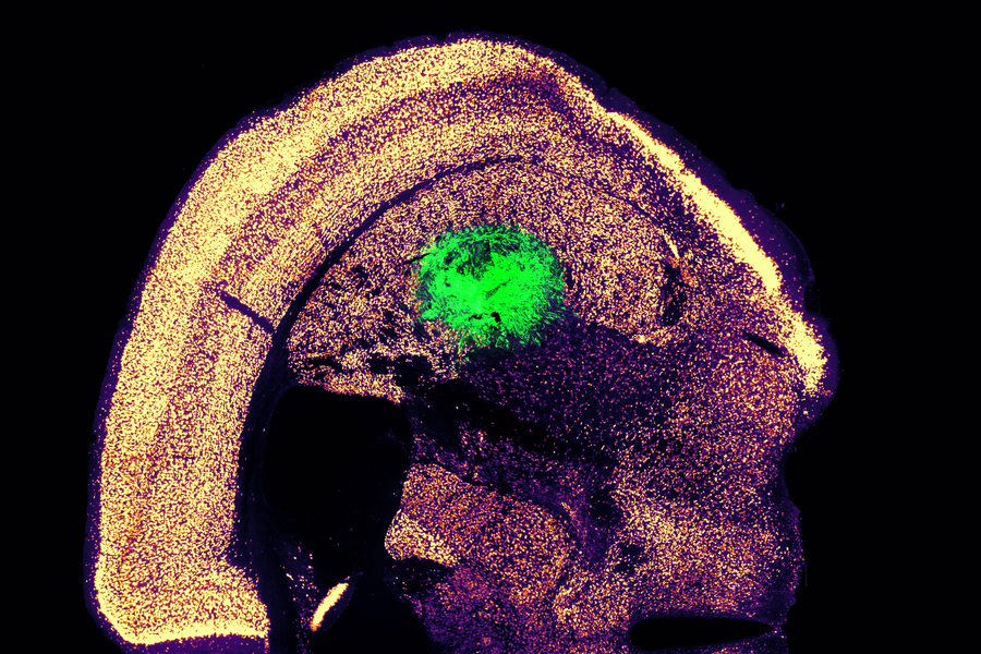
Researchers at MIT have devised a simplified process to convert a skin cell directly into a neuron. This image shows converted neurons (green) that have integrated with neurons in the brain’s striatum after implantation. Credits :Image: Courtesy of the researchers

Converting one type of cell to another – for example, a skin cell to a neuron – can be done through a process that requires the skin cell to be induced into a “pluripotent” stem cell, then differentiated into a neuron. Researchers at MIT have now devised a simplified process that bypasses the stem cell stage, converting a skin cell directly into a neuron.

Working with mouse cells, the researchers developed a conversion method that is highly efficient and can produce more than 10 neurons from a single skin cell. If replicated in human cells, this approach could enable the generation of large quantities of motor neurons, which could potentially be used to treat patients with spinal cord injuries or diseases that impair mobility.

“We were able to get to yields where we could ask questions about whether these cells can be viable candidates for the cell replacement therapies, which we hope they could be. That’s where these types of reprogramming technologies can take us,” says Katie Galloway, the W. M. Keck Career Development Professor in Biomedical Engineering and Chemical Engineering.

As a first step toward developing these cells as a therapy, the researchers showed that they could generate motor neurons and engraft them into the brains of mice, where they integrated with host tissue.

Galloway is the senior author of two papers describing the new method, which appear today in Cell Systems. MIT graduate student Nathan Wang is the lead author of both papers.

From skin to neurons

Nearly 20 years ago, scientists in Japan showed that by delivering four transcription factors to skin cells, they could coax them to become induced pluripotent stem cells (iPSCs). Similar to embryonic stem cells, iPSCs can be differentiated into many other cell types. This technique works well, but it takes several weeks, and many of the cells don’t end up fully transitioning to mature cell types.

“Oftentimes, one of the challenges in reprogramming is that cells can get stuck in intermediate states,” Galloway says. “So, we’re using direct conversion, where instead of going through an iPSC intermediate, we’re going directly from a somatic cell to a motor neuron.”

Galloway’s research group and others have demonstrated this type of direct conversion before, but with very low yields – fewer than 1 percent. In Galloway’s previous work, she used a combination of six transcription factors plus two other proteins that stimulate cell proliferation. Each of those eight genes was delivered using a separate viral vector, making it difficult to ensure that each was expressed at the correct level in each cell.

In the first of the new Cell Systems papers, Galloway and her students reported a way to streamline the process so that skin cells can be converted to motor neurons using just three transcription factors, plus the two genes that drive cells into a highly proliferative state.

Using mouse cells, the researchers started with the original six transcription factors and experimented with dropping them out, one at a time, until they reached a combination of three – NGN2, ISL1, and LHX3 — that could successfully complete the conversion to neurons.

Once the number of genes was down to three, the researchers could use a single modified virus to deliver all three of them, allowing them to ensure that each cell expresses each gene at the correct levels.

Using a separate virus, the researchers also delivered genes encoding p53DD and a mutated version of HRAS. These genes drive the skin cells to divide many times before they start converting to neurons, allowing for a much higher yield of neurons, about 1100 percent.

“If you were to express the transcription factors at really high levels in nonproliferative cells, the reprogramming rates would be really low, but hyperproliferative cells are more receptive. It’s like they’ve been potentiated for conversion, and then they become much more receptive to the levels of the transcription factors,” Galloway says.

The researchers also developed a slightly different combination of transcription factors that allowed them to perform the same direct conversion using human cells, but with a lower efficiency rate – between 10 and 30 percent, the researchers estimate. This process takes about five weeks, which is slightly faster than converting the cells to iPSCs first and then turning them into neurons.

Implanting cells

Once the researchers identified the optimal combination of genes to deliver, they began working on the best ways to deliver them, which was the focus of the second Cell Systems paper.

They tried out three different delivery viruses and found that a retrovirus achieved the most efficient rate of conversion. Reducing the density of cells grown in the dish also helped to improve the overall yield of motor neurons. This optimised process, which takes about two weeks in mouse cells, achieved a yield of more than 1000 percent.

Working with colleagues at Boston University, the researchers then tested whether these motor neurons could be successfully engrafted into mice. They delivered the cells to a part of the brain known as the striatum, which is involved in motor control and other functions.

After two weeks, the researchers found that many of the neurons had survived and seemed to be forming connections with other brain cells. When grown in a dish, these cells showed measurable electrical activity and calcium signaling, suggesting the ability to communicate with other neurons. The researchers now hope to explore the possibility of implanting these neurons into the spinal cord.

The MIT team also hopes to increase the efficiency of this process for human cell conversion, which could allow for the generation of large quantities of neurons that could be used to treat spinal cord injuries or diseases that affect motor control, such as ALS. Clinical trials using neurons derived from iPSCs to treat ALS are now underway, but expanding the number of cells available for such treatments could make it easier to test and develop them for more widespread use in humans, Galloway says.

The research was funded by the National Institute of General Medical Sciences and the National Science Foundation Graduate Research Fellowship Program.

Reprinted with permission of MIT News

‘Healthy’ Fatty Acid Not as Innocent as it Seems

Photo by Pexels on Pixabay

Eating a high-fat diet containing a large amount of oleic acid – a type of fatty acid commonly found in olive oil – could drive obesity more than other types of dietary fats, according to a study published in the journal Cell Reports.

The study found that oleic acid, a monounsaturated fat associated with obesity but also tentatively linked to cardiovascular benefits and often touted as a ‘healthy’ fatty acid, causes the body to make more lipid cells. By boosting a signalling protein called AKT2 and reducing the activity of a regulating protein called LXR, high levels of oleic acid resulted in faster growth of the precursor cells that form new lipid cells.

“We know that the types of fat that people eat have changed during the obesity epidemic. We wanted to know whether simply overeating a diet rich in fat causes obesity, or whether the composition of these fatty acids that make up the oils in the diet is important. Do specific fat molecules trigger responses in the cells?” said Michael Rudolph, PhD, assistant professor of biochemistry and physiology at the University of Oklahoma College of Medicine.

Rudolph and his team fed mice a variety of specialised diets enriched in specific individual fatty acids, including those found in coconut oil, peanut oil, milk, lard and soybean oil. Oleic acid was the only one that caused the precursor cells that give rise to fat cells to proliferate more than other fatty acids.

“You can think of the fat cells as an army,” Rudolph said. “When you give oleic acid, it initially increases the number of ‘fat cell soldiers’ in the army, which creates a larger capacity to store excess dietary nutrients. Over time, if the excess nutrients overtake the number of fat cells, obesity can occur, which can then lead to cardiovascular disease or diabetes if not controlled.”

Unfortunately, it’s not quite so easy to isolate different fatty acids in a human diet. People generally consume a complex mixture if they have cream in their coffee, a salad for lunch and meat and pasta for dinner. However, Rudolph said, there are increasing levels of oleic acid in the food supply, particularly when access to food variety is limited and fast food is an affordable option.

“I think the take-home message is moderation and to consume fats from a variety of different sources,” he said. “Relatively balanced levels of oleic acid seem to be beneficial, but higher and prolonged levels may be detrimental. If someone is at risk for heart disease, high levels of oleic acid may not be a good idea.”

Source: University of Oklahoma

Exercise Activates Cells that Protect Against Alzheimer’s

Photo by Barbara Olsen on Pexels

Using advanced single-nuclei RNA sequencing (snRNA-seq) and a widely used preclinical model for Alzheimer’s disease, researchers from Mass General Brigham and collaborators at SUNY Upstate Medical University have identified specific brain cell types that responded most to exercise. These findings, which were validated in samples from humans, shed light on the connection between exercise and brain health and point to future drug targets. Results are published in Nature Neuroscience.

“While we’ve long known that exercise helps protect the brain, we didn’t fully understand which cells were responsible or how it worked at a molecular level,” said senior author Christiane Wrann, DVM, PhD, a neuroscientist at Massachusetts General Hospital. “Now, we have a detailed map of how exercise impacts each major cell type in the memory centre of the brain in Alzheimer’s disease.”

Brain support cells—astrocytes enriched in the protein cadherin-4 (CDH4)
Scientists identified a distinct subtype of brain support cells—astrocytes enriched in the protein cadherin-4 (CDH4), shown in magenta, that seem to protect nerve cells against cell death. In Alzheimer’s disease, these cells become less abundant, but exercise seems to strengthen them. (Image credit: Luis Moreira)

The study focused on a part of the hippocampus – a critical region for memory and learning that is damaged early in Alzheimer’s disease. The research team leveraged single-nuclei RNA sequencing, a relatively new technologies that allow researchers to look at activity at the molecular level in single cells for an in-depth understanding of diseases like Alzheimer’s.

The researchers exercised a common mouse model for Alzheimer’s disease using running wheels, which improved their memory compared to the sedentary counterparts. They then analysed gene activity across thousands of individual brain cells, finding that exercise changed activity both in microglia, a disease-associated population of brain cells, and in a specific type of neurovascular-associated astrocyte (NVA), newly discovered by the team, which are cells associated with blood vessels in the brain. Furthermore, the scientist identified the metabolic gene Atpif1 as an important regulator to create new neurons in the brain. “That we were able to modulate newborn neurons using our new target genes set underscores the promise our study,” said lead author Joana Da Rocha, PhD, a postdoctoral fellow working in Dr Wrann’s lab.

To ensure the findings were relevant to humans, the team validated their discoveries in a large dataset of human Alzheimer’s brain tissue, finding striking similarities.

“This work not only sheds light on how exercise benefits the brain but also uncovers potential cell-specific targets for future Alzheimer’s therapies,” said Nathan Tucker, a biostatistician at SUNY Upstate Medical University and co-senior of the study. “Our study offers a valuable resource for the scientific community investigating Alzheimer’s prevention and treatment.”

Source: Mass General Brigham

Catch-up Sleep on the Weekend Can Improve Teens’ Anxiety

Photo by Eren Li

A new study presented at the SLEEP 2025 annual meeting found that teens who get moderate, but not excessive, catch-up sleep on weekends have fewer symptoms of anxiety symptoms.

Results show that teens who got up to two more hours of sleep on weekends than on weekdays exhibited fewer anxiety symptoms compared with those who did not sleep longer on weekends. However, longer durations of catch-up sleep on weekends were associated with slightly more internalising symptoms.

“The results show that both sleeping less on weekends than weekdays and sleeping substantially more on weekends were associated with higher anxiety symptoms,” said lead author Sojeong Kim, a doctoral candidate in the department of clinical psychology and psychology graduate advisor at the University of Oregon in Eugene. “In contrast, moderate catch-up sleep – defined as less than two hours – was associated with lower anxiety symptoms, suggesting that some weekend recovery sleep may be beneficial.”

The American Academy of Sleep Medicine recommends that teenagers 13 to 18 years of age should sleep 8 to 10 hours on a regular basis to promote optimal health. However, CDC data show that only 23% of high school students get sufficient sleep on an average school night.

“Many teens try to make up for lost sleep by sleeping in on weekends,” Kim said.

Consistently getting sufficient sleep is associated with better health outcomes including improved attention, behavior, learning, memory, emotional regulation, quality of life, and mental and physical health. In contrast, insufficient sleep in teenagers is associated with increased risks of problems such as depression and suicidal thoughts.

The study involved 1877 adolescents with a mean age of 13.5 years. Sleep duration was estimated using Fitbit devices, while internalising symptoms were assessed using the Child Behavior Checklist survey. Weekend catch-up sleep was calculated as the difference between weekend and weekday sleep duration.

Kim noted that it is important to identify the right amount of catch-up sleep that is beneficial to teens who restrict their sleep during the week.

“Too little or too much sleep variability from weekday to weekend may contribute to the symptoms someone is trying to combat, like physical or mental fatigue and feelings of anxiety,” she said.

Source: American Academy of Sleep Medicine

Psilocybin Dose Provides Two Years of Relief from Cancer Depression

Phase 2 trial reveals that a single dose of psilocybin offers long-term relief from symptoms of depression and anxiety.

Photo by Marek Piwnicki: https://www.pexels.com/photo/macro-photography-of-a-psilocybin-mushroom-13695325/

New results from a clinical trial reveal that a single dose of psilocybin can provide sustained reductions in depression and anxiety in individuals with cancer suffering from major depressive disorder. The findings are published by Wiley online in CANCER, a peer-reviewed journal of the American Cancer Society.

People with cancer often struggle with depression. In this phase 2 trial, 28 patients with cancer and major depressive disorder received psychological support from a therapist prior to, during, and following a single 25mg dose of psilocybin.

During clinical interviews conducted 2 years later, 15 (53.6%) patients demonstrated a significant reduction in depression, and 14 (50%) had sustained depression reduction as well as remission. Similarly, psilocybin reduced anxiety for 12 (42.9%) patients at 2 years.

An ongoing randomised, double-blind trial is currently evaluating up to two doses of 25mg of psilocybin versus placebo as treatment for depression and anxiety in patients with cancer. This study is building on the single-dose study in an effort to bring a larger majority of the patients into remission of depression and anxiety.

“One dose of psilocybin with psychological support to treat depression has a long-term positive impact on relieving depression for as much as 2 years for a substantial portion of patients with cancer, and we’re exploring whether repeating the treatment resolves depression for more than half of the patients,” said lead author Manish Agrawal, MD, of Sunstone Therapies. “If randomised testing shows similar results, this could lead to greater use of psilocybin to treat depression in patients with cancer.”

Source: Wiley