Tag: surgery

Human Instruction with AI Guidance Gives the Best Results in Neurosurgical Training

Study has implications beyond medical education, suggesting other fields could benefit from AI-enhanced training

Artificial intelligence (AI) is becoming a powerful new tool in training and education, including in the field of neurosurgery. Yet a new study suggests that AI tutoring provides better results when paired with human instruction.

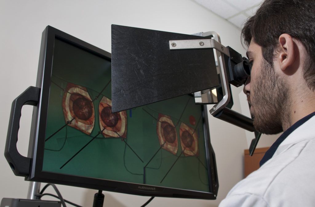
Researchers at the Neurosurgical Simulation and Artificial Intelligence Learning Centre at The Neuro (Montreal Neurological Institute-Hospital) of McGill University are studying how AI and virtual reality (VR) can improve the training and performance of brain surgeons. They simulate brain surgeries using VR, monitor students’ performance using AI and provide continuous verbal feedback on how students can improve performance and prevent errors. Previous research has shown that an intelligent tutoring system powered by AI developed at the Centre outperformed expert human teachers, but these instructors were not provided with trainee AI performance data.

In their most recent study, published in JAMA Surgery, the researchers recruited 87 medical students from four Quebec medical schools and divided them into three groups: one trained with AI-only verbal feedback, one with expert instructor feedback, and one with expert feedback informed by real-time AI performance data. The team recorded the students’ performance, including how well and how quickly their surgical skills improved while undergoing the different types of training.

They found that students receiving AI-augmented, personalised feedback from a human instructor outperformed both other groups in surgical performance and skill transfer. This group also demonstrated significantly better risk management for bleeding and tissue injury – two critical measures of surgical expertise. The study suggests that while intelligent tutoring systems can provide standardised, data-driven assessments, the integration of human expertise enhances engagement and ensures that feedback is contextualised and adaptive.

“Our findings underscore the importance of human input in AI-driven surgical education,” said lead study author Bianca Giglio. “When expert instructors used AI performance data to deliver tailored, real-time feedback, trainees learned faster and transferred their skills more effectively.”

While this study was specific to neurosurgical training, its findings could carry over to other professions where students must acquire highly technical and complex skills in high-pressure environments.

“AI is not replacing educators – it’s empowering them,” added senior author Dr Rolando Del Maestro, a neurosurgeon and current Director of the Centre. “By merging AI’s analytical power with the critical guidance of experienced instructors, we are moving closer to creating the ‘Intelligent Operating Room’ of the future capable of assessing and training learners while minimising errors during human surgical procedures.”

Source: McGill University

New Surgical Method for Groin Hernia in Women Without Laparoscopy

In a study conducted in Uganda and published in JAMA Surgery, researchers from Karolinska Institutet evaluated a new surgical method for treating groin hernias in women. The method could become an alternative in resource-limited settings where laparoscopic techniques are not generally available.

Groin hernia repair is the most common general surgical procedure in the world. Groin hernias are more common in men, but women are more likely to experience complications due to this condition.

Many women in low- and middle-income countries who need surgery for groin hernias lack access to laparoscopy (keyhole surgery). To evaluate a new method using open surgery, the researchers conducted a randomised clinical trial at two publicly funded hospitals in Uganda. The study included 200 women who underwent groin hernia surgery and were followed up after two weeks and after one year.

There are two main types of groin hernias, called inguinal and femoral hernias. The evaluation showed that the new surgical method was effective for both femoral and inguinal groin hernias.

Its effectiveness for both types is particularly important as the study also showed that nearly 45% of the women had femoral hernias, which carry a higher risk of complications.

“The fact that so many of the women had femoral hernias was unexpected and highlights the need to develop effective, safe and accessible methods,” says Alphonsus Matovu, PhD at the Department of Molecular Medicine and Surgery, Karolinska Institutet and first author of the article.

Millions of women affected

The results are promising as the new method could be developed into a viable alternative where access to advanced laparoscopic surgery is limited.

“Women with groin hernias can suffer serious and even fatal complications and therefore need access to effective surgical methods,” says Jenny Löfgren, docent at the same department and last author of the article. “The new method could become a valuable tool to improve care for millions of women”.

The method needs further evaluation, and the researchers will also follow up with the study participants five years after surgery to ensure long-term results. To improve treatment, the new method will also be compared with other surgical methods, both open and laparoscopic.

Source: Karolinska Institutet

Open Surgery for Lymph Node Removal is Still the Gold Standard in Testicular Cancer

Photo by cottonbro studio

A new study published in BJU International has found that the conventional, versatile open surgery approach to removal of the lymph nodes behind the intestines for patients whose testicular cancer has not advanced beyond the abdomen is the gold standard of care for men in this condition.

Open surgery involves making an incision on the abdomen for a direct view and access to the surgical area. In the appropriate patients, cancer cure rates are excellent with this surgery alone due to continued clinician experience and refinement of technique, particularly at high-volume centres.

The study, led by urologist and health services researcher Clint Cary, MD, MPH, MBA, of the Indiana University School of Medicine and the Regenstrief Institute, was conducted using information from the on 165 patients with clinical stage I or II testicular cancer and no prior chemotherapy. One of the highest-volume testicular cancer treatment groups in the US, IU School of Medicine’s Department of Urology is among those groups setting the bar for both better surgical results and fewer negative outcomes such as infertility. Study benchmarks included low blood loss, short hospital stay and rare major postoperative complications.

Dr Cary notes that the paper, which includes analysis of risk factors for complications, clearly indicates that prior to treatment, men and their families should have conversations with their local urologist about their experience level across risk factors, such as high body mass index (BMI), as well as have detailed discussions of complication rates. He adds that both patients and clinicians can use the findings of this paper as a bar for comparison as they make choices.

“In my clinical role at Indiana University in the operating room, I frequently perform lymph node dissections for men with a lower burden of metastatic cancer who have not had chemotherapy or may never receive it for this cancer. In my complementary role as a Regenstrief Institute health services researcher, I study the impact of clinical decisions,” said Dr Cary. “For this study, I wanted to know that this and other treatments I perform are making a difference and I want to know – whether we are continuing established treatments or introducing new approaches to surgery – how these decisions and the resulting care will impact patients’ quality of life and their cancer outcomes.

“This study is an example of how the benefit of providing clinical care and also being in a research environment enables us to learn from what we’re doing and to report it in a manner that is meaningful to both patients and physicians. We’re always looking for ways to improve upon surgical outcomes. Going beyond the scope of this paper, we are now expanding our work to begin a randomised trial comparing two surgical techniques for removing lymph nodes to understand if one is better than the other regarding postoperative recovery.”

The paper did not compare robot-assisted surgery with traditional open surgery, which may allow a more complete lymph node dissection. While robotic surgery in general offers potential benefits to both surgeons and patients, the authors note that the head to head comparison between these two approaches is limited due to small experiences with the robotic approach in testicular cancer.

The authors conclude that for patients undergoing removal of lymph nodes for testicular cancer and certainly more complex and challenging patients at higher risk of post-operative complications, the traditional open surgery remains the most effective approach and the gold standard.

Source: Regenstrief Institute

Hold the GLP-1 Agonists Before Surgery, New Advice Says

Photo by Natanael Melchor on Unsplash

Patients taking Glucagon-like peptide-1 (GLP-1) receptor agonists should stop taking them before they have surgery, due to the risk of aspirating while under general anaesthesia. This is the latest advice from the American Society of Anesthesiologists (ASA).

Initially approved by the Food and Drug Administration (FDA) for type 2 diabetes mellitus and cardiovascular risk reduction, GLP-1 agonists have shot up in popularity due to their effectiveness in weight loss. Despite having recent FDA approval, they have been used off-label for this purpose for quite some time.

When it comes to surgery, a number of organisations have recommended to hold these drugs either the day before or day of the procedure. For patients on weekly dosing, it is recommended to hold the dose for a week, the ASA notes.

GLP-1 agonists are associated with adverse gastrointestinal effects such as nausea, vomiting and delayed gastric emptying. The effects on gastric emptying are reported to be reduced with long-term use, most likely through rapid tachyphylaxis at the level of vagal nerve activation. Based on recent anecdotal reports, there are concerns that delayed gastric emptying from GLP-1 agonists can increase the risk of regurgitation and pulmonary aspiration of gastric contents during general anaesthesia and deep sedation. Patient taking GLP-1 agonists are more likely to have increased residual gastric contents as predicted by adverse gastrointestinal symptoms (nausea, vomiting, dyspepsia, abdominal distension).

The use of GLP-1 agonists in paediatrics has primarily been reported for the management of type 2 diabetes mellitus and obesity. The published literature on GLP-1 agonists in paediatrics is predominantly from paediatric patients 10 to 18 years old and concerns are similar to those reported in adults. During the conduct of general anaesthesia/deep sedation, children on GLP-1 agonists have similar gastrointestinal adverse events at a rate similar to adults.

In a review of the literature, the ASA Task Force on Preoperative Fasting found that, beyond a few case reports, there was little evidence for guidance on preoperative management of GLP-1 agonists. Nevertheless, they made recommendations for elective procedures. In the case of urgent or emergent procedures, they suggested treating the patient as ‘full stomach’.

If the patient’s GLP-1 agonists prescribed for diabetes management are held for longer than the dosing schedule, the guidelines urge surgeons to consider consulting an endocrinologist for bridging the antidiabetic therapy in order to avoid hyperglycaemia.

They further recommend that if gastrointestinal symptoms, such as severe nausea/vomiting/retching, abdominal bloating, or abdominal pain, are present, surgeons should consider delaying elective procedures. If the patient has no gastrointestinal symptoms and the GLP-1 agonists have been held as advised, the surgical team can carry on as normal.

Source: American Society of Anesthesiologists

Study Unravels the Mechanics of the Ideal Surgical Knot

Surgical knot tied on a rigid support. Credit: Alain Herzog / EPFL

Surgeons knot sutures intuitively. While simple square and granny sliding knots are often used in surgery, it takes years to master them so that they stay in place without loosening or breaking. Much mathematical research has been done on knot topology and geometry, but little is known about the physics of knot mechanics, like the material properties of knotted filaments. Now, in Science Advances, researchers have published the first physics-based study on the mechanics of surgical knots, and exactly what properties influence their strength.

“It’s astonishing to think how much we rely on knots, when we don’t really understand how they work,” says Pedro Reis, head of the Flexible Structures Lab in the School of Engineering (Institute of Mechanical Engineering). Reis and PhD student Paul Johanns teamed up with Lausanne-based plastic surgeon Samia Guerid to lead the study.

“Understanding surgical knot mechanics can raise awareness among experienced surgeons, be incorporated into training programs, and advance robotic surgery by enabling more effective knot-tying capabilities,” says Guerid. “Such knowledge could also influence the development of suture materials that enhance slippage resistance in sliding knots.”

The power of plasticity

Reis, an avid climber, has a personal interest in secure knots and has conducted several previous studies on knot mechanics. He explains that many knots can be described as free-ended structures that provide a holding force, with their functionality dictated by the variables of topology, geometry, elasticity, contact, and friction. But for the study of surgical knots, Reis and his colleagues considered a key sixth factor: polymer plasticity of the suturing filament.

The strength of sutures made from polypropylene filaments used in surgery depends on the tension applied during the tying of the knot (pretension). This pretension permanently deforms, or stretches the filament, creating a holding force. Too little pretension causes the knot to come undone; too much snaps the filament.

The team analyzed 50-100 knots tied by Guerid, and found that the surgeon was able, thanks to her years of experience, to intuitively target the pretension ‘sweet spot’. Using precision experiments, X-ray micro-computed tomography, and computer simulations, the scientists defined a threshold between ‘loose’ and ‘tight’ knots, and uncovered relationships between knot strength and pretension, friction, and number of throws.

“Surprisingly, despite the complex interplay between all six factors, we observed a simple, robust emergent behavior vis-à-vis knot strength. But we still don’t have a predictive model to fully explain the relationship between knot pretension and strength, which seems to be consistent, even outside surgical knots. We’re already looking into this question.”

A training tool for surgeons…and robots

The team’s findings could be a valuable tool for training surgeons, as they could allow the parameters of a secure knot to be translated into practical guidelines. While experience would remain important, the idea is that safe knot-tying could be taught using predictive models, rather than intuition gained only through years of practice.

“Our data gives us a recipe for determining the ideal pretension and number of throws, for example, depending on the type of filament used,” Reis says

“The lack of physics-based analysis has been a limitation,” Guerid adds. “Quantifiable data on knot mechanics could be integrated into training programs to assess the tensile strength of each knot, ensuring trainees acquire necessary skills for successful surgeries. The data could also facilitate development of robotic surgery via the programming of robotic systems.”

Source: EurekAlert!

The Three Global Challenges Surgeons Need to Tackle

Photo by Jafar Ahmed on Unsplash

Despite significant advances over the last 30 years, surgical research is still limited to comparing the benefit of one technique over another. It can be founded on assumptions that a new device or approach is always better – leading to poorly evaluated devices and procedures having negative effects on patients.

Writing in The Lancet, experts from the NIHR Global Health Research Unit for Global Surgery GlobalSurg Collaborative – a programme backed by funding from the NIHR (National Institute for Health and Care Research) – propose three priority areas for surgery:

Access, equity, and public health must be recognised as crucial issues for surgery.

In 2015, five billion people did not have access to safe and affordable surgical care. Of those who did, 33 million individuals faced catastrophic health expenditure in payment for surgery and anaesthesia. During the COVID-19 pandemic, over 28 million cases of elective surgery are likely to have been cancelled. Surgery has a key role in addressing the most important and growing global health challenges, such as trauma, congenital anomalies, safe childbirth, and non-communicable diseases.

Inclusion and diversity must improve in both surgical research and the profession.

Women, minoritised groups, and patients from low-income and middle-income countries remain under-represented in clinical practice and major research work. Advancing inclusion and diversity will ensure a research agenda that delivers pragmatic, simple, and context-specific research that reflects the needs of all patients.

Climate change is the greatest global health threat facing the world.

Surgical theatres are some of the most energy and resource intense areas of a hospital. Surgical practice relies on many single-use, non-biodegradable products as well as anaesthetic gases that have a large environmental footprint. Moving towards net-zero operating practices could reduce health-sector carbon emissions and allow surgeons and policy makers to reassess how surgery fits into a wider health system.

Comment co-author Dmitri Nepogodiev, from the University of Birmingham, said: “Richard Horton, Editor-in-Chief of The Lancet, once described surgical research as ‘a comic opera performance’. That was in 1996 and things have changed significantly since then.

“However, truly improving lives requires surgical researchers to use the next quarter of a century to tackle the most pressing questions on equity and access, the role of surgery in public health, and sustainability.

“Despite the problems of large waiting lists and an economic squeeze on health systems, surgeons must focus on these priority areas — placing surgery as a leader in medical specialties and demonstrating its value as a fundamental element of universal health care.”

The experts note that large, randomised controlled trials with well-defined endpoints are now more usual in surgical research, whilst exploration into the placebo effect, has led to a fundamental re-examination of the benefits of some surgical procedures and whether they benefit patients at all.

Surgeons and anaesthetists have developed successful international collaborative research efforts that have enabled rapid recruitment of participants and globally relevant studies and trials, while following internationally set standards of clinical trial practice. Surgeons can now provide reliable answers to crucial questions in operative surgery, and their research has improved patient care and resource use in health systems.

A Molecular Mechanism for Hydrocephalus may Enable a Non-surgical Treatment

MRI images of the brain
Photo by Anna Shvets on Pexels

Researchers at Massachusetts General Hospital have discovered a novel molecular mechanism behind the most common forms of acquired hydrocephalus – which could lead to the first non-surgical treatments for the life-threatening disease. Research in animal models uncovered a pathway through which infection or bleeding in the brain triggers inflammation, causing increased production of cerebrospinal fluid (CSF) by the choroid plexus and lead to swelling of the brain ventricles.

“Finding a nonsurgical treatment for hydrocephalus, given the fact neurosurgery is fraught with tremendous morbidity and complications, has been the holy grail for our field,” says Kristopher Kahle, MD, PhD, a paediatric neurosurgeon at MGH and senior author of the study in the journal Cell. “We’ve identified through a genome-wide analytical approach the mechanism that underlies the swelling of the ventricles which occurs after a brain bleed or brain infection in acquired hydrocephalus. We’re hopeful these findings will pave the way for approval of an anti-inflammatory drug to treat hydrocephalus, which could be a game-changer for populations in the US and around the world that don’t have access to surgery.”

Occurring in about 0.2% of births, acquired hydrocephalus is the most common cause of brain surgery in children, though it can affect people at any age. In underdeveloped regions where bacterial infection is the most prevalent form, hydrocephalus is often deadly for children due to the lack of surgical intervention. Brain surgery, where a shunt is implanted to drain fluid from the brain, is the only known treatment. But about half of all shunts in paediatric patients fail within two years of placement, according to the Hydrocephalus Association, requiring repeat neurosurgical operations and a lifetime of brain surgeries.

Pivotal to the process is the choroid plexus, the brain structure that routinely pumps cerebrospinal fluid into the four ventricles of the brain to keep the organ buoyant and injury-free within the skull. An infection or brain bleed, however, can create a dangerous neuroinflammatory response where the choroid plexus floods the ventricles with cerebral spinal fluid and immune cells from the periphery of the brain in a cytokine storm, swelling the brain ventricles.

“Scientists in the past thought that entirely different mechanisms were involved in hydrocephalus from infection and from haemorrhage in the brain,” explains co-author Bob Carter, MD, PhD, chair of the Department of Neurosurgery at MGH. “Dr Kahle’s lab found that the same pathway was involved in both types and that it can be targeted with immunomodulators like rapamycin, a drug that’s been approved by the US Food and Drug Administration for transplant patients who need to suppress their immune system to prevent organ rejection.”

MGH researchers are continuing to explore how rapamycin and other drugs which quell the inflammation seen in acquired hydrocephalus could be repurposed. “What has me most excited is that this noninvasive therapy could provide a way to help young patients who don’t have access to neurosurgeons or shunts,” says Kahle. “No longer would a diagnosis of hydrocephalus be fatal for these children.”

Source: Massachusetts General Hospital

Inaccurate Anaesthesia Start Times Leading to Lost Revenue

Photo by Anna Shvets on Pexels

Inaccurately recording the start of anaesthesia care during a procedure is common and results in significant lost billing time for anaesthesia practices and medical centres, suggests a study being presented at the American Society of Anesthesiologists’ ADVANCE 2023, the Anesthesiology Business Event.

The anaesthesia start time (AST) must be documented from a computer logged into the electronic health record (EHR), and typically occurs once the patient is in the operating room (OR). However, the anaesthesiologist meets with the patient prior to their arrival in the OR and begins tasks that are vital to the procedure, such as administering pre-medication and attaching monitors, time which is is not typically recorded. Depending on the patient and procedure, adding two to five minutes to the AST when logging it would account for the preparation and transit time, researchers say.

“These seemingly minor inaccuracies of recorded AST can cost medical centres and anaesthesia practices hundreds of thousands of dollars in lost revenue,” said Nicholas Volpe Jr, MD, MBA, lead author of the study and an anaesthesiology resident physician at Northwestern University McGaw Medical Center, Chicago. “We suspect most anaesthesiologists are unaware that they aren’t recording AST accurately. It’s not a result of negligence, but rather reflects that workflow hasn’t been optimised for accuracy.”

For the study, the researchers analysed 40 312 procedures with anaesthesia over 12 months at a single academic centre. In 68.74% of cases , AST was recorded as starting once the patient was in the OR, without factoring in the preparation time. Using the national average charge for anesthaesia time, the missing time translated to over $600 000 in lost revenue for the year, the researchers determined.*

“Logging AST is one of the many new tasks that anaesthesiologists learn when starting a new role,” said Dr Volpe. “Transitioning from an internship to clinical anaesthesia practice involves learning a significant amount of new information, and understanding the importance of an accurately recorded AST may seem like a relatively minor issue compared to important patient-care information.”

Several approaches could help address inaccurate AST documentation, including educating anaesthesiologists on how to improve their AST recording practices and providing visual reminders such as signs in the OR, Dr Volpe said. Also, an AST capture function could be built into the EHR mobile application so that AST can be noted by anaesthesiologists on the way to the OR, or the EHR could automatically add two minutes to the AST log time, he said. The researchers plan to roll out some of those initiatives in the spring and determine if they are effective.

*The projected savings are theoretical and not linked to billing at the institution where the study was conducted. 

Source: American Society of Anesthesiologists

Better Outcomes with Bypass Surgery in Chronic Limb-threatening Ischaemia

Photo by Natanael Melchor on Unsplash

Performing open bypass surgery to restore circulation for people with a severe form of peripheral artery disease (PAD) resulted in better outcomes for specific patients compared to a less-invasive procedure, according to findings published in the New England Journal of Medicine.

PAD is a condition in which blood flow to one or both legs is reduced by a buildup of fatty plaque in the arteries. One in 10 of patients with this condition develop a severe form of PAD called chronic limb-threatening ischaemia (CLTI), a painful and debilitating condition that can lead to amputation if untreated. Up to about 22 million people worldwide have CLTI, which is also associated with an increased risk of heart attack, stroke, and death.

“Given the projected rise in the number of patients with chronic limb-threatening ischaemia, it is critically important that we understand the full impact of our interventions for this disease,” said Matthew Menard, MD, a study author and associate professor of surgery and co-director of the endovascular surgery program at Brigham and Women’s Hospital, Boston. “These findings help do that and also can assist clinicians and caregivers in providing the best possible care to patients.”

The Best Endovascular versus Best Surgical Therapy for Patients with CLTI (BEST-CLI) trial is a landmark study supported by the National Heart, Lung, and Blood Institute (NHLBI).

To compare effectiveness of two common treatments for CLTI, researchers enrolled 1830 adults who were planning to have revascularisation, a procedure used to restore blood flow in their blocked arteries, and who were eligible for both treatment strategies.

One treatment strategy was an open bypass surgery, in which blood is redirected around the blocked leg artery by using a segment of a healthy vein. The other strategy was an endovascular procedure, where a balloon is dilated and/or a stent is placed in the blocked segment of the artery to improve blood flow. To compare the surgical strategy to the less-invasive endovascular approach, researchers randomised participants into one of two parallel trials between 2014–2021.

The first trial, defined as cohort 1, included 1434 adults who were judged to be the best candidates for the bypass surgery because they had an adequate amount of an optimal vein (the single-segment great saphenous vein) preferred for the procedure. Participants were then randomly assigned to have either a surgical bypass or endovascular procedure. Researchers followed the trial participants for up to seven years.

The second trial, defined as cohort 2, included 396 adults who were not the best candidates for the open bypass because they did not have an adequate amount of the preferred saphenous vein. They were randomised to have either an endovascular procedure or a bypass that used alternate graft material instead of the saphenous vein. Participants were followed-up for up to three years.

At the end of the trial, the researchers found that participants in cohort 1 who received the bypass were 32% less likely to have major medical events related to CLTI than those who had an endovascular procedure. This result was driven by a 65% reduction in major repeat surgeries or procedures to retain blood flow in the lower leg and a 27% reduction in major amputations. No differences were found in death rates between the participants who received the bypass surgery and those who received an endovascular procedure.

Adults in cohort 2 – those who did not have the optimal vein for the bypass – had no major differences in outcomes based on having had an open bypass or an endovascular procedure.

“Our findings support complementary roles for these two treatment strategies and emphasise the need for preprocedural planning to assess patients and inform what treatment is selected,” said co-principal investigator Alik Farber, MD, at Boston Medical Center.

Common symptoms of CLTI include leg and foot pain, foot infections, and open sores on the leg and foot that don’t fully heal. Without having a procedure to redirect or open blocked blood flow to the lower body, about 4 in 10 adults with CLTI have a lower leg or foot amputation.

BEST-CLI is the largest CLTI clinical trial to date and builds on prior research that aims to answer questions about the risks and benefits of revascularisation strategies for CLTI.

Source: NIH/National Heart, Lung and Blood Institute

Pair of Studies Reveal Ways to Improve Surgical Care in Countries like South Africa

Photo by Natanael Melchor on Unsplash

Surgical care experts published two important studies in The Lancet that will help to provide safer surgery for thousands of patients around the world – particularly in Low- and Middle-income Countries (LMIC) such as South Africa.

Researchers found that routinely changing gloves and instruments just before closing wounds could significantly reduce Surgical Site Infection (SSI), the most common post-operative complication. This switch could prevent as many as 1 in 8 cases of SSI.

Secondly, they tested a new toolkit that can make hospitals better prepared for pandemics, heatwaves, winter pressures and natural disasters that could reduce cancellations of planned procedures around the world.

Surgical infections

Patients in LMICs are disproportionately affected by wound infections. The ChEETAh trial was run in Benin, Ghana, India, Mexico, Nigeria, Rwanda and South Africa. With the publication of their findings in The Lancet, researchers are calling for the practice to be widely implemented – particularly in LMICs.

Co-author Mr Aneel Bhangu, from the University of Birmingham, commented: “Surgical site infection is the world’s most common postoperative complication – a major burden for both patients and health systems. Our work demonstrates that routine change of gloves and instruments is not only deliverable around the world, but also reduced infections in a range of surgical settings. Taking this simple step could reduce SSIs by 13% – simply and cost-effectively.”

Patients who develop SSI experience pain, disability, poor healing with risk of wound breakdown, prolonged recovery times and psychological challenges. In health systems where patients have to pay for treatment this can be a disaster and increases the risk of patients being plunged into poverty after their treatment. The simple and low-cost practice of changing your gloves and instruments just before closing the wound is something which can be done by surgeons in any hospital around, meaning a huge potential impact.

Surgical Preparedness Index

Experts from the NIHR Global Research Health Unit on Global Surgery also unveiled their ‘Surgical Preparedness Index’ (SPI) in The Lancet. This is a key study assessing the extent to which hospitals around the world were able to continue elective surgery during COVID.

Researchers identified different features of hospitals that made them more or less ‘prepared’ for times of increased pressure. Using COVID as an important example, they highlighted that health systems are put under stress for all sorts of reasons each year – from seasonal pressures to natural disasters, and warfare. A team of clinicians from 32 countries designed the SPI which scores hospitals based on their infrastructure, equipment, staff, and processes used to provide elective surgery. The higher the resulting SPI score, the more prepared a hospital is for disruptions.

After creating the SPI tool, the experts asked 4714 clinicians in 1632 hospitals across 119 countries to assess the preparedness of their local surgical department. Overall most hospitals around the world were poorly prepared, and suffered a big drop in the number of procedures they were able to provide during COVID. A 10-point increase in the SPI score corresponded to four more patients that had surgery per 100 patients on the waitlist.

Lead author Mr. James Glasbey, from the University of Birmingham, commented: “Our new tool will help hospitals internationally improve their preparation for external stresses ranging from pandemics to heatwaves, winter pressures and natural disasters. We believe it help hospitals to get through their waiting lists more quickly, and prevent further delays for patients. The tool can be completed easily by healthcare workers and managers working in any hospital worldwide – if used regularly, it could protect hospitals and patients against future disruptions.”

Professor Dion Morton, Barling Chair of Surgery at the University of Birmingham and Director of Clinical Research at the Royal College of Surgeons of England commented: “Although not all postoperative deaths are avoidable, many can be prevented by increasing investment in research, staff training, equipment, and better hospital facilities. We must invest in improving the quality of surgery around the world.”

Source: University of Birmingham