Researchers have developed a new method to precisely and rapidly correct genetic alterations in cultured patient cells.
The genetically corrected stem cells are produced from a 2–3 mm skin biopsy taken from patients with different genetic diseases. The corrected stem cells are essential in the research and for the development of new therapies for the diseases in question.
The scientists based the new method on previous groundbreaking research in the fields of stem cells and gene editing; the first technique is the invention of induced pluripotent stem cells, iPSCs from differentiated cells, which won the Nobel in 2012. The other technique is the CRISPR-Cas9 ‘gene scissors’, which got the prize in 2020. The new method combines these techniques to correct gene alterations that cause inherited diseases, creating fully functional new stem cells.
The researchers aim to eventually produce autologous cells with therapeutic properties. The use of the patient’s own corrected cells could help in avoiding the immunological challenges hampering the organ and tissue transplantation from a donor. The new method was developed by PhD student Sami Jalil and is published in Stem Cell Reports.
More than 6000 inherited diseases are known to exist, which are caused by various gene alterations. Currently, some are treated with a cell or organ transplant from a healthy donor, if available.
“Our new system is much faster and more precise than the older methods in correcting the DNA errors, and the speed makes it easier and diminishes also the risk of unwanted changes,” commented adjunct professor Kirmo Wartiovaara, who supervised the work.
“In perfect conditions, we have reached up to 100 percent efficacy, although one has to remember that the correction of cultured cells is still far away from proven therapeutic applications. But it is a very positive start” Prof Wartiovaara added.
Studies of balding male mice have uncovered a possible cause of hair loss in men and women as well. The findings, published in Nature Aging, provide new insight into how hair and tissues age.
The study shows as hair stem cells age, they lose the adhesion that keeps them lodged inside the hair follicle. As their adhesiveness wanes, the stem cells escape from their location, called the bulge, into the dermis. Once outside their delicate microenvironment, they generally can’t survive.
“The result is fewer and fewer stem cells in the hair follicle to produce hair,” said lead author Rui Yi, the Paul E. Steiner Research Professor of Pathology at Northwestern University Feinberg School of Medicine. “This results in thinning hair and ultimately baldness during ageing.”
This finding could be applicable to older men and women with thinning hair as mice and humans share hair and stem cell similarities, Prof Yi said.
By labelling individual stem cells with a fluorescent marker, the researchers were able, for the first time, to track hair follicle ageing in real time in live animals. Scientists also discovered two key genes responsible for enhancing adhesiveness of the stem cells. They are now trying to reinstate these genes to see if that will reverse hair loss.
During follicles’ normal cycles of life and death, a large number of stem cells remain permanently lodged in the stem cell compartment of hair follicles to keep producing hair follicle cells.
“We believe this stem cell escape mechanism has never been reported before, because nobody could track the aging process in live animals,” Yi said.
Though scientists knew hair follicles become miniaturised during aging, how it happened was unclear. Many thought it was due to cell death or the inability of cells to divide as they age.
“We discovered, at least in part, it is due to hair follicle stem cells migrating away from their niche,” Prof Yi said. “Cell death also occurs during our observation. So, our discovery doesn’t dispute existing theories but provides a new mechanism.”
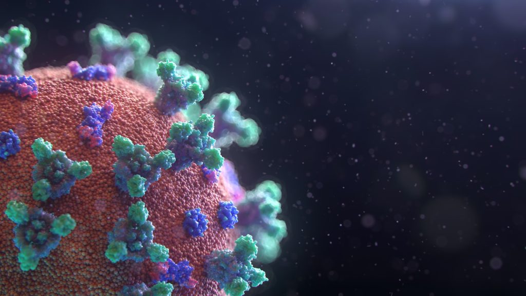
SARS-CoV-2 virus. Source: Fusion Medical Animation on Unsplash
The global race to develop new stem cell-based COVID treatments during the pandemic was filled with violations of government regulations, inflated medical claims and distorted public communication, according to an article appearing in Stem Cell Reports.
While stem cell therapy has treatment applications for a limited range of diseases and conditions, at present no clinically tested or government-approved cell therapies are available for the treatment or prevention of COVID or long COVID.
Despite this, some clinics have started offering unproven and unsafe “stem cell” therapies that promise to prevent COVID by strengthening the immune system or improving overall health, according to lead author Laertis Ikonomou, PhD, associate professor of oral biology in the University at Buffalo School of Dental Medicine.
The article explores the negative effects that misinformation about cell therapies has on public health, as well as the roles that researchers, science communicators and regulatory agencies should play in curbing the spread of inaccurate information and in promoting responsible, accurate communication of research findings.
“Efforts to rapidly develop therapeutic interventions should never occur at the expense of the ethical and scientific standards that are at the heart of responsible clinical research and innovation,” said Prof Ikonomou.
Other investigators include Megan Munsie, PhD, professor of ethics, education and policy in stem cell science at the University of Melbourne; and
Many of the studies on possible stem cell-based COVID treatments are at an early stage of investigation and further evaluation on larger sample sizes is required, says Munsie. However, the findings from preliminary studies are frequently exaggerated through press releases, social media and uncritical news media reports.
“Given the urgency of the ongoing pandemic, even the smallest morsel of COVID science is often deemed newsworthy and rapidly enters a social media landscape where—regardless of its accuracy – it can be widely shared with a global audience,” said Aaron Levine, PhD, associate professor of public policy at Georgia Institute of Technology..
Clinics selling such treatments sometimes use these findings and news reports to exploit the fears of vulnerable patients by unethically advertising unproven stem cell treatments benefits of boosting the immune system, regenerating lung tissue and preventing transmission of COVID, said co-author Leigh Turner, PhD, professor of health, society and behaviour at the University of California, Irvine.
Reportedly some harm to patients resulted from unproven stem cell therapies, including blindness and death. Patients suffer financially as well, said Prof Ikonomou, as the products range in price from a few thousand to tens of thousands of dollars, and people are often encouraged to receive the expensive treatments every few months.
Patients who COVID may decline vaccines, stop wearing masks and stop other COVID safety measures, Prof Turner warned. They may also be less likely to participate in ethically conducted clinical trials.
“The premature commercialisation of cell-based therapeutics will inevitably harm the field of regenerative medicine, increase risks to patients and erode the public’s trust,” said Prof Ikonomou.
Despite warnings, many offending companies continue to make false claims. The authors recommend that regulatory agencies consider implementing stronger measures.
They also suggest that scientific and professional societies lobby regulatory agencies to increase enforcement of laws and regulations. The authors recommended that science communicators and journalists can combat misinformation by not engaging in hyperbolic coverage of research results and conveying study limitations.
For the first time, mice have been born from gametes that have been created entirely from stem cells, marking the beginning of a revolutionary new reproductive option.
The experiment is the brainchild of Dr Katsuhiko Hayashi of Kyushu University, who has led the pursuit of making gametes outside of a living body. If adapted for humans, these wild reproductive pursuits are bound to shake up our entire conception of the beginning of life, similar to the way “test-tube” babies did when in vitro fertilisation (IVF) was first introduced.
Dr Hayashi dreams of even bigger possibilities; since stem cells can be rapidly created from skin or other cells, they are an endless source of raw material to make sperm and egg cells. These gametes, if fully functional, can merge into a zygote inside a test tube, be transplanted into a surrogate, and birth a new generation without ever seeing testes or ovaries.
Though still far off for humans, in vitro gametogenesis, or IVG, has great potential. Researchers can use these lab-grown models to better understand how reproductive cells form and mature. For couples struggling to conceive, or people who’ve lost reproductive function due to diseases like cancer, IVG would offer a new route towards pregnancy. Same-sex couples could also potentially conceive children with their own genetic makeup. There are many possibilities, and a wide range of ethical problems.
The basis of the technology uses induced pluripotent stem cells (iPSCs), which can be nudged in any direction, including sperm and egg. Back in 2011, Dr Hayashi showed that by bathing stem cells in a particular chemical soup, his team was able to produce sperm cell precursors, with the capacity to turn into functional sperm.
In 2016, the team achieved the same with eggs in mice, mimicking the entire process of how ovaries make eggs – which were used to produce healthy pups. However, eggs made in a test tube couldn’t develop naturally outside the ovary. Fresh ovarian tissue from mice was needed, creating an obvious challenge for fertility treatments in humans.
In the current study, the team focused on the support cells that normally encapsulate a developing egg. These support cells thrive inside the ovary, secreting hormones and nutrients that help support the metabolic needs of an egg – a crucial step, which includes forming ovarian follicles for the eggs to mature in.
These ovary-supporting cells can also be made from stem cells if the right chemical keys are used, and so after five years Dr Hayashi figured out those keys. Many of them sport fanciful names like ‘sonic hedgehog‘ (SHH), but most of these proteins belong to the morphogen family, in that they can morph the physical structure and identity of a tissue.
After dousing stem cells with this soup, the cells differentiated into foetal ovary supporting cells, with a gene expression profile closely mimicking that of their natural counterparts.
Next, the researchers added precursor immature egg cells, also made from stem cells. Together, the cells coalesced into tiny ovarian follicles, with support cells forming a bubble wrapping the developing egg. The eggs were then fertilised with sperm, transplanted into surrogate mouse mothers, and after normal pregnancies, resulted in about a dozen healthy pups. Those mice eventually gave birth to babies of their own.
The artificial ovary produces mature eggs less effectively than its natural counterpart, suggesting there’s still much to be learned about this stage of reproduction.
Application of this technology to assisted reproduction in humans is still decades away: human reproductive cells take far longer to mature than those in mice, and likely require different supporting nutrients for the sperm, egg, and surrounding tissue.
The team is now testing their chemical soup in marmosets, to be followed by primates.
Currently no laws or ethical frameworks deal with IVG, since the technology is so new.
Dr Hayashi is taking it step by step, and welcoming public discourse before even considering any clinical use. The first step, he said, is verifying the quality of the stem-cell derived eggs, adding, “That could take a long, long time.”
Researchers in Japan have developed a new, noninvasive way to monitor the tricky art of stem cell production.
The current era of ethical stem cell research was ushered in by the 2012 Nobel prize-winning discovery that ordinary cells could be coaxed to revert to their earliest pluripotent stage ushered in. Suddenly, scientists could have an ethical, near-inexhaustible supply of pluripotent stem cells — the most versatile of stem cells — that can become any type of cell much like how embryonic stem cells function.
These reprogrammed cells called induced pluripotent stem cells (or iPS cells) hold great promise for regenerative medicine, where they can be used to develop tissue or organ replacement-based treatments for life-threatening diseases.
One key challenge is that it is a lengthy and delicate process to artificially induce ordinary cells to reset back to pluripotency. Obtaining iPS cells therefore is a matter of chance. However, knowing all they can about the complex chemical changes happening inside during reprogramming can help scientists increase the chances of successfully obtaining viable iPS cells for clinical applications. Current methods that track reprogramming status, however, use destructive and costly techniques.
A study led by Dr Tomonobu Watanabe, professor at Hiroshima University’s Research Institute for Radiation Biology and Medicine, showed that Raman spectroscopy could be a low-cost, simpler, and non-intrusive technique to monitor the cell’s internal environment as it transitions.
Dr Watanabe explained: “The quality evaluation and sorting of existing cells have been carried out by investigating the presence or absence of expression of surface marker genes. However, since this method requires a fluorescent antibody, it is expensive and causes a problem of bringing the antibody into the cells.”
He added that the “solution of these problems can accelerate the spread of safe and low-cost regenerative medicine using artificial tissues. Through our method, we provide a technique for evaluating and sorting the quality of iPS cells inexpensively and safely, based on scattering spectroscopy.”
Raman spectroscopy is an alternative to invasive approaches that require dyes or labels to extract biochemical information. It instead makes use of vibration signatures produced when light beams interact with chemical bonds in the cell. Since each chemical has its own distinct vibration frequency, scientists can use it to identify the cell’s molecular makeup.
The team used this spectroscopic technique to get the “chemical fingerprints” of mouse embryonic stem cells, the neuronal cells they specialised into, and the iPS cells formed from those neuronal cells. These data were then used to train an AI model to can track the reprogramming is progressing, and verify iPS cell quality by checking for a “fingerprint” match with the embryonic stem cell.
To measure the progress, they assigned the “chemical fingerprint” of neuronal cells as the transformation starting point and the embryonic stem cell’s patterns as the desired end goal. Along the axis, they used “fingerprint” samples collected on days 5, 10, and 20 of the neuronal cells’ reprogramming as reference points on how the process is advancing.
“The Raman scattering spectrum contains comprehensive information on molecular vibrations, and the amount of information may be sufficient to define cells. If so, unlike gene profiling, it allows for a more expressive definition of cell function,” Dr Watanabe said.
“We aim to study stem cells from a different perspective than traditional life sciences.”
Journal information: Germond, A., et al. (2020) Following Embryonic Stem Cells, Their Differentiated Progeny, and Cell-State Changes During iPS Reprogramming by Raman Spectroscopy. Analytical Chemistry doi.org/10.1021/acs.analchem.0c01800.
A new study revealed surprising insights into how specialised drugs that regenerate immune cells lost to chemotherapy actually work.
In cancer patients following chemotherapy, there is a decrease in immune cells because chemotherapy also impacts the stem cells in bone marrow, which were meant to develop into new immune cells. This means that the immune system is then left short of immune cells to fight new infections.
Certain drugs exist, such as plerixafor, that can stimulate the release of stem cells from the bone marrow into the blood stream, so that they can be harvested and then reintroduced into the patients after treatment. These stem cells develop into new immune cells, bolstering the immune system. However, there was a lack of detailed knowledge of how these drugs actually worked.
Now, a study conducted in mice by researchers at the University of Copenhagen demonstrates how the medicine works at the cell level—and, surprisingly, how plerixafor, one of the two applied and tested drugs, is more effective than the other, despite the fact that the other drug, on paper, appears to be the most effective of the two. This discovery may not just help improve stem cell transplantation; it may also lead to improved drugs in the future.
“We have tested two drugs for stem cell transplantation which appear to have the same effect. What they do is block a receptor, causing the bone marrow to release stem cells into the blood. What the new study shows, though, is that they do not just block the receptor; one of the two drugs also affects other signaling pathways in the cell. And in short, that makes it more effective than the other of the two drugs,” explained PhD student Astrid Sissel Jørgensen from the Department of Biomedical Sciences at the University of Copenhagen.
“We used to believe that all we had to do was block the receptor, and that the two drugs had the same effect. It now appears that there is more to it,” she said.
The drugs tested by the researchers mobilise stem cells by acting as CXCR4 receptor antagonists. There are several drugs that target this receptor, including drugs inhibiting HIV replication.
“The drugs not only block the receptor’s normal signaling. One of the two drugs we have tested also affect some of the other cell pathways and even make the receptor withdraw into the cell and disappear from the surface,” explained corresponding author Professor Mette Rosenkilde. The study results revealed that one of the two drugs makes the bone marrow release more stem cells into the blood.
These findings on how the drugs affect cell pathways differently is also known as biased signalling. Mechanisms like these are what make the one drug more effective in practice than on paper, and they challenge the current view of these drugs.
“The results of our study directly influence our view of drugs used for stem cell transplantation. In the long term, though, it may also affect our view of future drugs, and how new drugs should be designed to have the best possible effect, both in connection with stem cell mobilisation, but also for treating HIV infections, where this particular receptor also plays a main role,” said Prof Rosenkilde.
Journal information: Astrid S. Jørgensen et al, Biased action of the CXCR4-targeting drug plerixafor is essential for its superior hematopoietic stem cell mobilization, Communications Biology (2021). DOI: 10.1038/s42003-021-02070-9
Researchers at Johns Hopkins Medicine have come up with a method that uses adipose cells, better known as fat, as a practical and plentiful source of stem cells for use in spinal fusion surgeries.
Spinal fusion ‘welds’ two or more vertebrae together so that they heal into a single, solid bone. Unfortunately, the surgery — using bone taken from other parts of the patient’s body — fails in up to a fifth of procedures. Stem cells, harvested from a patient’s marrow and allowed to mature into bone cells, can improve the outcome of spinal fusions. However, the aspiration method for getting stem cells out of the marrow carries an infection risk and often is painful.
In a study published in the journal Spine, researchers sought to try out adipose cells rather than bone marrow as a source for the stem cells.
Performing spinal fusion procedures in rats, the researchers found that freshly isolated stem cells from fat worked just as well as the more commonly used bone marrow stem cells. The researchers say this suggests the technique could be a candidate for human clinical trials.
“Bone marrow stem cells are isolated in human patients from the hip,” said Christina Holmes, PhD, a former Johns Hopkins Medicine postdoctoral fellow now at Florida State University. “But using a huge needle to take out bone marrow is a painful procedure, and we can only get a limited number of cells, so we’ve found an alternative source by using stem cells from fat.”
Alexander Perdomo-Pantoja, MD, a postdoctoral fellow at the Johns Hopkins University School of Medicine, said spinal fusion procedures are used to treat many different conditions.
“Spinal fusions are used for anything that causes spinal instability, which usually produces significant mechanical pain,” he says. “You see it frequently when we get older as the intervertebral discs, ligaments and muscles in the spine deteriorate. But these procedures can also be used to treat instability when it is caused by tumors, fractures, deformities or trauma.”
The researchers isolated stem cells from fat and bone marrow, and implanted them into rat spines. For the adipose-derived stem cells, the researchers used freshly isolated cells to see if they could speed up and simplify the procedure.
Stem cells are currently sourced either from bone marrow or fat and are often grown in a lab culture to mature them enough for spinal fusion. During culturing, there is a risk of contamination or transformation into unusable bone. Holmes said that freshly isolating cells avoids these problems, along with reducing labour and material costs.
While stem cells from fat are commonly used in cosmetic procedures, they are not often used in spinal fusions, she adds.
“We feel that fat cells are a logical alternative to bone marrow cells because most patients have an adequate supply of fat cells,” Tsaid imothy Witham, MD, director of the Johns Hopkins Neurosurgery Spinal Fusion Laboratory. ”Fat also is much more accessible during surgery and can be harvested with less stem cell death than bone marrow. Spinal fusion is a very common procedure, and we feel this approach could be applied across a wide cohort of spinal fusion patients.”
The researchers found significantly more bone formation and blood supply in the fresh adipose-derived stem cells compared with what they saw in previous studies with cultured cells from both fat and bone marrow.
The next step for Witham and his team is identifying which cells are the most advantageous for spinal fusions and then characterising them.
Journal information: Alexander Perdomo-Pantoja et al. Comparison of Freshly Isolated Adipose Tissue-derived Stromal Vascular Fraction and Bone Marrow Cells in a Posterolateral Lumbar Spinal Fusion Model, Spine (2020). DOI: 10.1097/BRS.0000000000003709
As we age, neural stem cells lose the ability to divide and create new neurons, resulting in a decline in memory. Now, research led by Sebastian Jessberger, a professor at the Brain Research Institute of the University of Zurich, explains why this happens.
The new neurons are used all over the brain, including the hippocampus which is responsible for memory. Declines here from age and Alzheimer’s mean fewer neurons are produced here, impacting memory functions.
“As we get older, stem cells throughout the body gradually lose their ability to proliferate. Using genetic engineering and cutting-edge microscope technology, we were able to identify a mechanism that is associated with this process,” explained doctoral candidate and first author Khadeesh bin Imtiaz. The results were published in the journal Cell Stem Cell.
The study used a mouse model to show that as organisms age, neurons’ ability to divide becomes impaired. Protein structures ensure that accumulated harmful proteins are laid out unequally among the two daughter neurons, important for the longevity of neurons. As the neurons age, the amount of nucleic proteins changes, resulting in impaired distribution of proteins, reducing the number of newly generated neurons in the brains of older mice.
The researchers identified a nuclear protein called lamin B1, levels of which decrease as people age. When lamin B1 was increased in aged mice, there was an improvement in stem cell division and the number of neurons increased.
The study was part of wider research into ageing and stem cells. “While our study was limited to brain stem cells, similar mechanisms are likely to play a key role when it comes to the ageing process of other stem cells,” said Prof Jessberger.
The latest findings represent an important step in understanding how brain stem cells change with age. “We now know that we can reactivate aging stem cells in the brain. Our hope is that these findings will one day help increase levels of neurogenesis, for example in older people or those suffering from degenerative diseases such as Alzheimer’s. Even if this may still be many years in the future,” concluded Prof Jessberger.