Tag: radiation

No Increased Childhood Cancer Risk near UK Nuclear Sites, Study Finds

Photo by Vladyslav Cherkasenko on Unsplash

Children living near nuclear power stations in the UK are not at increased risk of childhood cancers, according to a new analysis.

The research was led by scientists at Imperial College London and University of Bristol and commissioned by the UK Committee on the Medical Aspects of Radiation in the Environment (COMARE). The results, published in International Journal of Epidemiology, found no evidence of increased risk of childhood cancers among children living near 28 nuclear installations between 1995 and 2016.

Researchers analysed cancer incidence data for nearly 50 000 cases of childhood leukaemia, non-Hodgkin’s lymphoma (LNHL), central nervous system (CNS) tumours, and other solid tumours in children aged 0–14 years.

They looked at data for communities living within 25 kilometres of installations, including those which have been linked to historical concerns about potential health impacts – such as Sellafield in Cumbria and Dounreay in northern Scotland.

The analysis found no evidence of increased risk of childhood cancers among children living near 28 nuclear installations between 1995 and 2016.
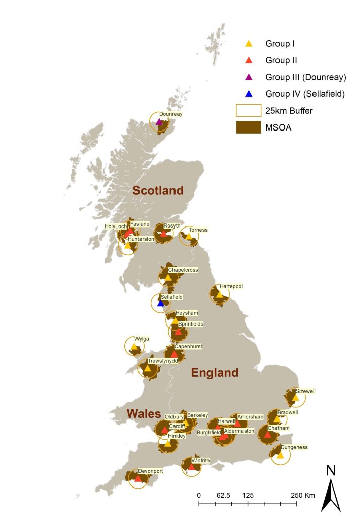
(Credit: Davies, B. et al. Int J Epidemiol, 2025)

Using these data and advanced statistical modelling, they found no increased incidence of childhood cancers in these areas compared to national averages.[1] They also found no evidence that cancer risk increased the closer children lived to the nuclear sites.

Dr Bethan Davies, from Imperial’s School of Public Health and lead author of the study, said: “For many years there have been public concerns about the potential health impacts of living near nuclear installations. Our analysis suggests that children living near these sites today are not at increased risk.”

The latest study builds on decades of research following reports in the 1980s of clusters of cancer cases near nuclear facilities in England, Scotland and Germany[2] – following which, the UK Government set up COMARE to advise on the health effects of radiation.

Early investigations confirmed clusters of cases of some cancers near nuclear installations, particularly LNHL.

However, subsequent studies failed to show any direct link between these cases and radiation exposure from nuclear facilities.

In 2016, a COMARE report[3] suggested other potential explanations for these case clusters, including infections introduced due to population mixing in the areas.

The new findings come at a time of renewed interest in nuclear energy as part of the UK’s strategy to meet net-zero carbon targets and the government committing £14.2bn to build a new nuclear power station in Suffolk and develop small modular reactors.

The researchers say that while their study offers reassurance, they support COMARE’s recommendations for ongoing surveillance of cancer incidence near nuclear sites.

The authors acknowledge a number of limitations with their study, including the use of residential address at diagnosis as a proxy for exposure.

They were also unable to account for individual-level risk factors – such as genetic or medical conditions. However, they emphasise that the study’s design and comprehensive data make it one of the most detailed assessments to date.

Dr Davies added: “As the UK government announces a multibillion-pound investment for new nuclear energy infrastructure, our findings should provide reassurance that the historical clusters of childhood cancers reported near sites such as Sellafield and Dounreay are no longer evident.”

Professor Mireille Toledano, Mohn Chair in Population Child Health in Imperial’s School of Public Health, said: “These findings are both timely and important. As the UK and other countries expand their nuclear energy capacity, it’s vital that public health remains a central consideration. It’s reassuring that our study found that the historic case clusters have resolved, but it remains important we continue to monitor public health data around such sites across the UK for any emerging trends of concern.”

The full study, published today in the International Journal of Epidemiology, was supported by funding from the National Institute for Health and Care Research (NIHR), Health Data Research UK (HDRUK) and the UK Medical Research Council (UK Research and Innovation (UKRI)).

The work was carried out through the NIHR Health Protection Research Unit in Chemical and Radiation Threats and Hazards – a partnership between the UK Health Security Agency (UKHSA) and Imperial College London.

The work was also supported by the NIHR Imperial Biomedical Research Centre, a translational research partnership between Imperial College Healthcare NHS Trust and Imperial College London.

[1] Researchers obtained national incident cases of cancer diagnosed between 1995 and 2016 in children under 15 years of age from NHS England (formerly Public Health England), Welsh Cancer Intelligence and Surveillance Unit and Health Protection Scotland.

[2] A cluster of cases of leukaemia in children living close to the Sellafield nuclear plant was reported in 1983. An Independent Advisory Group confirmed the cluster and the UK government established COMARE to advise on the health effects of radiation. Subsequent studies identified increased risks of cancers in children and young adults living near Sellafield, Dounreay (Scotland), and Hamburg (Germany) nuclear installations.

[3] Committee on Medical Aspects of Radiation in the Environment (COMARE) – Seventh report (2016) https://assets.publishing.service.gov.uk/media/5a7f70ed40f0b6230268f83c/COMARE_17th_Report.pdf

Source: Imperial College London

Cancer Risk from CT Scans up to Four Times Higher than Previous Estimates

Photo by Mart Production on Pexels

Computed tomography (CT) scans may account for 5% of all cancers annually, according to a new study out of UC San Francisco that cautions against overusing and overdosing CTs. For children, the greatest risk comes from scans of the head.

The danger is greatest for infants, followed by children and adolescents. But adults are also at risk, since they are the most likely to get scans. In the U.S., nearly 103 000 cancers are predicted to result from the 93 million CT scans that were performed in 2023 alone. This is 3 to 4 times more than previous assessments, the authors said.

“CT can save lives, but its potential harms are often overlooked,” said first author Rebecca Smith-Bindman, MD, a UCSF radiologist and professor of epidemiology and biostatistics and obstetrics, gynaecology and reproductive sciences.

“Given the large volume of CT use in the United States, many cancers could occur in the future if current practices don’t change,” said Smith-Bindman.

“Our estimates put CT on par with other significant risk factors, such as alcohol consumption and excess body weight,” she said. “Reducing the number of scans and reducing doses per scan would save lives.”

Benefits and potential dangers

CT is both indispensable and widely used to detect tumours and diagnose many illnesses. Since 2007, the number of annual CT exams has surged by 30% in the U.S. But the ionising radiation dose from CT is a known cancer risk.

To assess the public health impact of current CT use, the study estimates the total number of lifetime cancers associated with radiation exposure in relation to the number and type of CT scans performed in 2023.

“Our approach used more accurate and individualised CT dose and utilisation data than prior studies, allowing us to produce more precise estimates of the number of radiation-induced cancers,” said co-author Diana Miglioretti, PhD, a breast cancer researcher and division chief of biostatistics at UC Davis. “These updated estimates suggest the excess risks – particularly among the youngest children – are higher than previously recognised.”

Researchers analysed 93 million exams from 61.5 million patients in the U.S. The number of scans increased with age, peaking in adults between 60 to 69 years old. Children accounted for 4.2% of the scans. The researchers excluded testing in the last year of a patient’s life because it was unlikely to lead to cancer.

Future cancers from radiation exposure

Adults 50 to 59 had the highest number of projected cancers: 10 400 cases for women, 9300 for men. The most common adult cancers were lung, colon, leukaemia, bladder and breast. The most frequently projected cancers in children were thyroid, lung and breast.

The largest number of cancers in adults would come from CTs of the abdomen and pelvis, while in children they came from CTs of the head. Projected cancer risks were highest among those who underwent CT when they were under 1 year old. They were 10 times more likely to get cancer compared to others in the study.

The researchers said some CT scans are unlikely to help patients, and are overused, such as those for upper respiratory infections or for headaches without concerning signs or symptoms. They said patients could lower their risk by getting fewer of these scans or by getting lower dose scans.

“There is currently unacceptable variation in the doses used for CT, with some patients receiving excessive doses,” Smith-Bindman said.

Co-author Malini Mahendra, MD, a UCSF assistant professor of Pediatric Critical Care, said it was important that families understand the risk of developing cancer from paediatric scans.

“Few patients and their families are counselled about the risk associated with CT examinations,” she said. “We hope our study’s findings will help clinicians better quantify and communicate these cancer risks, allowing for more informed conversations when weighing the benefits and risks of CT exams.”

Source: University of California – San Francisco

Can Space Radiation Affect Astronauts’ Long-term Cognition?

Photo: Pixabay CC0

During missions into outer space, galactic cosmic radiation (GCR) will penetrate current spacecraft shielding and thus pose a significant risk to human health. Previous studies have shown that GCR can cause short-term cognitive deficits in male rodents. Now a study published in the Journal of Neurochemistry reveals that GCR exposure can also cause long-lasting learning deficits in female rodents.

The impact of GCR on cognition was lessened when mice were fed an antioxidant and anti-inflammatory compound called CDDO-EA.

Beyond its immediate implications for space exploration, the findings contribute to a broader understanding of radiation’s long-term impact on cognitive health.

“Our study lays the groundwork for future causal delineation of how the brain responds to complex GCR exposure and how these brain adaptations result in altered behaviours,” said co-corresponding author Sanghee Yun, PhD, of the Children’s Hospital of Philadelphia Research Institute and the University of Pennsylvania Perelman School of Medicine.

Source: Wiley

Radon Gas Contributing to Rise in Lung Cancer among Young Adults

Photo by Vladyslav Cherkasenko on Unsplash

Although lung cancer is traditionally thought of as a “smoker’s disease,” a surprising 15–20% of newly diagnosed lung cancers occur in people who have never smoked, many of whom are in their 40s or 50s.

This concerning rise in non-smoking lung cancer cases is likely linked to long-term, high exposures of radon gas. This colourless, odourless gas is emitted from the breakdown of radioactive material naturally occurring underground that then seeps through building foundations. The gas can linger and accumulate in people’s homes and lungs silently unless they know to test for it.

Although the U.S. Environmental Protection Agency (EPA) recommends regular radon testing and corrective measures to lower exposure levels in homes, a new consumer survey conducted on behalf of The Ohio State University Comprehensive Cancer Center (OSUCCC) showed that a stunning 75% of Americans have not had their homes tested for radon, and over half (55%) are not concerned about radon exposure in their homes, community or schools.

“Anyone with lungs can develop lung cancer, and as a community we should be aware and concerned about radon exposure because it’s thought to be one of the leading causes of lung cancer in never-smokers – and there is something we can do reduce our risk,” said David Carbone, MD, PhD, a thoracic medical oncologist. “There are relatively simple tests to measure radon in the home and actions to reduce radon exposure.”

This includes installing outside the home a radon remediation system that sucks air from the basement, where radon gas typically lingers. Increasing air flow by opening windows and using fans/venting in your home, and sealing cracks in the floors, walls and foundation is also important.

Lung cancer rising in young non-smokers

The No. 1 risk factor for lung cancer is long-term cigarette smoking; however, rates of lung cancer among non-smokers continue to rise. The symptoms of the disease are the same regardless of whether the person has smoked: generally not feeling well or feeling tired all the time, frequent cough, chest pain, wheezing, shortness of breath or coughing up blood. These symptoms happen with other illnesses too, but Carbone notes anyone – regardless of age – who has a lingering symptom that doesn’t resolve despite initial treatment should insist on having it checked out.

Lung cancer screening is currently available only to people at the highest risk for the disease – older adults with a history of heavy smoking.

If detected in its earliest stages, the cure rate for lung cancer can be 90–95%. The bulk of cases, however, are not detected until the disease has spread throughout the lung or to other parts of the body, when treatments aren’t as effective. It is important that anyone deemed at risk for lung cancer get timely screening, and that people who might be at increased risk due to secondhand smoke, radon or occupational exposures (like firefighting) talk to their doctors about testing.

“Your health and the health of your family are the most important things you have. Really push to get your concerns addressed if your symptoms aren’t resolving, even if you don’t fit the typical ‘picture’ of lung cancer. It could truly save your life,” said Carbone.

Requiring radon testing in homes, schools and workplaces

Carbone noted that having high levels of radon exposure at school or work is just as much a health hazard as having high-level exposure in your basement.

He says he strongly supports potential legislation to require radon testing at schools, at places of business and during home sales to help reduce community risk. The effects of radon on your lungs is cumulative and can be delayed by decades.

“So your children playing in your basement or going to school today, exposed to unknown levels of radon, could be at risk for developing lung cancer 10, 20, 30 years from now,” Carbone said. “And because the gas is totally colourless and odourless, you would have no idea you were being exposed unless you knew the importance of proactively testing.”

Source: Ohio State University Wexner Medical Center

Radon Gas: Ubiquitous, Carcinogenic – and Possible Stroke Risk

Photo by Vladyslav Cherkasenko on Unsplash

A new study has found that exposure to radon, the second leading cause of lung cancer, is also linked to an increased risk of stroke. The study, which examined exposures in middle age to older female participants, found an increased risk of stroke among those exposed to high and even moderate concentrations of the gas compared to those exposed to the lowest concentrations. The study is published in Neurology®, the medical journal of the American Academy of Neurology.

Radon is a naturally occurring radioactive gas produced in certain rocks and soils which contain uranium or radium. In South Africa, some areas such as in the Western Cape have higher concentrations of radon due to underlying granite geology. It is also a concern near gold mine dumps, which have higher levels of uranium.

The gas can make its way into homes through cracks in basement walls and floors, construction joints and gaps around pipes.

“Radon is an indoor air pollutant that can only be detected through testing that measures concentrations of the gas in homes,” said study author Eric A. Whitsel, MD, MPH, of the University of North Carolina in Chapel Hill.

“Our research found an increased risk of stroke among participants exposed to radon above – and as many as two picocuries per litre (pCi/L) below – concentrations that usually trigger Environmental Protection Agency recommendations to install a home radon mitigation system.”

The study involved 158 910 female participants with an average age of 63 who did not have stroke at the start of the study.

They were followed for an average of 13 years. During the study, there were 6979 strokes among participants.

To determine radon exposures, researchers linked participants’ home addresses to radon concentration data from the U.S. Geological Survey and the U.S. Environmental Protection Agency (EPA).

The EPA recommends that average indoor radon concentrations do not exceed four picocuries per liter (pCi/L). For concentrations this high, the EPA recommends installing a radon mitigation system to lower radon levels in the home.

Participants were divided into three groups. The highest group had homes in areas where average radon concentrations were more than four pCi/L. The middle group lived in areas with average concentrations between two and four pCi/L. The lowest group lived in areas with average concentrations of less than two pCi/L.

In the group with the highest radon exposures, there were 349 strokes per 100 000 person-years compared to 343 strokes in the middle group and 333 strokes in group with the lowest exposure.

Person-years represent both the number of people in the study and the amount of time each person spends in the study.

After adjusting for factors such as smoking, diabetes and high blood pressure, researchers found participants in the highest group had a 14% increased risk of stroke compared to those in the lowest group.

Those in the middle group had a 6% increased risk.

“It’s important to note that we found an increased stroke risk among those exposed to radon concentrations as much as two pCi/L below the current lung cancer-based threshold for recommending radon mitigation,” said Whitsel.

“More studies are needed to confirm our findings. Confirmation would present an opportunity to improve public health by addressing an emerging risk factor for stroke.”

A limitation of the study was that it included only female participants who were middle age or older and primarily white, so the results may not be the same for other populations.

Source: American Academy of Neurology

A New Way to Prevent Cognitive Decline from Radiotherapy

Photo by National Cancer Institute on Unsplash

Microglia, the brain’s immune cells, can trigger cognitive deficits after radiation exposure and may be a key target for preventing these symptoms, University of Rochester researchers have found. Their work, published in the International Journal of Radiation Oncology Biology Biophysics, builds on previous research showing that after radiation exposure microglia damage synapses, the connections between neurons that are important for cognitive behaviour and memory.

“Cognitive deficits after radiation treatment are a major problem for cancer survivors,” M. Kerry O’Banion, MD, PhD, professor of Neuroscience, member of the Wilmot Cancer Institute, and senior author of the study said.

“This research gives us a possible target to develop therapies to prevent or mitigate against such deficits in people who need brain radiotherapy.”

Using several behavioural tests, researchers investigated the cognitive function of mice before and after radiation exposure.

Female mice performed the same throughout, indicating a resistance to radiation injury but Male mice could not remember or perform certain tasks after radiation exposure.

This cognitive decline correlates with the loss of synapses and evidence of potentially damaging microglial over-reactivity following the treatment.

Researchers then targeted the pathway in microglia important to synapse removal. Mice with these mutant microglia had no cognitive decline following radiation. And others that were given the drug, Leukadherin-1, which is known to block this same pathway, during radiation treatment, also had no cognitive decline.

“This could be the first step in substantially improving a patient’s quality of life and need for greater care,” said O’Banion. “Moving forward, we are particularly interested in understanding the signals that target synapses for removal and the fundamental signaling mechanisms that drive microglia to remove these synapses. We believe that both avenues of research offer additional targets for developing therapies to help individuals receiving brain radiotherapy.”

O’Banion also believes this work may have broader implications because some of these mechanisms are connected to Alzheimer’s and other neurodegenerative diseases.

Source: University of Rochester Medical Center

Study Confirms Link between CT Scans in Young People and Increased Risk of Cancer

Credit: Pixabay CC0

A multinational study of almost one million individuals confirms a strong and clear association between radiation exposure from CT scans in young people and an increased risk of blood cancers. These results, published in Nature Medicine, highlight the importance of continuing to apply strict radiological protection measures, particularly in paediatric populations. 

The benefits of computed tomography (CT) for imaging in patient management (including diagnostic efficacy, treatment planning and disease follow-up) are undisputed. But in recent decades the extensive use of this procedure has raised concerns about the potential cancer risks associated with exposure to ionising radiation, particularly in young patients.

“The exposure associated with CT scans is considered low (less than 100mGy), but it is still higher than for other diagnostic procedures,” says Elisabeth Cardis, Head of the Radiation Group at ISGlobal and senior author of the study. Previous studies have suggested an increased risk of cancer in in children exposed to CT scans, but they had several methodological limitations.

To address these limitations, clinicians, epidemiologists and dosimetrists from nine European countries (Belgium, Denmark, France, Germany, Netherlands, Norway, Spain, Sweden, and UK) came together to conduct a multinational, European-funded study, EPI-CT, coordinated by the International Agency for Research on Cancer (IARC).

“Implementing this large, multinational study was challenging – it involved extracting data from radiology records of 276 hospitals and linking them to population-based registries in nine countries, all while maintaining the confidentiality of the individuals’ data,” says Cardis.

A dose-dependent association

The study analysed data from almost one million people, who underwent at least one CT scan before the age of 22. The dose of radiation delivered to the bone marrow, where blood cells are produced, was estimated for each person. By linking this information to national cancer registries, EPI-CT researchers were able to identify those who developed a blood cancer over time. Individuals were followed for an average of 7.8 years, although for those who had CT scans in the early years of the technology, researchers were able to monitor cancer incidence for more than 20 years after the first scan.

The results show a clear association between the total radiation doses to the bone marrow from CT scans and the risk of developing both myeloid and lymphoid malignancies. A dose of 100mGy approximately tripled the risk of developing a blood cancer. These results suggest that a typical scan today (with an average dose of about 8mGy) increases the risk of developing these malignancies by about 16%. “In terms of absolute risk, this means that, for every 10 000 children who have a CT scan, we can expect to see about 1–2 cases of cancer in the 12 years following the examination,” says first author Magda Bosch de Basea, ISGlobal researcher at the time of the study.

The authors point out that more work is needed to ensure that doses and technical parameters are systematically and adequately collected in the clinics in real time to further improve risk estimates in the future.

Public health implications

Today, more than one million children in Europe undergo CT scans every year. Although radiation doses from CT scans have decreased substantially in recent years, the findings of this study underline the need to raise awareness among the medical community and to continue to apply strict radiation protection measures, especially in the youngest patients. “The procedure must be properly justified – taking into account possible alternatives – and optimised to ensure that doses are kept as low as possible while maintaining good image quality for the diagnosis,” Cardis explains. 

Source:

US to Trial Anti-radiation Pills That Could Protect Against Fallout from ‘Dirty Bombs’

The US National Institutes of Health is funding a new clinical trial of an oral anti-radiation treatment. So-called dirty bombs are nuclear weapons that release a cloud of radioactive isotopes, and are thought to be an attractive option for terrorists because they are far easier to build than true nuclear explosives.

In such a situation, as well as a nuclear accident, the danger is not so much from direct radiation in the form of X-rays, gamma rays and alpha and beta particles, but from radioactive materials which are absorbed into the body via contaminated air, food or water. Certain radioisotopes from radioactive fallout are readily absorbed by the body, and cause damage to DNA and cellular structures. Current treatment contain chemicals that bind to these radioisotopes, preventing them from being taken up by the body and instead rapidly excreted.

The Food and Drug Administration has approved two products for removing internal radioactive contamination. These drugs, both based on diethylenetriamine pentaacetate (DTPA), are administered intravenously by a healthcare provider and can remove three radioactive elements: plutonium, americium, and curium. 

In contrast, HOPO 14-1 is an oral capsule, which would be easier than an intravenous drug to stockpile and to deploy and administer during an emergency. Preclinical research has shown that HOPO 14-1 can effectively remove many radioactive contaminants, including uranium and neptunium in addition to plutonium, americium and curium. These studies also have found that HOPO 14-1 is up to 100 times more effective than DTPA at binding and removing these radioactive elements.

The trial will seek 42 healthy volunteers aged 18 to 65, who will of course not be exposed to radioactive fallout. They will be assigned into seven groups of six. Each participant in the first group will receive a 100-milligram (mg) dose of HOPO 14-1. The subsequent groups will receive increasingly higher doses of the study drug up to 7500 mg in the final group, if lower doses are deemed safe. Participants will undergo intensive safety monitoring and will be followed for 14 days to measure the absorption, distribution and elimination of the study drug. Results are expected in 2024.

Source: National Institutes of Health

Genetic Radiation Damage Passed down Through Fathers

Chromosomes. Source: NIH

Whether radiation exposure of fathers can have consequences on their children is one of the most long-standing questions in radiation biology. Using the nematode Caenorhabditis elegans as a model, University of Cologne researchers reported in the journal Nature that radiation damage to mature sperm cannot be repaired but is instead passed on to the offspring.

Female eggs with radiation damage either accurately repair it or, if the damage is too severe, are eliminated and no damage is passed on. However, when the egg is fertilised with a radiation-damaged sperm, the maternal repair proteins that are provided by the egg try to repair the paternal DNA.

For this purpose, a highly error-prone repair mechanism is used and fuses the broken DNA pieces randomly. These random fusions of the breaks then lead to structural changes in the paternal chromosomes. The resulting offspring now carry the chromosome damage and in turn their offspring show severe developmental defects. The work done on C. elegans lays the foundation for a better understanding of the mechanisms for the heritable effects of paternal radiation exposure.

This work has now been published under the title ‘Inheritance of paternal DNA damage by histone-mediated repair restriction’ in 

The offspring that results from male animals that have been exposed to radiation and healthy female worms carry on the so-called structural variations – random connections of chromosome parts. In the offspring, these aberrations lead to recurrent breaks but this damage can no longer be repaired. Instead, the damaged chromosomes are shielded from accurate repair by proteins, so-called histones, that densely pack the long strands of DNA. In the densely packed DNA, the breaks can no longer be reached by the repair proteins. The packed DNA structures are held tightly together by the specific histone proteins, HIS-24 and HPL-1. When those histone proteins are removed, the paternally inherited damage is completely eliminated and viable offspring can be produced. The finding that histone proteins govern the accessibility of DNA for repairs could provide effective therapeutic targets for treating radiation damage.

Adding to the work on nematodes, the team detected the same structural variants, or randomly assembled chromosomes, in humans. Also here, the chromosome aberrations were specifically passed on from the fathers but not the mothers. For this, the scientists analysed various data sets from the 1000 Genome Project that contains genetic data from more than a thousand people and the Islandic deCODE project with genetic data from the respective mothers, fathers and children.

“Genome aberrations, especially structural variations in chromosomes, which develop in the paternal germline, are thought to increase the risk of disorders like autism and schizophrenia,” study leader Professor Dr Björn Schumacher said. This means that also in humans, mature sperm needs to be especially protected from radiation damage, and damaged mature sperm should not be used for conception. He added, “Such damage could potentially be inflicted during radiotherapy or chemotherapy and thus pose a risk in the two months that it takes to generate new sperm to replace the damaged one.” This is because in contrast to mature sperm, newly generated sperm have the capacity to accurately repair the damage.

Interestingly, the scientists found those structural variations in the chromosomes also in nematodes in the wild and in the human population. These results suggest that damage to mature sperm and the inaccurate repair of paternal DNA in the zygote could be major drivers for genetic diversity during evolution and might be responsible for genetic diseases in humans.

Source: University of Cologne

New Radiation Shielding is a Weight off Cath Lab Shoulders

Radiation warning sign
Photo by Vladyslav Cherkasenko on Unsplash

Testing has shown that a new radiation shielding system offered equivalent radiation protection to the standard lead gowns worn in the catheterisation lab. By using such independent, adjustable shields instead of wearing shielding, the occupational shoulder and back strain inherent to wearing those heavy gowns can be eliminated.

The Rampart shielding system consists of an adjustable stand made of lead equivalent acrylic shielding – was found to block 96% to 98% of radiation scattered to the operator’s head, torso, and waist during an average week of cardiac angiography, according to medical radiation specialist Glenn Ison.

This was equivalent or better than shielding provided by lead gowns and a ceiling-mounted lead shield, Ison said in a presentation at the EuroPCR meeting.

“We found it’s like walking on the moon, doing cases without a lead gown. It’s quite strange,” he remarked.

The researchers had operators wear radiation monitors to measure operator scatter dose to the head, torso, and waist (both under and over their lead gowns) with and without the Rampart.

Dr Ison also pointed out that head and face protection in particular was substantially better with the Rampart shield (radiation exposure -96% vs -70% with standard practice).

Indeed, the shielding of the cumbersome lead gowns — which can weigh up to 13.6kg — only extends to major areas of the torso and waist. Adding on protective glasses and shin guards further adds to this weight.

Ison cited a survey showing that half of interventional cardiologists reported neck or back pain during their careers.

“The ability to maintain protection from scatter radiation whilst lowering or even removing the weight of current lead gowns is a game changer for operators with current back or neck troubles and a way to prevent such problems in the future,” he said. “The longer you’ve been in the game, the more this appeals.”

He added that anaesthetic and nursing staff also had a new highly shielded area to stand in, thanks to the new system.

In response to a panellist question of whether the Rampart would restrict the operator’s arm movements and impede emergency CPR, Ison noted that other companies are working on different shielding types, perhaps some with ceiling-mounted designs making it easier for operators to move around.

“Any benefit to reduce radiation exposure to the operators and staff is an important step forward. The occupational hazards for interventional cardiology are not small, and we need to focus on ways to improve this in the future. To me this is a good step forward,” commented Roxana Mehran, MD, interventional cardiologist at Mount Sinai Health System in New York City.

Ison cautioned that the shield’s effectiveness depends on being correctly positioned. “You must make sure it’s locked in correctly” and test it according to table size and shape, he advised. “Use of real-time audible alarm radiation monitors would be advised.”

Source: MedPage Today