Tag: multiple sclerosis

Two Treatments for Progressive Multiple Sclerosis Fail to Show Benefit

This is a pseudo-colored image of high-resolution gradient-echo MRI scan of a fixed cerebral hemisphere from a person with multiple sclerosis. Credit: Govind Bhagavatheeshwaran, Daniel Reich, National Institute of Neurological Disorders and Stroke, National Institutes of Health

In people with primary progressive multiple sclerosis (MS), a new study has found no difference in the amount of time before disability worsened between people taking certain medications and those not receiving treatment. The study is published in Neurology®, the medical journal of the American Academy of Neurology.

With MS, the body’s immune system attacks the myelin sheaths of nerves. People with primary progressive MS experience a steady decline in symptoms. About 10 to 15% of people with the disease have this type of MS.

The study looked at rituximab and ocrelizumab, anti-CD20 infusion therapies that target a protein called CD20 found on some white blood cells called B-cells. Removing these cells from the bloodstream is believed to reduce inflammation and damage that can occur to the myelin.

Ocrelizumab is approved by the US Food and Drug Administration (FDA) for primary progressive MS and for people with relapses, but rituximab is not. Rituximab is FDA approved for other diseases like rheumatoid arthritis and prescribed off label for MS in the US.

“MS is a disabling disease, so treatments that slow the progression to worse disability are sorely needed,” said study author Laure Michel, MD, PhD, of Rennes University in France. “Anti-CD20 therapies are widely prescribed, in part because there are few alternate treatments. However, our study suggests they may not slow disability from worsening for people with primary progressive MS.”

The study involved 1184 people with primary progressive MS who had an average age of 56. They did not take MS medications in the two years prior to the study. For the study, 295 people were treated with rituximab, 131 were treated with ocrelizumab and 728 were untreated.

They were followed for an average of four years. Participants’ level of disability was measured on a scale with scores ranging from zero, meaning no symptoms, to 10 points, meaning death due to MS. At the start of the study, all participants had a score of 6.5 or less. Researchers then measured how long it took for people to advance to their first confirmed disability progression.

For those whose score was less than 5.5 at the start of the study, advancing one point on the scale was considered progressing in disability. If their score was 5.5 or more, advancing 0.5 points on the scale was disability progression.

After adjusting for possible differences between the treated and untreated groups, researchers found there was no difference in the time it took to progress to the next level of disability between those taking a medication and those taking no medication. “

Medications for MS can be expensive and come with risks of side effects,” said Michel. “Our results indicate that there should be a constant evaluation of MS therapies to determine if the benefits outweigh the risks for people with primary progressive MS.”

A limitation of the study was that it was retrospective and did not follow people in real time. Also, among those taking medications, most were taking rituximab with fewer people taking ocrelizumab. More research is needed in larger groups of people to confirm the findings.

Source: American Academy of Neurology

New Paper Suggests that MS Protects Against Alzheimer’s Disease

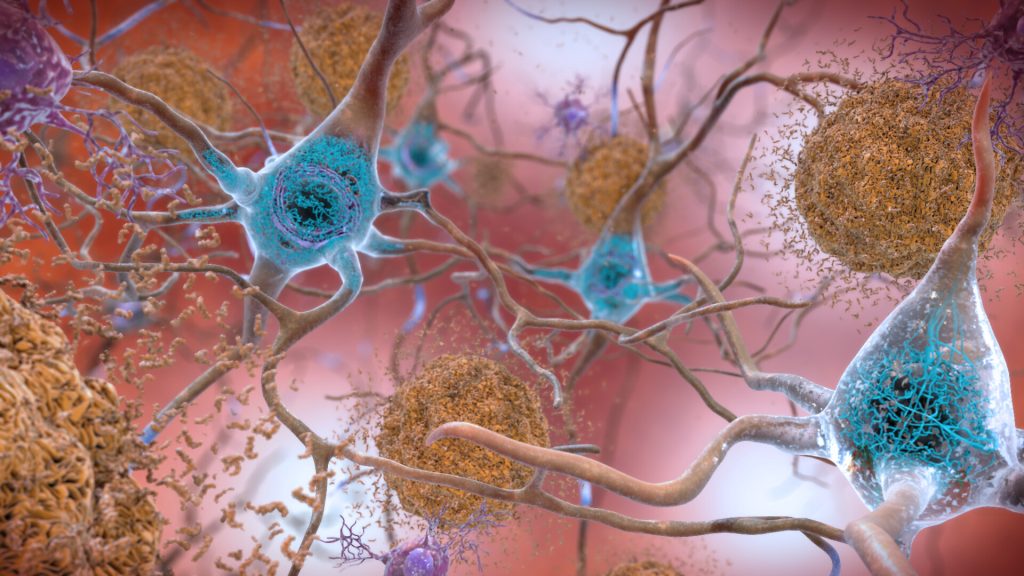
Neurons in the brain of an Alzheimer’s patient, with plaques caused by tau proteins. Credit: NIH

People with multiple sclerosis (MS) are far less likely than those without the condition to have the molecular hallmarks of Alzheimer’s disease, according to a paper published in the Annals of Neurology.

The study from Washington University School of Medicine in St. Louis, suggests a new direction for researching Alzheimer’s treatments, said Matthew Brier, MD PhD, an assistant professor of neurology and radiology and the study’s first author.

“Our findings imply that some component of the biology of multiple sclerosis, or the genetics of MS patients, is protective against Alzheimer’s disease,” Brier said. “If we could identify what aspect is protective and apply it in a controlled way, that could inform therapeutic strategies for Alzheimer’s disease.”

A collaboration between WashU Medicine experts in Alzheimer’s and MS, the study was prompted by a suspicion Brier’s mentor and collaborator Anne Cross, MD, had developed over decades of treating patients with MS, an immune-mediated disease that attacks the central nervous system. Although her patients were living long enough to be at risk of Alzheimer’s or had a family history of the neurodegenerative disease, they weren’t developing the disease.

“I noticed that I couldn’t find a single MS patient of mine who had typical Alzheimer’s disease,” said Cross, the Manny and Rosalyn Rosenthal and Dr. John Trotter MS Center Chair in Neuroimmunology. “If they had cognitive problems, I would send them to the memory and aging specialists here at the School of Medicine for an Alzheimer’s assessment, and those doctors would always come back and tell me, ‘No, this is not due to Alzheimer’s disease.’”

Cognitive impairment caused by MS can be confused with symptoms of Alzheimer’s disease; Alzheimer’s can be confirmed with blood and other biological tests.

To confirm Cross’ observation, the research team used a new, FDA-approved blood test that was developed by Washington University researchers. Known as PrecivityAD2, the blood test is highly effective at predicting the presence of amyloid plaques in the brain. Such plaques are an indicator of Alzheimer’s disease and previously only could be verified with brain scans or spinal taps.

Brier, Cross and their colleagues recruited 100 patients with MS to take the blood test, 11 of whom also underwent PET scans at the School of Medicine’s Mallinckrodt Institute of Radiology. Their results were compared with the results from a control group of 300 individuals who did not have MS but were similar to those with MS in age, genetic risk for Alzheimer, and cognitive decline.

“We found that 50% fewer MS patients had amyloid pathology compared to their matched peers based on this blood test,” Brier said. This finding supported Cross’ observation that Alzheimer’s appeared to be less likely to develop among those with MS. It is not clear how amyloid accumulation is linked to the cognitive impairment typical of Alzheimer’s, but the accumulation of plaques is generally understood to be the first event in the biological cascade that leads to cognitive decline.

The researchers also found that the more typical the patient’s MS history was, in terms of age of onset, severity and overall disease progression, the less likely they were to have amyloid plaque accumulation in that patient’s brain compared with those with atypical presentations of MS. This suggests there is something about the nature of MS itself that is protective against Alzheimer’s disease, which Brier and Cross are planning to investigate.

MS patients generally have multiple flare-ups of the illness over the course of their lifetimes. During these flare-ups, the immune system attacks the central nervous system, including within the brain. It’s possible that this immune activity also reduces amyloid plaques, the researchers said.

“Perhaps when the Alzheimer’s disease amyloid pathology was developing, the patients with MS had some degree of inflammation in their brains that was spurred by their immune responses,” Brier said. Referring to work by co-author David M. Holtzman, MD, Brier noted that activated microglia, which are part of the brain’s immune response in MS, have been shown to clear amyloid from the brain in animal models.

Brier and Cross have begun the next steps of this research, both to tease out the possible human genetics involved, as well as to test amyloid plaque development in animal models representing MS.

Source: Washington University School of Medicine

Visualising Multiple Sclerosis with a New MRI Procedure

This is a pseudo-colored image of high-resolution gradient-echo MRI scan of a fixed cerebral hemisphere from a person with multiple sclerosis. Credit: Govind Bhagavatheeshwaran, Daniel Reich, National Institute of Neurological Disorders and Stroke, National Institutes of Health

A key feature of multiple sclerosis (MS) is that it causes the patient’s own immune system to attack and destroy the myelin sheaths in the central nervous system. To date, it hasn’t been possible to visualise the myelin sheaths well enough to use this information for the diagnosis and monitoring of MS.  Now researchers have developed a new magnetic resonance imaging (MRI) procedure that maps the condition of the myelin sheaths more accurately than was previously possible.

The researchers successfully tested the procedure on healthy people for the first time, and published their results in Magnetic Resonance in Medicine.

In the future, the MRI system with its special head scanner could help doctors to recognise MS at an early stage and better monitor the progression of the disease.

This technology, developed by the researchers at ETH Zurich and University of Zurich, led by Markus Weiger and Emily Baadsvik from the Institute for Biomedical Engineering, could also facilitate the development of new drugs for MS. But it doesn’t end there: the new MRI method could also be used by researchers to better visualise other solid tissue types such as connective tissue, tendons and ligaments.

Quantitative myelin maps

Conventional MRI devices capture only inaccurate, indirect images of the myelin sheaths because these devices typically work by reacting to water molecules in the body that have been stimulated by radio waves in a strong magnetic field.

But the myelin sheaths, which wrap around the nerve fibres in several layers, consist mainly of fatty tissue and proteins. That said, there is some water – known as myelin water – trapped between these layers.

Standard MRIs build their images primarily using the signals of the hydrogen atoms in this myelin water, rather than imaging the myelin sheaths directly.

The ETH researchers’ new MRI method solves this problem and measures the myelin content directly.

It puts numerical values on MRI images of the brain to show how much myelin is present in a particular area compared to other areas of the image.

A number 8, for instance, means that the myelin content at this point is only 8 percent of a maximum value of 100, which indicates a significant thinning of the myelin sheaths.

Essentially, the darker the area and the smaller the number in the image, the more the myelin sheaths have been reduced.

This information ought to enable doctors to better assess the severity and progression of MS.

Measuring signals within millionths of a second

It is difficult however to image the myelin sheaths directly, since the signals that the MRI triggers in the tissue are very short-lived; the signals that emanate from the myelin water last much longer.

“Put simply, the hydrogen atoms in myelin tissue move less freely than those in myelin water. That means they generate much briefer signals, which disappear again after a few microseconds,” Weiger says, adding: “And bearing in mind a microsecond is a millionth of a second, that’s a very short time indeed.” A conventional MRI scanner can’t capture these fleeting signals because it doesn’t take the measurements fast enough.

To solve this problem, the researchers used a specially customised MRI head scanner that they have developed over the past ten years together with the companies Philips and Futura.

This scanner is characterised by a particularly strong gradient in the magnetic field.

“The greater the change in magnetic field strength generated by the three scanner coils, the faster information about the position of hydrogen atoms can be recorded,” Baadsvik says.

Generating such a strong gradient calls for a strong current and a sophisticated design.

As the researchers scan only the head, the magnetic field is more contained and concentrated than with conventional devices.

In addition, the system can quickly switch from transmitting radio waves to receiving signals; the researchers and their industry partners have developed a special circuit for this purpose.

The researchers have already successfully tested their MRI procedure on tissue samples from MS patients and on two healthy individuals. Next, they want to test it on MS patients themselves. Whether the new MRI head scanner will make its way into hospitals in the future now depends on the medical industry. “We’ve shown that our process works,” Weiger says. “Now it’s up to industry partners to implement it and bring it to market.”

Source: ETH Zurich

Explaining the Parallels between Vitamin B12 Deficiency and Multiple Sclerosis

This is a pseudo-colored image of high-resolution gradient-echo MRI scan of a fixed cerebral hemisphere from a person with multiple sclerosis. Credit: Govind Bhagavatheeshwaran, Daniel Reich, National Institute of Neurological Disorders and Stroke, National Institutes of Health

For decades, scientists have noted an intriguing similarity between a deficiency in vitamin B12 – an essential nutrient that supports healthy development and functioning of the central nervous system (CNS) – and multiple sclerosis (MS), a chronic disease in which the body’s immune system attacks the CNS and which can produce neurodegeneration.

Both vitamin B12 deficiency and MS produce similar neurological symptoms, including numbness or tingling in hands and feet, vision loss, difficulty walking or speaking normally and cognitive dysfunction, such as problems with memory.

In a new study, published in Cell Reports, researchers at Sanford Burnham Prebys, with collaborators elsewhere, describe a novel molecular link between vitamin B12 and MS that takes place in astrocytes – important non-neuronal glial cells in the brain.

The findings by senior study author Jerold Chun, MD, PhD, professor and senior vice president of neuroscience drug discovery, and Yasuyuki Kihara, PhD, research associate professor and co-corresponding author, and colleagues suggest new ways to improve the treatment of MS through CNS-B12 supplementation.

“The shared molecular binding of the brain’s vitamin B12 carrier protein, known as transcobalamin 2 or TCN2, with the FDA-approved MS drug fingolimod provides a mechanistic link between B12 signaling and MS, towards reducing neuroinflammation and possibly neurodegeneration,” said Chun.   

“Augmenting brain B12 with fingolimod or potentially related molecules could enhance both current and future MS therapies.”

In their paper, the team at Sanford Burnham Prebys, with collaborators at University of Southern California, Juntendo University in Japan, Tokyo University of Pharmacy and Life Sciences and State University of New York, focused on the molecular functioning of FTY720 or fingolimod (Gilenya®), a sphingosine 1-phosphate (S1P) receptor modulator that suppresses distribution of T and B immune cells errantly attacking the brains of MS patients.

Working with an animal model of MS as well as human post-mortem brains, the researchers found that fingolimod suppresses neuroinflammation by functionally and physically regulating B12 communication pathways, specifically elevating a B12 receptor called CD320 needed to take up and use needed B12 when it is bound to TCN2, which distributes B12 throughout the body, including the CNS.  This known process was newly identified for its interactions with fingolimod within astrocytes. Importantly, the relationship was also observed in human MS brains.

Of particular note, the researchers reported that lower levels of CD320 or dietary B12 restriction worsened the disease course in an animal model of MS and reduced the therapeutic efficacy of fingolimod, which occurred through a mechanism in which fingolimod hitchhikes by binding to the TCN2-B12 complex, allowing delivery of all to the astrocytes via interactions with CD320, with component losses disrupting the process and worsening disease.

These new findings further support to the use of B12 supplementation – especially in terms of delivering the vitamin to astrocytes within the brain – while revealing that fingolimod can correct the impaired astrocyte-B12 pathway in people with MS. 

The scientists said it is possible that other S1P receptor modulators on the market, such as Mayzent®, Zeposia® and Ponvory®, may access at least parts of this CNS mechanism.  The study supports B12 supplementation with S1P receptor modulators with the goal of improving drug efficacy for this class of medicines.

The study also opens new avenues on how the B12-TCN2-CD320 pathway is regulated by sphingolipids, specifically sphingosine, a naturally occurring and endogenous structural analogue of fingolimod, toward improving future MS therapies, Chun said. 

“It supports creating brain-targeted B12 formulations. In the future, this mechanism might also extend to novel treatments of other neuroinflammatory and neurodegenerative conditions.”

Source: Sanford-Burnham Prebys

New Drug with a Different Approach Holds Promise as a Treatment for Multiple Sclerosis

This is a pseudo-colored image of high-resolution gradient-echo MRI scan of a fixed cerebral hemisphere from a person with multiple sclerosis. Credit: Govind Bhagavatheeshwaran, Daniel Reich, National Institute of Neurological Disorders and Stroke, National Institutes of Health

Researchers have found in pre-clinical studies of a small molecule drug that it has promise as a potential new treatment for multiple sclerosis (MS). The results from the Centre for Addiction and Mental Health-led study have been published in the journal Science Advances.

Expanding on Dr Fang Liu’s earlier work that identified a novel drug target for the treatment of MS, she and her team have now created a small molecule compound that is effective in two different animal models of MS. This represents a key advancement that brings this MS research closer to the clinic to impact patient care.

MS is a progressive neurological disease that currently has no cure.

It is associated with a wide-range of debilitating symptoms, including problems with coordination, cognition, muscle weakness and depression. For unknown reasons, it is more common in northern latitudes and more than twice as common in women.

It is known that MS damages myelin, a protective sheath that forms around nerves in the brain and spinal cord. As the myelin damage is triggered by inflammation in the immune system, up until now all current drug treatments for MS target the immune system.

In this study, CAMH Senior Scientist Dr Fang Liu and her team treated MS in a completely different way – targeting the glutamate system. Study results showed that the newly synthesised lead compound not only reduced MS-like symptoms, it also may repair the damaged myelin in two different pre-clinical models of MS.

“Our compound had a stunning effect on rescuing myelin and motor function in the lab models, and I hope these effects will translate to the clinic to add to current treatments and bring new hope to patients with MS,” said Dr Liu.

“As with cancer chemotherapy drug cocktails, simultaneous targeting of the MS disease pathway at multiple points can have synergistic effects and result in better outcomes.”

Dr Iain Greig, Reader in Medicinal Chemistry at the University of Aberdeen, alongside his team, are working to turn the molecules identified by Dr Liu into advanced “drug-like” molecules suitable for continued development towards clinical use in patients.

He added: “In all my years as a medicinal chemist, I have never seen a more promising starting point for a drug development project. It has been a huge pleasure to be involved in this program and I am looking forward to continuing to drive it towards to the clinic.”

Much of the funding for this novel treatment for MS, which Dr. Fang and her team have been investigating for over a decade, has come from the Multiple Sclerosis Society of Canada and the National Multiple Sclerosis Society USA’s Fast Forward commercial research program.

“We are pleased to have helped enable the early development of a novel neuroprotective strategy for MS, and look forward to seeing it progress through the critical next stages needed to determine its potential benefits for people living with MS,” said Walt Kostich, PhD, head of the National MS Society (USA)’s Fast Forward commercial research programme.

Dr. Liu believes that the evidence of efficacy and tolerability generated in this study for the small molecule drug makes it a good candidate to be developed for human trials. The next steps in drug development will involve some further pre-clinical research, including investigating safety and stability of the compound. CAMH and the University of Aberdeen have already filed patent applications to protect this research and are actively seeking industry partners to further advance this work towards clinical trials over the next few years.

Source: Centre for Addiction and Mental Health

Thinning of the Retina is an Early Marker of MS

Retina and nerve cells. Credit: NIH

For the first time, a study has shown that diagnosis of multiple sclerosis (MS) can be significantly improved by additionally measuring the thickness of retinal layers in the eye in a currently existing procedure. Use of the procedure helps to detect the condition at an earlier stage and predict its progression more accurately, which can help to improve patient outcomes. The findings have been published in the journal Neurology.

As part of their investigation, the research team headed by Gabriel Bsteh and Thomas Berger collaborated with ophthalmology colleagues examine 267 MS patients over five years. Their research built on study results published in 2022, which showed that MS relapse-related damage to the retina reflects the degree of damage caused to the patient’s brain. The previous study also demonstrated that a 5µm reduction in the thickness of the retinal layer following optic neuritis indicated a doubling of the risk of permanent disability after the next relapse. Thanks to the latest research with the large cohort of MS patients, the research team has confirmed that the thickness of the retinal layer can be used as a precise biomarker to assist early diagnosis.

Diagnostic procedure already available

The researchers used a procedure known as optical coherence tomography (OCT) to measure the thickness of the retinal layer. An imaging method that uses infrared light, OCT allows for the generation of high-resolution, three-dimensional images of extremely thin layers of tissue measuring just a few µm. OCT is also a tool for diagnosing and evaluating the progression of eye diseases such as glaucoma. “So we already have this procedure at our disposal,” commented Gabriel Bsteh, first author of the study. He added: “If we use optical coherence tomography alongside the current criteria to diagnose MS, we obtain significantly more accurate results at a much earlier stage. This means we can initiate treatment measures sooner, which considerably improves the long-term prognosis for patients.”

The retina: a window to the brain

Multiple sclerosis is an autoimmune, chronic inflammatory disease that causes inflammation and loss of nerve cells throughout the nervous system. For the most part, patients are unable to feel the consequences of this damage to begin with, so the condition often goes undiagnosed until a late stage, meaning that valuable time is lost during which effective treatment could have been administered. Given that early detection and prognosis of the disease’s progression play a decisive role in MS cases, medical researchers have been trying to find improved detection methods for some time now to help avert serious consequences such as impaired mobility and blindness as far as possible. “We have identified a new biomarker for MS diagnosis, namely the retinal layer thickness, which can be likened to a window to the brain,” said Gabriel Bsteh, summing up the study’s key finding. In the next phases of research, the focus will turn to the importance of retinal layer thickness in measuring responses to MS treatment.

Source: Medical University of Vienna

Forming Supportive Connections for Multiple Sclerosis Sufferers

This is a pseudo-colored image of high-resolution gradient-echo MRI scan of a fixed cerebral hemisphere from a person with multiple sclerosis. Credit: Govind Bhagavatheeshwaran, Daniel Reich, National Institute of Neurological Disorders and Stroke, National Institutes of Health

Multiple sclerosis (MS), an unpredictable, often disabling disease of the central nervous system with symptoms ranging from numbness and tingling to blindness and paralysis,1 is estimated to affect 2.8 million people around the world.2 Most people with MS are diagnosed between the ages of 20 and 50 years, with at least two to three times more women than men being diagnosed with the disease.1

Non Smit, Chairperson of Multiple Sclerosis South Africa, stresses the importance of generating extensive awareness to reach individuals with MS as well as healthcare providers and therapists. “This inclusive approach aims to establish a support system and platform that addresses crucial issues such as treatment accessibility, advocacy, epidemiology, and financial assistance,” says Smit.

The progress, severity, and specific symptoms of MS in any one person cannot yet be predicted; the disease varies greatly from person to person, and from time to time in the same person.1 Although MS can be very debilitating, it is estimated that about two-thirds of affected persons are still able to walk, although many may need an aid such as a cane or crutches.1

Dr Andile Mhlongo, Medical Advisor, Specialty Care at Sanofi South Africa, says: “There are no hard and fast rules about what life with MS will mean for each patient, because everybody experiences MS differently, depending on which part of the brain is affected. Symptoms range from problems with mobility to problems with vision, extreme tiredness and thinking – but these are just a few examples. It mostly affects young people, and if untreated can have a devasting impact on the lives of patients and their families.”

In terms of diagnosis, in early MS elusive symptoms that come and go might indicate any number of possible disorders. Some people have symptoms that are very difficult to interpret. While no single laboratory test is yet available to prove or rule out MS, magnetic resonance imaging (MRI) is a great help in reaching a definitive diagnosis.2

MS comes in several forms, including clinically isolated syndrome, relapsing-remitting MS, secondary progressive MS and primary progressive MS.The course is difficult to predict: some people may feel and seem healthy for many years after diagnosis, while others may be severely debilitated very quickly. Most people fit somewhere in-between.3

Clinically isolated syndrome (CIS) is the first episode of neurological symptoms experienced by a person, lasting at least 24 hours. They may experience a single sign or symptom, or more than one at the same time. CIS is an early sign of MS, but not everyone who experiences CIS goes on to develop MS.3

In relapsing-remitting MS (RRMS) people experience attacks or exacerbations of symptoms, which then fade or disappear. The symptoms may be new, or existing ones that become more severe. About 85% of people with MS are initially diagnosed with RRMS.3

Secondary progressive MS (SPMS) is a secondary phase that may develop years or even decades after diagnosis with RRMS. Most people who have RRMS will transition to SPMS, with progressive worsening of symptoms and no definite periods of remission.3

Primary progressive MS (PPMS) is diagnosed in about 10–15% of people with MS. They have steadily worsening symptoms and disability from the start, rather than sudden attacks or relapses followed by recovery.3

While there is no medicine that can cure MS, treatments are available which can modify the course of the disease. Sanofi has been a partner in the MS community since 2012, through the introduction of two treatments. One of these is an oral formulation for patients with relapsing forms of MS and the other is an infusion therapy for patients with rapidly evolving, severe relapsing-remitting MS.

“Sanofi continues to be a partner through research and development to bring about therapies to improve the management of this disease. Sanofi also supports various initiatives that bring education to patients and healthcare providers and the MS community in general,” says Mhlongo.

Advances in treating and understanding MS are being achieved daily and progress in research to find a cure is encouraging. In addition, many therapeutic and technological advances are helping people with MS to manage symptoms and lead more productive lives.2

For further information on MS, visit: https://www.sanofi.com/en/our-science/rd-focus-areas/neurology-rd or https://www.multiplesclerosis.co.za

References

  1. Multiple Sclerosis SA. What is multiple sclerosis? Available from: https://www.multiplesclerosis.co.za/ms-information/what-is-ms, accessed 29 May 2023.
  2. MS International Federation. About World MS Day. Available from: https://worldmsday.org/about/, accessed 29 May 2023.
  3. MS International Federation. Types of MS. Available from: https://www.msif.org/about-ms/types-of-ms/?gclid=Cj0KCQjw4NujBhC5ARIsAF4Iv6fOHSQYim5KoJidw7_9ig8HBcC3FRKWBmXViloS6H-__GPuavAsTgoaAuJjEALw_wcB, accessed 31 May 2023.

Oestriol Shown to Reverse Cortex Damage from MS in Mouse Model

Source: Pixabay CC0

Treating a mouse model of multiple sclerosis with the pregnancy hormone oestriol reversed the breakdown of myelin in the brain’s cortex, a key region affected in multiple sclerosis, according to a new UCLA Health study.

In multiple sclerosis, inflammation spurs the immune system to strip away the protective myelin coating around nerve fibres in the brain’s cortex, hampering electrical signals sent and received by the brain. Atrophy of the cortex in MS patients is associated with permanent worsening of disability, such as cognitive decline, visual impairment, weakness and sensory loss.

No currently available treatments for MS can repair damage to myelin. Instead, these treatments target inflammation to reduce symptom flare-ups and new nerve tissue scarring. Previous UCLA-led research found that oestriol, a type of oestrogen hormone produced in pregnancy, reduced brain atrophy and improved cognitive function in MS patients.

In the new study, researchers treated a mouse model of MS with oestriol and found that it prevented brain atrophy and induced remyelination in the cortex, indicating that the treatment can repair damage caused by MS, rather than just slow the destruction of myelin.

This is the first study to identify a treatment that could repair myelin in the cortex, undoing some of the damage caused by MS.

Source: University of California – Los Angeles Health Sciences

Biomarkers Suggest That an Old Antihistamine Could Reverse MS

Source: CC0

A decade after scientists identified an over-the-counter antihistamine as a treatment for multiple sclerosis, researchers have developed an approach to measure the drug’s effectiveness in repairing the brain, making it possible to also assess future therapies for the devastating disorder. Their study is published in PNAS.

The UC San Francisco researchers, led by physician-scientist Ari Green, MD, who together with neuroscientist Jonah Chan, PhD, first identified clemastine as a potential MS therapy, used MRI scans to study the drug’s impact on the brains of 50 participants in a clinical study.

In the brain, water trapped between the thin layers of myelin that wrap nerve fibers cannot move as freely as water floating between brain cells. This unique property of myelin allowed imaging experts to develop a technique to compare the difference in myelin levels before and after the drug was administered, by measuring the so-called myelin water fraction, or the ratio of myelin water to the total water content in brain tissue.

In their study, the researchers found that patients with MS who were treated with clemastine experienced modest increases in myelin water, indicating myelin repair. They also proved that the myelin water fraction technique, when focused on the right parts of the brain, could be used to track myelin recovery.

“This is the first example of brain repair being documented on MRI for a chronic neurological condition,” said Green. “The study provides the first direct, biologically validated, imaging-based evidence of myelin repair induced by clemastine. This will set the standard for future research into remyelinating therapies.”

Myelin increased even after medication was stopped

In the study, patients with MS who enrolled in the ReBUILD trial were divided into two groups: the first group received clemastine for the first three months of the study and the second group received clemastine only in months three to five. Using the myelin water fraction as a biomarker, the researchers found that myelin water increased in the first group after participants received the drug and continued to increase after clemastine was stopped. In the second group, the myelin water fraction showed decreases in myelin water in the first portion of the study, under the placebo, and a rebound after participants received clemastine.

The findings corroborate the results of a previous study with the same 50 patients that had found the allergy medication reduced delayed nerve signalling, potentially alleviating symptoms.

In the current study, researchers looked at the corpus callosum, a region of the brain with a high myelin content that connects the right and left hemispheres. They found that significant repair occurred outside the visible lesions typically associated with MS. This underscores the need to focus on myelin repair beyond these lesion sites.

Clemastine works in this setting by stimulating the differentiation of myelin-making stem cells. This places the medication a generation ahead of existing MS drugs that work by dampening the activity of the immune system, calming inflammation and reducing the risk of relapse. It still isn’t ideal, though, making the water fraction measurement an important tool in developing better therapeutics.

“Clemastine can only be partially effective at the doses we can use,” said Green, who is also a neuro-ophthalmologist and chief of the Division of Neuroimmunology and Glial Biology in the UCSF Department of Neurology. “It can be sedating, which may be especially undesirable in patients with MS. We are hopeful better medications will be developed, but clemastine has proven to be the tool to show remyelination is possible.”

Proposed future research will examine clemastine’s potential in treating brain injury in premature infants, who often experience myelin damage. 

Source: University of California San Francisco

Epstein–Barr Virus Antibodies may Trigger Multiple Sclerosis

Source: CC0

Researchers at Karolinska Institutet have found further links between Epstein–Barr virus and multiple sclerosis. A study published in Science Advances shows that some individuals have antibodies against the virus that mistakenly attack a protein in the brain and spinal cord.

Many years ago, the Epstein–Barr virus (EBV), which infects most people early in life and then usually lies dormant was linked to multiple sclerosis (MS) but the reason remained a mystery. Increasing evidence, including two papers published in Science and Nature last year, suggests that EBV infection precedes MS and that antibodies against the virus may be involved. However, the molecular mechanisms seem to vary between patients and remain largely unknown.

“MS is an incredibly complex disease, but our study provides an important piece in the puzzle and could explain why some people develop the disease,” says Olivia Thomas, postdoctoral researcher at the Department of Clinical Neuroscience, Karolinska Institutet and shared first author of the paper. “We have discovered that certain antibodies against the Epstein-Barr virus, which would normally fight the infection, can mistakenly target the brain and spinal cord and cause damage.”

The researchers analysed blood samples from more than 700 patients with MS and 700 healthy controls. They found that antibodies that bind to a certain protein in the Epstein-Barr virus, EBNA1, can also bind to a similar protein in the brain and spinal cord called CRYAB, whose role is to prevent protein aggregation during conditions of cellular stress such as inflammation. These misdirected, cross-reactive antibodies may damage the nervous system and cause severe symptoms in MS patients, including problems with balance, mobility and fatigue. The antibodies were present in about 23 percent of MS patients and 7% of control individuals.

“This shows that, whilst these antibody responses are not required for disease development, they may be involved in disease in up to a quarter of MS patients,” says Olivia Thomas. “This also demonstrates the high variation between patients, highlighting the need for personalised therapies. Current therapies are effective at reducing relapses in MS but unfortunately, none can prevent disease progression.”

“We are now expanding our research to investigate how T cells fight EBV infection and how these immune cells may damage the nervous system in multiple sclerosis and contribute to disease progression,” says joint first author of the paper Mattias Bronge, affiliated researcher at the Department of Clinical Neuroscience, Karolinska Institutet.

Source: Karolinska Institutet