Using female zebrafish as a model, researchers have found that aerobic exercise may influence various connections in the body to lessen the damaging health impacts of environmental nanoplastics. In the study, which is published in The FASEB Journal, adult female zebrafish were exposed to polystyrene nanoplastics for 21 days, with or without moderate aerobic exercise.
“Once ingested, nanoplastics may cross epithelial barriers and accumulate in multiple organs, including the liver, heart, brain, and ovary, eliciting oxidative stress, inflammation, and endocrine disruption,” the authors wrote. “Among these targets, the ovary appears particularly susceptible, yet the mechanisms underlying nanoplastic-induced ovarian accumulation and toxicity remain poorly characterized.”
Exposure to nanoplastics alone caused significant ovarian accumulation of particle-like structures, elevated oxidative stress, increased follicular cell death, and disrupted reproductive hormones. It also induced anxiety‑ and depression-like behaviors in tank and shoaling tests, accompanied by elevated stress hormone levels. In contrast, concurrent aerobic exercise lessened these effects.
Investigators also found that aerobic exercise counteracted gut microbe imbalances caused by nanoplastics. Analyses linked these microbial shifts to enhanced fatty acid and tryptophan metabolism, which correlated with improved neuroendocrine health.
The findings indicate that aerobic exercise may mitigate nanoplastic-induced neuroendocrine dysfunction via gut–ovary–brain connections.
New research findings from the University Medical Center Göttingen (UMG) show that both atria undergo profound changes in cases of persistent atrial fibrillation. Until now, the left atrium was considered the primary site of the disease. The results of the international study were published in the journal Cardiovascular Research.
Atrial fibrillation is the most common persistent heart rhythm disorder worldwide, and is caused by chaotic electrical activity in the atria. As a result, the heart beats irregularly and often too fast. Many patients suffer from palpitations, shortness of breath, reduced physical performance, or exhaustion. The so-called persistent form of atrial fibrillation is particularly problematic, as the arrhythmia no longer resolves on its own. Over time, this leads to structural and functional changes in the heart tissue. The condition significantly increases the risk of stroke, heart failure, and premature mortality. Until now, research and treatment have focused primarily on the left atrium and the pulmonary veins that drain into it, which are considered major triggers of atrial fibrillation.
A research team at the University Medical Center Göttingen (UMG) investigated whether and to what extent the right atrium is also affected by long-term atrial fibrillation. The study shows that the right atrium also undergoes profound remodelling processes and increasingly resembles the left atrium.
“The results suggest that persistent atrial fibrillation must be understood as a disease of both atria,” says Dr Aiste Liutkute, the study’s first author. “This could also explain why established therapies are often not permanently successful in cases of long-standing atrial fibrillation.”
The Approach
The research team analysed tissue samples from the right and left atria of patients with persistent atrial fibrillation, which had been collected during heart surgery. Samples from non-transplanted donor hearts with no known cardiac arrhythmias served as the control group.
Using state-of-the-art mass spectrometry techniques, the scientists examined thousands of proteins simultaneously to identify disease-related changes in heart tissue. Mass spectrometry is a high-resolution analytical method that allows molecules to be precisely identified and quantified based on their mass. This makes it possible to determine which proteins are present in heart muscle cells and how their composition changes in the presence of disease. For the study, the researchers first created a comprehensive reference library of the human heart proteome. The proteome refers to the totality of all proteins in a tissue or cell. Proteins perform central functions in the body and provide insight into which biological processes are active in the cells. In addition to the proteome analyses, the team examined the scarring of heart tissue under a microscope, confirmed notable protein changes using biochemical methods, and identified blood markers that indicate stress on the heart.
The Results
The analyses show that, in cases of persistent atrial fibrillation, the right atrium exhibits pathological changes similar to those in the left atrium. In both atria, the researchers found increased tissue scarring, a breakdown of important heart muscle structures, and clear signs of cellular stress and remodeling processes. These changes impair normal electrical signal transmission in the heart and may contribute to the persistence of atrial fibrillation. At the same time, many of the molecular differences that normally exist between the right and left atria disappeared. For instance, the right atrium lost typical protein markers that normally characterize its specific function and increasingly took on features of the left atrium, including proteins associated with altered energy metabolism and structural remodeling of heart muscle cells.
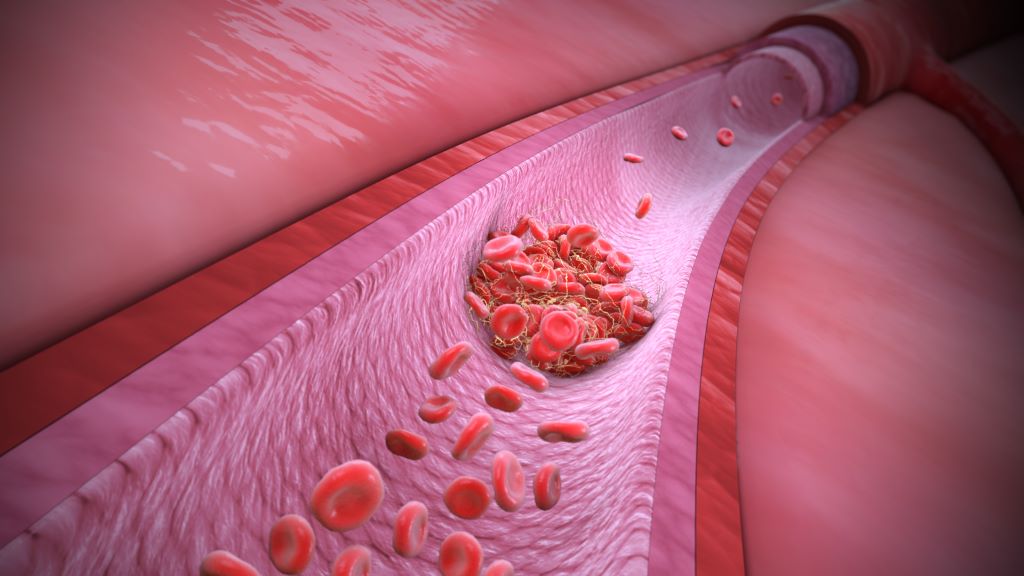
Researchers at McGill University have developed a rapid way to engineer blood clots that stop severe bleeding and support tissue healing more effectively. Their technique, called “click clotting,” links red blood cell surface proteins through a chemical reaction, resulting in a biocompatible clot that is 13 times more resistant to fracturing and four times more adhesive than natural blood clots. The team said the method, described in Nature, could be used to develop life-saving biomaterials to help control severe bleeding, as well as benefit people with clotting disorders.
“Natural blood clots can be slow to form and mechanically fragile, which limits their ability to stop severe bleeding and can compromise healing,” said Jianyu Li, senior author and Professor of Mechanical Engineering and Canada Research Chair in Tissue Repair and Regeneration. “Our work shows that, when engineered appropriately, red blood cells can play a central structural role, enabling the design of stronger and more functional biomaterials.”
Shuaibing Jiang led the research during his PhD studies at McGill. He is now a Postdoctoral Associate at Mass General Brigham and Women’s Hospital, Harvard Medical School.
Researchers at the University of British Columbia, the Medical College of Wisconsin, the University of Colorado Boulder, the University of Toronto, and the Versiti Blood Research Institute also contributed.
Connected by chemical reaction
Previous efforts to crosslink red blood cells used chitosan, a polymer derived from crustacean shells, but these led to brittle clots, ruptured cells and inconsistent clotting. In “click clotting,” the clot structure is fundamentally strengthened through a fast, bio-safe chemical reaction that connects proteins on the red blood cell surface, forming a solid gel in just five seconds.
Because the “click” reaction doesn’t interfere with normal blood chemistry, it can work alongside the body’s natural clotting process. As a result, the artificial cell‑based gel, called a “cytogel,” can be added to whole blood, where it becomes embedded within the body’s own fibrin clot.
“The technology enables both autologous clots (using the patient’s own blood) and allogeneic clots (using type-matched donor blood). Autologous clots can be prepared in approximately 20 minutes, while allogeneic clots can be prepared within about 10 minutes. Given typical clinical time constraints, this approach has strong potential for in-patient emergency care, wound management and related settings,” Li said.
The results were confirmed through in vitro testing, as well as by testing on rodents. A highlight was the effective healing and regeneration observed in the injured liver, with performance exceeding that of the clinically used product tested in this study. Analysis showed minimal evidence of immune reactivity and no toxicity in major organs.
Further research required
The researchers say that while further study is required before the cytogel can be used in clinical settings, the research establishes a foundation for its design and application.
“Engineered blood clots have strong potential for broad clinical use and could improve outcomes across many medical situations,” Li said.