Tag: 1/9/25

Clinical Trial Shows a Constipation Drug Aids Kidney Function

Human kidney. Credit: Scientific Animations CC0

Chronic kidney disease (CKD) is a major health issue worldwide. Many patients end up requiring regular dialysis to avoid kidney failure and stay alive. Despite the severity of the condition, there are currently no drugs available that improve kidney function. A research group led by Tohoku University Graduate School of Medicine’s Professor Takaaki Abe has found a remarkable solution to treat patients with CKD by co-opting a drug typically used for constipation. This is the first time that this drug (lubiprostone) was shown to prevent the decline of renal function in patients with CKD.

“We noticed that constipation is a symptom that often accompanies CKD, and decided to investigate this link further,” explains Abe. “Essentially, constipation disrupts the intestinal microbiota, which worsens kidney function. Working backwards, we hypothesised that we could improve kidney function by treating constipation.”

To address this issue, the group conducted a multicentre Phase II clinical trial (LUBI-CKD TRIAL) at nine Japanese medical institutions, enrolling 150 patients with moderate CKD. This study evaluated the effects of lubiprostone on kidney function. The results showed that, compared to the placebo group, the decline in kidney function (defined as the estimated glomerular filtration rate: eGFR) was suppressed in a dose-dependent manner in patients treated with 8µg or 16µg of lubiprostone.

The researchers also investigated the mechanism underlying how this effect occurred. They found that lubiprostone increases spermidine production, which improves mitochondrial function by promoting bacterial growth in the gut. The improved mitochondrial function was seen to exert a renoprotective effect – suppressing further kidney damage.

Going forward, the research team has plans to validate the trial results in a larger population (Phase 3 clinical trial) and advance the exploration of biomarkers that predict treatment efficacy. Their goal is to provide each patient with CKD the optimal treatment plan tailored to their needs. This discovery has the potential to significantly transform the conventional approach to CKD treatment, which primarily focuses on reducing uremic toxins.

These findings suggest a new therapeutic strategy in which laxatives suppress renal function decline. This strategy is expected to contribute to the development of treatments for not only CKD, but also mitochondrial dysfunction disorders. The results of this study were published in Science Advances on August 30, 2025.

Source: Tohoku University

Test Detects Brain Cancers in Cerebrospinal Fluid with High Accuracy

Photo by Cottonbro on Pexels

A novel, multi-analyte test developed by researchers at Johns Hopkins Medicine can accurately identify brain cancers using small samples of cerebrospinal fluid (CSF), offering a promising new tool to guide clinical decision-making.

The findings, supported by funding from the National Institutes of Health, were published in Cancer Discovery and demonstrate that combining multiple biological markers, including tumour-derived DNA and immune cell signatures, is more effective for diagnosing central nervous system cancers than using any one marker alone.

“This study highlights how much more information we can gain when we evaluate several analytes together,” says senior study author Chetan Bettegowda, MD, PhD, Professor and Director of the Department of Neurosurgery at the Johns Hopkins University School of Medicine. “The ability to detect cancers with high specificity and also gain insight into the immune environment of the brain could be an important advance in the care of patients with brain tumours.”

To evaluate the potential of a multi-analyte approach, investigators analysed 206 CSF samples, including samples from patients with high-grade gliomas, medulloblastomas, metastases and central nervous system lymphomas. Their test, called CSF-BAM (cerebrospinal fluid–B/T cell receptor, aneuploidy and mutation), measured chromosomal abnormalities, tumour-specific mutations, and T and B cell receptor sequences. In combination, these markers identified brain cancers with more than 80% sensitivity (ability to detect cancer) and 100% specificity (correctly identified those who were cancer-free) in the validation cohort. The 100% specificity means no false positives were recorded among individuals with noncancerous conditions.

The study also showed that the assay could distinguish between the immune cell populations present in cancer and noncancer cases, offering additional biological context that could be helpful in more-challenging clinical scenarios. Investigators say this ability to categorize T and B cell populations in the CSF provides insights into both disease presence and immune response.

“Many patients with brain lesions face invasive diagnostic procedures to confirm a cancer diagnosis,” says Christopher Douville, MD, assistant professor of oncology and a senior study author. “A tool like this could help us make better-informed decisions about who really needs a biopsy and who doesn’t.”

Researchers say the test could be particularly useful for cases in which conventional imaging or cytology is inconclusive, or in situations when obtaining tissue for diagnosis is risky or not possible. The multi-analyte approach, they say, enables clinicians to better detect cancer and better understand the disease status, supporting a more tailored approach to patient care.

Source: Johns Hopkins Medicine

Experiments Add to Evidence of Links Between Amyloid Deposits in Brain and Bone Marrow

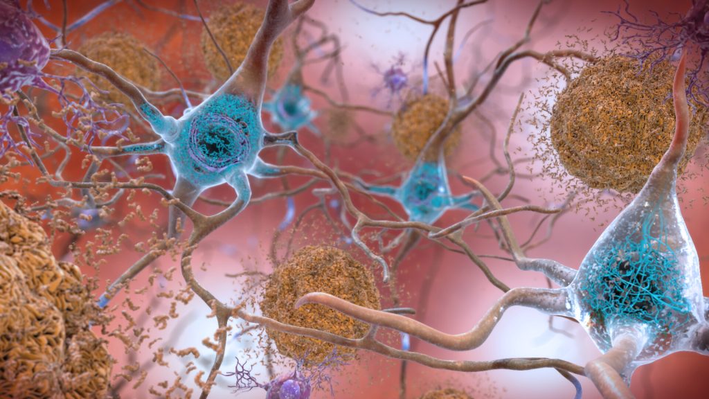
Neurons in the brain of an Alzheimer’s patient, with plaques caused by tau proteins. Credit: NIH

A recent study led by a team of researchers at The Johns Hopkins University School of Medicine examining aging mice has provided what is believed to be the first evidence that particles of amyloid beta protein, found in people with Alzheimer’s disease (AD), build up in the bone marrow of the animals, although not in the exact same form as the large, dense plaques found in the brains of people with Alzheimer’s disease.

“Although amyloid buildup has been found in organs outside the brain – such as the heart, kidneys, and nerves – it remains unclear whether similar deposits form in bone or bone marrow with aging or in Alzheimer’s disease,” says contributing study author Mei Wan, PhD, professor of the department of Orthopaedic Surgery. While brain amyloid has been extensively studied for its role in memory loss and neurodegeneration, far less is known about amyloid elsewhere in the body. In fact, almost nothing is known about whether amyloid forms in the skeleton or how it might contribute to age-related bone loss.”

AD is primarily associated with excessive amyloid plaques in the brain. Osteoporosis is a bone disease marked by low bone mineral density with an increased risk of fractures. Recent research suggests these two age-related conditions may be connected, and scientists are beginning to uncover common underlying causes.

Funded by the National Institutes of Health, the study findings, published in Nature Aging, advance scientific understanding of long-suspected similar biological processes that may be at work in osteoporosis – a form of bone loss – and Alzheimer’s dementia, the researchers say. The work may also offer potential new targets for preventing or treating bone loss.  

The buildup of amyloid is triggered by fat cells in the bone marrow, known as bone marrow adipocytes (BMAds), and a protein they release called SAP/PTX2 in aged mice and mice with AD. These amyloid deposits impair bone-building cells (osteoblasts) and activate bone-resorbing cells (osteoclasts), leading to bone loss. In previous mouse models, removing senescent BMAds or blocking SAP/PTX2 have shown to significantly reduce amyloid buildup and restored bone health.

In this study, male and female mice ranging from 4 to 24 months were kept in a temperature-controlled room provided with ongoing access to food and water and exposed to a 12-hour light-dark cycle. Researchers put a concentration of 5mg/ml in the drinking water of the mice aged 18 months and examined the effects CPHPC had on their age-related bone loss. CPHPC (also named Miridesap) is a small molecule compound originally designed to treat amyloidosis which is a rare disease marked by the buildup of amyloid proteins. A control group of mice aged 4, 9, 22 and 24 months were given the same dosage of water without the CPHPC drug  

High-resolution imaging of thigh and shin bones revealed amyloid fibrils forming ring-like structures around BMAds in aged mice and mice genetically engineered to have a form of AD. SAP/PTX2-driven amyloid clumps were found to enhance bone loss.

Study results also showed that CPHPC successfully depleted SAP/PTX2 and reversed bone deterioration in the older mice, suggesting a promising new therapeutic strategy for osteoporosis in the elderly, a strategy that would seek to eliminate aging fat cells or amyloid-promoting proteins.  

Wan adds, “Our study is what we believe to be the first to show that harmful amyloid fibres (Aβ fibrils) build up in the bone marrow of aged mice. We also found that fat cells in the bone marrow release a protein called SAP/PTX2, which plays a major role in triggering this amyloid buildup and damaging bone. These findings uncover a new connection between bone loss and dementia risk and may open the door to new research on how protecting bone health could also help protect brain function.”

This discovery provides an opportunity for new treatments targeting bone aging and Alzheimer’s-associated osteoporosis by focusing on the elimination of senescent fat cells or amyloid-promoting proteins.

Source: Johns Hopkins Medicine

SAHPRA Approves Mpox Test Using African Medicines Regulatory Harmonisation (AMRH) Continental EUL Procedure

Mpox (monkeypox) virus. Source: NIH

Pretoria, 27 August 2025 – The South African Health Products Regulatory Authority (SAHPRA) has approved a molecular test kit for Mpox , Cobas MPXV, for use on the Cobas 6800/8800 Systems, within nine working days through a collaborative and harmonised review process under the African Medicines Regulatory Harmonisation (AMRH). Rapid and accurate testing is essential for early detection and to enable timely treatment, and effective containment of the virus.

SAHPRA’s Medical Device Unit, utilising both the World Health Organization (WHO) Emergency Use Listing (EUL) report and the African Medicines Regulatory Harmonisation (AMRH) Continental EUL Procedure, has successfully leveraged reliance mechanisms to facilitate the review and approval of Mpox diagnostic tools. This approach has proven instrumental in accelerating access to critical health technologies, particularly in public health emergencies.

The emergency use approval of the Cobas MPXV for use on the Cobas 6800/8800 Systems, developed by Roche Molecular Systems, Inc. and licensed to Roche Diagnostics South Africa (Pty) Ltd, represents a critical advancement in expanding diagnostic capacity amid Mpox outbreaks. Rapid and accurate testing is essential for early detection, timely treatment, and effective containment of the virus.

 “As part of our efforts to promote regulatory efficiency and responsiveness, SAHPRA has successfully leveraged reliance mechanisms to facilitate the review and approval of Mpox diagnostic tools. This marks a significant milestone in supporting regional and continental harmonisation of the regulatory processes for medical devices, including In-Vitro Diagnostics (IVDs),” says Dr Boitumelo Semete-Makokotlela, SAHPRA’s Chief Executive Officer.

For more information on the AMRH continental EUL listing publication, see here: https://www.nepad.org/news/public-notice-amrh-steering-committee-approves-emergency-use-listing-of-two-mpox

Source: SAHPRA

Clinical Trial Challenges 40-year-old Standard of Care for Heart Attack Patients

Human heart. Credit: Scientific Animations CC4.0

Beta-blocker therapy showed no evidence of an effect on all-cause death, reinfarction or heart failure admission in patients with myocardial infarction (MI) managed invasively who had left ventricular ejection fraction (LVEF) ≥ 40%, according to late-breaking research from the REBOOT trial presented in a Hot Line session today at ESC Congress 20251 and simultaneously published in the New England Journal of Medicine

Explaining the rationale for the REBOOT trial, Principal Investigator, Professor Borja Ibáñez from the Centro Nacional de Investigaciones Cardiovasculares Carlos III (CNIC) and Fundación Jiménez Díaz University Hospital, Madrid, Spain, said: “Beta-blockers have long been a foundational treatment after acute MI; however, supporting evidence is derived from trials that predate modern standards of care − before the time of routine reperfusion, invasive management, potent antiplatelet therapies and statins. Re-examining the role of beta-blockers is warranted, particularly among patients with uncomplicated MI and LVEF > 40% in whom the benefits of beta-blockers are not well established, unlike with reduced LVEF (≤ 40%).”  

The investigator-initiated randomised open blinded-endpoint REBOOT trial was conducted at 109 centres across Spain and Italy. Patients with MI (with or without ST-segment elevation) were eligible for enrolment if they underwent invasive management during the index hospitalisation and had a predischarge LVEF > 40%, with no history or signs of heart failure. Patients were randomised 1:1 to beta-blocker or no beta-blocker therapy. The primary endpoint was a composite of all-cause mortality, nonfatal reinfarction or heart failure admission. 

Among 8505 patients who underwent randomisation, the mean age was 61 years and 19.3% were women. A total of 10% had a prior MI and 12% were on beta-blocker treatment before the index hospitalisation. 

After a median follow-up of 3.7 years, the primary composite outcome of all-cause death, nonfatal reinfarction or heart failure admission occurred in a similar proportion of patients in each group: 22.5/1000 patient-years in beta-blocker group and 21.7/1000 patient-years in the no beta-blocker group (hazard ratio [HR] 1.04; 95% confidence interval [CI] 0.89 to 1.22; p=0.63). 

All-cause mortality occurred in 11.2 and 10.5/1000 patient-years on beta-blocker therapy and no-beta blocker therapy, respectively (HR 1.06; 95% CI 0.85 to 1.33). Nonfatal reinfarction occurred in 10.2 and 10.1/1000 patient-years, respectively (HR 1.01; 95% CI 0.80 to 1.27), while heart failure admission occurred in 2.7 and 3.0/1,000 patient-years, respectively (HR 0.89; 95% CI 0.58 to 1.38).  

Regarding safety, admission for stroke occurred in 2.6/1000 patient-years in the beta-blocker group and 1.7/1000 patient-years in the no beta-blocker group (HR 1.50; 95% CI 0.90 to 2.49). Admission for symptomatic advanced atrioventricular block occurred in 0.5 of patients in the beta-blocker group and 0.4/1000 patient-years of patients in the no beta-blocker group (HR 1.18; 95% CI 0.40 to 3.50). 

There appeared to be an absence of benefit with beta-blockers across the prespecified subgroups. However, fewer events were noted in patients with mildly reduced LVEF (40−49%) on beta-blockers vs no beta-blockers, although low patient numbers limit interpretability. Women experienced overall more events, especially when on beta-blockers. 

Professor Ibáñez concluded: “Beta-blocker therapy showed no evidence of benefit across the study population of patients with MI managed invasively who had LVEF > 40%. However, as also presented today at ESC Congress, a meta-analysis of data from four trials, including REBOOT, suggest there may be a positive signal in patients with mildly reduced LVEF (40−49%).2” 

Source: European Society of Cardiology