The World’s First Precision Institute to Redefine the Healthcare Industry as We Know It

Blending 4IR technology and holistic approaches to health, the future of personalised and predictive ‘Wellcare’ comes to Cape Town and Johannesburg

InUversal Group, a health and biotech market disruptor in Africa and the Middle East that is transforming the way we think about healthcare, medicine and hospitals, is set to open next-gen medical and wellness hubs in Cape Town this December 2023 and a monumental R1 Billion Development in Sandton, Johannesburg, to follow in 2024. These visionary, state-of-the-art health and wellness facilities are designed to embrace the holistic nature of individuals’ wellbeing, emphasising the intricate interplay of biological, social and psychological facets.

Comprising a team of esteemed medical experts working collaboratively to transform disease treatment through innovative and holistic strategies, the InUversal Group is committed to alleviating South Africa and Africa’s healthcare challenges through the application of 4IR technology that is set to improve healthcare accessibility and standards for individuals across the continent. As an increasing number of international visitors travel the globe in search of medical treatments, the InUversal Group is committed to making South Africa’s major metropolises, including Johannesburg, Cape Town, and Durban, the go-to destinations for personalised Wellcare – a term coined by the group that is anticipatory in nature and requires a holistic approach to health.

Wellcare harnesses proven strategies to attain an optimal and healthy balance between individuals’ health, time, and finances, ensuring that they can lead healthier, happier and more fulfilling lives. This ambitious endeavour aligns with South Africa’s reputation as a hub for medical tourism, offering world-class medical services, competitive pricing, and a rich cultural and immersive experience.

The Institute of Universal Wellcare (InUWell) will be based in the heart of Cape Town at the prestigious V&A Waterfront Mall and is the first of its kind – a digitally-immersed, multidisciplinary institute of holistic health and wellbeing in a warm and welcoming retail environment. InUWell’s versatile multifunctional design, and forward-thinking commitment to radical sustainability, offers an unparalleled experience that is a seamless blend of physical and digital realms. The Institute is set over 2000 square metres and is considered to be the heart of “Wellcare.”

This festive season, InUWell is opening its doors to immersive health and wellness experiences where individuals are invited to learn more about health and well-being, while exploring and having fun in an engaging, euphoric, multi-sensory environment as they connect and share memorable moments with friends and family.

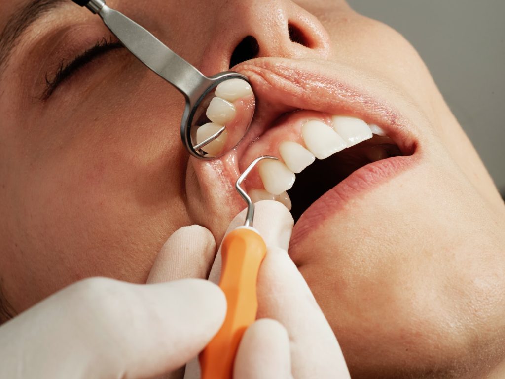
InUWell provides a diverse range of services including DNA genetic testing, comprehensive health screenings and diagnostics, specialised treatments and therapies, Wellcare lifestyle products and services, active health studios, multisensory immersive experiences and a digital health bank with evidence-based healthcare insights.

“The InUversal Group is an ecosystem shifting and stretching boundaries, creating connections, taking complex intricate life decisions and making them SIMPLE,” says Dr Kamlen Pillay, Founder and CEO of the InUversal Group and Plastic Surgeon. “InUWell, under the InUversal Group, is a single destination for all your health and wellness needs. It is the perfect place to learn about your body and how to take care of it, to access the latest technology and treatments and therapies, and to connect with other people who are on the same journey,” says Dr Pillay.

“The InUversal Group’s WellCare Programmes empower individuals of all generations to take precise, proactive, and preventative measures, not only to extend the quantity of years in our lives but also to infuse more vitality and quality into those years,” says Dr Pillay.

The Group is launching several innovative health technology products which will enter the market early next year, including the Johannesburg facility called SIM Sandton, that is unique in Africa and will host a 5* hotel, InUWell Precinct, Step-Down Facility as well as a multi-disciplinary Surgical Theatre Complex with more than 20 of Johannesburg’s top specialists. 

Working with esteemed medical specialists, leading MedTech equipment and companies, and lifestyle and wellness retail brand partners, the InUversal Group invites potential collaborators to join the vanguard of companies and brands helping to shape the future of health and Wellcare practices in Africa and globally, with the shared mission of enhancing the well-being of countless individuals.

“Imagine a world where every man, woman and child has the agency over three valuable assets – their health, time and money. A world where every person has the dignity of choice where they live, work and play. A world where hospitals are not places we go to when we are sick but rather to stay healthy. Imagine a world where hospitals are for profits but not for profiteering. A world where each day, each and every one of us, uses our energy collectively to leave the world in a slightly better place than we found it, the day before,” concludes Dr Pillay.

To get involved or find out more information, visit: inuwell.global or contact experience@inuwell.global to book an appointment. InUWell Cape Town will officially be opening its doors on 19 December 2023.

Tissue Regeneration might One Day Replace Root Canals

Photo by Caroline Lm on Unsplash

Tissue regeneration might one day replace the pain and discomfort of a root canal for most people. ADA Forsyth scientists are testing a novel technology to treat endodontic diseases (diseases of the soft tissue or pulp of the teeth) more effectively. The technology may also even be applicable to other parts of the body, such as helping to regrow bones.

The study, published in The Journal of Dental Research, demonstrates regenerative properties of resolvins, specifically Resolvin E1 (RvE1), when applied to dental pulp. Resolvins are part of a greater class of Specialised Proresolving Mediators (SPMs). This class of molecule is naturally produced by the body and is exquisitely effective in the control of excess inflammation associated with disease.

“Pulpitis (inflammation of dental pulp) is a very common oral health disease that can become a serious health condition if not treated properly,” said Dr Thomas Van Dyke, Vice President at the Center for Clinical and Translational Research at ADA Forsyth, and a senior scientist leading the study.

“Root canal therapy (RCT) is effective, but it does have some problems since you are removing significant portions of dentin, and the tooth dries out leading to a greater risk of fracture down the road. Our goal is to come up with a method for regenerating the pulp, instead of filling the root canal with inert material.”

Inflammation of this tissue is usually caused by damage to the tooth through injury, cavities or cracking, and the resulting infection can quickly kill the pulp and cause secondary problems if not treated.

The study applied RvE1 to different levels of infected and damaged pulp to explore its regenerative and anti-inflammatory capacities.

There were two major findings. First, they showed RvE1 is very effective at promoting pulp regeneration when used in direct pulp-capping of vital or living pulp (replicating conditions of reversible pulpitis). They were also able to identify the specific mechanism supporting tissue regeneration.

Second, the scientists found that placing RvE1 on exposed and severely infected and necrotic pulp did not facilitate regeneration.

However, this treatment did effectively slow down the rate of infection and treat the inflammation, preventing the periapical lesions (abscesses) that typically occur with this type of infection.

Previous publications have shown that if the infected root canal is cleaned before RvE1 treatment, regeneration of the pulp does occur.

While this study focused on this technology in treating endodontic disease, the potential therapeutic impact is far reaching.

Dr Van Dyke explained, “because application of RvE1 to dental pulp promotes formation of the type of stem cells that can differentiate into dentin (tooth), bone, cartilage or fat, this technology has huge potential for the field of regenerative medicine beyond the tissues in the teeth. It could be used to grow bones in other parts of the body, for instance.”

Source: Forsyth Institute

New Compound Restores Lost Brain Function in Mice after Stroke

Photo by Kanashi ZD on Unsplash

An international study published recently in the journal Brain has reported promising results in restoring function lost in mice and rat models of stroke. Researchers were able to restore lost brain function using small molecules that in the future could potentially be developed into a stroke recovery therapy.

“Communication between nerve cells in large parts of the brain changes after a stroke and we show that it can be partially restored with the treatment,” says Tadeusz Wieloch, senior professor of neurobiology at Lund University in Sweden.

“Concomitantly, the rodents regain lost somatosensory functions, something that around 60 per cent of all stroke patients experience today. The most remarkable result is that the treatment began several days after a stroke,” Wieloch continues.

In an ischaemic stroke, lack of blood flow to affected parts of the brain lead to loss of function such as paralysis, sensorimotor impairment and vision and speech difficulties, but also to pain and depression.

There are currently no approved drugs that improve or restore the functions after a stroke, apart from clot-dissolving treatment in the acute phase (within 4.5 hours of the stroke). Some spontaneous improvements occur, but many stroke patients suffer chronic loss of function.

For example, about 60% of stroke sufferers, experience lost somatosensory functions such as touch and position sense.

The new study shows that rats that were treated with a class of substances that inhibit the metabotropic glutamate receptor (mGluR5), a receptor that regulates communication in the brain’s nerve cell network.

“Rodents treated with the GluR5 inhibitor regained their somatosensory functions,” says Tadeusz Wieloch, who led the study.

Two days after the stroke, ie when the damage had developed and function impairment was most prominent, the researchers started treating the rodents that exhibited the greatest impaired function.

“A temporary treatment effect was seen after just 30 minutes, but treatment for several weeks is needed to achieve a permanent recovery effect. Some function improvement was observed even when the treatment started 10 days after a stroke,” says Tadeusz Wieloch.

Importantly, sensorimotor functions improved, even though the extent of the brain damage was not diminished.

This, explains Tadeusz Wieloch, is due to the intricate network of nerve cells in the brain, known as the connectome – the way brain areas are inter connected and communicate form the basis for various brain functions.

“Impaired function after a stroke is due to cell loss, but also because of reduced activity in large parts of the connectome in the undamaged brain. The receptor mGluR5 is apparently an important factor in the reduced activity in the connectome, which is prevented by the inhibitor which therefore restores the lost brain function,” says Tadeusz Wieloch.

The results also showed that sensorimotor function was further improved if treatment with the mGluR5 inhibitor is combined with somatosensory training by housing several rodents in cages enriched with toys, chains, grids, and plastic tubes.

The researchers hope that in the future their results could lead to a clinical treatment that could be initiated a few days after an ischaemic stroke.

“Combined with rehabilitation training, it could eventually be a new promising treatment. However, more studies are needed. The study was conducted on mice and rats, and of course needs to be repeated in humans. This should be possible since several mGluR5 inhibitors have been studied in humans for the treatment of neurological diseases other than stroke, and shown to be tolerated by humans,” says Tadeusz Wieloch.

Source: Lund University

New Therapy Eliminates ‘Problematic’ T Cells in Skin Autoimmune Diseases

Photo: CC0

In a groundbreaking study published in Science, researchers discovered distinct mechanisms controlling different types of immune cells, and found that, by precisely targeting these mechanisms, they could selectively eliminate ‘problematic cells’ and reshape the skin’s immune landscape.

The skin is packed with specialised immune cells that protect against infections and cancer and promote healing. These cells, called tissue-resident T cells or TRM cells, stay in place to fight infections and cancerous cells in the skin.

However, when not controlled properly, some of these skin TRM cells can contribute to autoimmune diseases, such as psoriasis and vitiligo.

Researchers, led by University of Melbourne’s Professor Laura Mackay, a Laboratory Head and Immunology Theme Leader at the Peter Doherty Institute of Infection and Immunity (Doherty Institute), found a way to redress this imbalance.

University of Melbourne’s Dr Simone Park, an Honorary Research Fellow and former Postdoctoral Fellow in the Mackay Lab at the Doherty Institute, and lead first author of the study, said that this research is the first to describe the unique elements that control various types of skin TRM cells in animal models, offering precise targets for potential treatment strategies.

“Specialised immune cells in our skin are diverse: many are critical to prevent infection and cancer, but others play a big role in mediating autoimmunity,” said Dr Park.

“We discovered key differences in how distinct types of skin T cells are regulated, allowing us to precisely edit the skin’s immune landscape in a targeted way.”

University of Melbourne’s Dr Susan Christo, Senior Research Officer in the Mackay Lab at the Doherty Institute and co-first author of the study, explained how these discoveries could advance efforts to treat skin disease.

“Most autoimmune therapies treat the symptoms of the disease rather than addressing the cause. Conventional treatments for skin disorders often impact all immune cells indiscriminately, meaning that we could also be wiping out our protective T cells,” said Dr Christo.

“Until now, we didn’t know how to pick apart ‘bad’ T cells in the skin from the ‘good’ protective ones. Through this research, we discovered new molecules that allow us to selectively remove disease-causing T cells in the skin.”

The research team harnessed this new knowledge to eliminate ‘problematic’ cells that can drive autoimmune disorders, while preserving the ‘good’ ones that are essential to maintain protective immunity.

University of Melbourne’s Professor Laura Mackay, senior author of the study, explained that these findings could pave the way for more precise and long-lasting therapies for skin disease.

“Skin conditions like psoriasis and vitiligo are difficult to treat long-term. The T cells driving disease are hard to remove, so patients often need life-long treatment. Our approach has the potential to revolutionise the way we treat these skin disorders, significantly improving outcomes for people dealing with challenging skin conditions,” said Professor Mackay.

With the study demonstrating successful removal of specific skin T cells in animal models, further research is necessary to validate the efficacy of these strategies in human subjects.

Dr Park hopes the study will inspire the development of new treatments for skin disease.

“These discoveries bring us one step closer to developing new drugs that durably prevent autoimmune skin disorders without compromising immune protection,” said Dr Park.

Source: The Peter Doherty Institute for Infection and Immunity

Is Stem Cell Therapy for Knee Osteoarthritis Worthwhile?

Photo by Towfiqu barbhuiya: https://www.pexels.com/photo/person-feeling-pain-in-the-knee-11349880/

Cell therapy has been explored as a new regenerative treatment for osteoarthritis, but the efficacy of stem cell transplantation from different sources for the treatment of knee osteoarthritis (KOA) remains controversial. A recent analysis of all relevant published studies indicates that stem cell transplantation from different sources is effective for treating knee osteoarthritis, the most prevalent chronic joint disease.

The review and meta-analysis, which is published in the Journal of Orthopaedic Research, included 16 studies involving 875 patients with knee osteoarthritis (441 in the stem cell transplantation group and 434 in the control group). Stem cell treatment was associated with significant reductions in patient-reported pain from the third month onwards. The most significant pain relief at different postoperative months came from fat-derived and umbilical cord–derived stem cells. A patient’s own fat-derived stem cells resulted in better pain alleviation compared with those from other donors. Also, a patient’s own fat-derived stem cells led to the most effective recovery of knee joint function.

“Stem cell transplantation proved safe and effective for knee osteoarthritis treatment,” the authors wrote. “Different sources stem cells have a good effect on alleviating knee joint pain, restoring knee joint function, and minimising patient trauma.”

Source: Wiley

Embracing the Power of Collective Action on International Volunteer Day 2023

Photo by Zach Vessels on Unsplash

The United Nations International Volunteer Day, celebrated annually on 5 December, is a unique opportunity for volunteers and organisations worldwide to celebrate their contributions, share their values, and promote their work within their communities. The theme for 2023 is: “The Power of Collective Action: If Everyone Did.”

The United Nation’s (UN) fourth State of the World’s Volunteerism Report (SWVR 2022), titled Building Equal and Inclusive Societies, shows that the ways in which volunteers and entities interact, collaborate and partner are vital for the achievement of the 2030 Agenda for Sustainable Development and the Sustainable Development Goals.

Volunteerism plays a crucial role in promoting a culture of collaborative decision-making and reshaping power dynamics. Volunteers can act as connectors, bridging the gap between different groups and facilitating better understanding and cooperation.

“At Sanofi South Africa, we understand the immense value of collective efforts in building a healthier, more resilient world for patients, communities, partners, and employees,” says Prudence Selani, Head of Corporate Affairs at Sanofi South Africa. “Our dedication to addressing some of the world’s most pressing challenges is evident in our comprehensive social impact strategy, which is now an integral part of our business, influencing every level of our organisation.”

Sanofi’s ‘Play to Win’ strategy, intensifies the pharmaceutical company’s focus on improving healthcare access, reducing its environmental footprint, and building an inclusive workplace. This commitment extends to ensuring a sustainable planet for future generations.

The organisation is making a lasting impact through its corporate social responsibility (CSR) legacy project in Mamelodi, in the City of Tshwane, which forms part of Sanofi’s global In and Beyond the Workplace social impact strategy pillar – “to reflect the diversity of its communities, unleash the full potential of its employees, and transform healthcare to be more inclusive and equitable.”

After engagement with numerous organisations, Sanofi partnered with Entokozweni Resource Centre in Mamelodi to make a real difference in people’s lives. The non-profit organisation is dedicated to uplifting the Mamelodi community through a variety of programs and initiatives, focusing on long-term developmental goals and immediate community needs​.

“The launch of our Legacy Project on 15 September 2023, at Entokozweni was a resounding success, drawing extensive support from our staff,” says Selani. “We dedicated the day to connecting with the people of Entokozweni and the broader community, marking the beginning of a long-lasting relationship.”

Support for the community includes:

  • Donating essential items such as mattresses, toiletries, stationery, uniforms, and clothing.
  • Providing 250 Coursera licences to empower Mamelodi’s youth with skills for employment and helping to transform the lives of their families and community.
  • Hosting the Mamelodi Community Health Centre’s (CHC) Purpose Day at Entokozweni, focusing on educating the community about gut health and the importance of hygiene.
  • Converting an illegal dumping site into a vibrant indigenous garden during the Sanofi Legal Ethics & Business Integrity department (LEBI) Giving Week, through collaborating with employees, local communities, and various stakeholders.

“As we celebrate International Volunteer Day, we are reminded of the importance of working together to effect real change,” says Selani. “Volunteerism is one of the most vital delivery mechanisms for social, environmental and economic transformation, ensuring a lasting impact because it can change people’s mindsets, attitudes, and behaviours. The power of collective action is not just a theme for a day but a guiding principle in transforming lives through united efforts.”

Join the UN in recognising volunteers all over the world using the hashtags #IfEveryoneDid and #IVD2023. SanofiVolunteers #InternationalVolunteerDay2023 #CollectiveAction #SocialImpact

Abnormally High Levels of HDL-C Linked to Dementia in Older Adults

Photo by Matteo Vistocco on Unsplash

Abnormally high levels of high density lipoprotein cholesterol (HDL-C), are associated with an increased risk of dementia in older adults, according to study led by Monash University. Researchers said very high levels of the ‘good cholesterol’ HDL-C linked to dementia risk in this study were uncommon and not diet related, but more likely to reflect a metabolic disorder. The findings may help doctors to recognise a group of older patients potentially at risk of dementia, particularly in those aged 75 and older.

Published in The Lancet Regional Health – Western Pacific, this is one of the largest studies of elevated HDL-C levels and dementia in initially healthy older people aged mostly over 70, enrolled in the ASPREE* study.

Over an average 6.3 years, participants with very high HDL-C (> 80mg/dL or > 2.07mmol/L) at study entry were observed to have a 27% higher risk of dementia compared to participants with optimal HDL-C levels, while those aged 75 years and older also showed a 42% increased risk compared to those with optimal levels.

Very high HDL-C levels were categorised as 80mg/dL (> 2.07mmol/L) or above.

The optimal level of HDL-C of 40 to 60mg/dL (1.03–1.55mmol/L) for men and 50 to 60mg/dL (1.55–2.07mmol/L) for women was generally beneficial for heart health.

Among 18 668 participants included in this analysis, 2709 had very high HDL-C at study entry, with 38 incidents of dementia in those aged less than 75 years with very high levels, and 101 in those aged 75 and more with very high levels.

First author and Monash University School of Public Health and Preventive Medicine senior research fellow Dr Monira Hussain said that further research was needed to explain why a very high HDL cholesterol level appeared to affect the risk of dementia.

Dr Hussain said these study findings could help improve our understanding of the mechanisms behind dementia, but more research was required.

“While we know HDL cholesterol is important for cardiovascular health, this study suggests that we need further research to understand the role of very high HDL cholesterol in the context of brain health,” she said.

“It may be beneficial to consider very high HDL cholesterol levels in prediction algorithms for dementia risk.”

*The Aspirin in Reducing Events in the Elderly (ASPREE) trial is a double-blind, randomised, placebo-controlled trial of daily aspirin in healthy older people. 

Source: Monash University

At Last, Objective Evidence for the Involvement of Neck Muscles in Headaches

Source: CC0

Researchers have identified objective evidence of how the neck muscles are involved in primary headaches. The study findings, being presented at the annual meeting of the Radiological Society of North America (RSNA), could lead to better treatments.

The distinct underlying causes of primary headaches, comprising tension-type headaches and migraines, are still not fully understood.

“Our imaging approach provides first objective evidence for the very frequent involvement of the neck muscles in primary headaches, such as neck pain in migraine or tension-type headache, using the ability to quantify subtle inflammation within muscles,” said Nico Sollmann, MD, PhD, resident at University Hospital Ulm and University Hospital Rechts der Isar in Munich, Germany.

In tension-type headaches there is often the perception of a tightening in the head and mild to moderate dull pain on both sides of the head. While these headaches are typically associated with stress and muscle tension, their exact origin is not fully understood.

Migraines are characterised by a severe throbbing pain and generally occur or are worse on one side of the head. Migraines may also cause nausea, weakness and light sensitivity.

Neck pain is commonly associated with primary headaches but there are no objective biomarkers for myofascial involvement. Myofascial pain is associated with inflammation or irritation of muscle or of the connective tissue, known as fascia, that surrounds the muscle.

For the study, Dr Sollmann and colleagues aimed to investigate the involvement of the trapezius muscles in primary headache disorders by quantitative magnetic resonance imaging (MRI) and to explore associations between muscle T2 values and headache and neck pain frequency.

The prospective study recruited 50 participants, mostly women, ranging in age from 20 to 31 years old. Of the participants, 16 had tension-type headache, and 12 had tension-type headache plus migraine episodes. The groups were matched with 22 healthy controls.

All participants underwent 3D turbo spin-echo MRI. The bilateral trapezius muscles were manually segmented, followed by muscle T2 extraction.

Associations between muscle T2 values and the presence of neck pain, number of days with headache, and number of myofascial trigger points as determined by manual palpation of the trapezius muscles were analysed (adjusting for age, sex and body mass index).

The tension-type headache plus migraine group demonstrated the highest muscle T2 values. Muscle T2 was significantly associated with the number of headache days and the presence of neck pain.

The increased muscle T2 values could be interpreted as a surrogate of inflammation arising from the nervous system and increased sensitivity of nerve fibres within myofascial tissues.

“The quantified inflammatory changes of neck muscles significantly correlate with the number of days lived with headache and the presence of subjectively perceived neck pain,” Dr Sollmann said.

“Those changes allow us to differentiate between healthy individuals and patients suffering from primary headaches.”

Muscle T2 mapping could be used to stratify patients with primary headaches and to track potential treatment effects for monitoring.

“Our findings support the role of neck muscles in the pathophysiology of primary headaches,” Dr Sollmann said. “Therefore, treatments that target the neck muscles could lead to a simultaneous relief of neck pain, as well as headache.”

Dr Sollmann pointed out that non-invasive treatment options that directly target the site of pain in the neck muscles could be highly effective and safer than systemic drugs.

“Our imaging approach with delivery of an objective biomarker could facilitate therapy monitoring and patient selection for certain treatments in the near future,” he added.

Source: Radiological Society of North America

New Device Uses an Eye-safe Laser to Detect Traumatic Brain Injury

Photo: Unsplash

Researchers from the University of Birmingham have designed and developed a novel diagnostic device to detect traumatic brain injury (TBI) by shining a safe laser into the eye.

The technique is radically different from other diagnostic methods and is expected to be developed into a hand-held device for use in the critical ‘golden hour’ after traumatic brain injury, when life critical decisions on treatment must be made.

The device, described in Science Advances, incorporates a class 1, CE marked, eye-safe laser and a unique Raman spectroscopy system, which uses light to reveal the biochemical and structural properties of molecules by detecting how they scatter light, to detect the presence and levels of known biomarkers for brain injury.

There is an urgent need for new technologies to improve the timeliness of TBI diagnosis. TBI is caused by sudden shock or impact to the head, which can cause mild to severe injury to the brain, and rapid intervention is necessary to prevent further irreversible damage.

Diagnosis at the point of injury is difficult. Moreover, radiological investigations such as X-ray or MRI are very expensive and slow to show results.

Birmingham researchers, led by Professor Pola Goldberg Oppenheimer from the School of Chemical Engineering, designed and developed the novel diagnostic hand-held device to assess patients as soon as injury occurs.

It is fast, precise and non-invasive for the patient, causing no additional discomfort, can provide information on the severity of the trauma, and will be suitable to be used on-site to assess TBI.

Professor Pola Goldberg Oppenheimer said: “Early diagnosis of TBI is crucial, as life-critical decisions on treatment must be made with the first ‘golden hour’ after injury. However current diagnostic procedure relies on observation by ambulance crews, and MRI or CT scans at a hospital – which may be some distance away.”

The device works by scanning the retina where the optic nerve sits. Since the optic nerve is so closely linked to the brain, it carries the same biological information in the form of protein and lipid biomarkers.

These biomarkers exist in a very tightly regulated balance, meaning even the slightest change may have serious effects on the ‘brain-health’. TBI causes these biomarkers to change, indicating that something is wrong.

Previous research has demonstrated the technology can accurately detect the changes in animal brain and eye tissues with different levels of brain injuries — picking up the slightest changes.1,2,3

The device detailed in the current paper detects and analyses the composition and balance of these biomarkers to create ‘molecular fingerprints’.

The current study details the development, manufacture, and optimisation of a proof-of-concept prototype, and its use in reading biochemical fingerprints of brain injury on the optic nerve, to see whether it is a viable and effective approach for initial ‘on the scene’ diagnosis of TBI.

The researchers constructed a phantom eye to test its alignment and ability to focus on the back of the eye, used animal tissue to test whether it could discern between TBI and non-TBI states, and also developed decision support tools for the device, using AI, to rapidly classify TBIs.

The device is now ready for further evaluation including clinical feasibility and efficacy studies, and patient acceptability.

The researchers expect the diagnostic device to be developed into a portable technology which is suitable for use in point-of-care conditions capable to rapidly determine whether TBI occurs as well as classify whether it is mild, moderate or severe, and therefore, direct triage appropriately and in timely manner.

Source: University of Birmingham

No-aspirin Regimen Benefits Heart Failure Patients with LVADs

Photo by cottonbro studio

A recent clinical trial published in JAMA found that excluding aspirin for advanced heart failure (HF) patients with a ventricular assist device saw a reduction in bleeding events while maintaining their survival rates.

The ARIES-HM3 Randomised Clinical Trial assessed the safety and efficacy of excluding aspirin from the antithrombotic regimen in patients with advanced HF who have undergone implantation of a fully magnetically levitated left ventricular assist device (LVAD).

“We can now safely say that not giving aspirin is not only safe from a thromboembolic risk profile but results in improved adverse event rate by a significant reduction in non-surgical bleeding which is a well-known complication related to LVAD therapy,” said Mirnela Byku, MD, P.D, MBA, co-author of the study and director of the UNC Durable Mechanical Circulatory Device Program at the UNC School of Medicine.

“Improving not only longevity but also reducing morbidity and improving quality of life is a big focus in the field of MCS.”

Until this study, there had been no consensus in the field about use of or dose of aspirin in the LVAD population.

The international clinical trial followed a randomised, double-blind, placebo-controlled design and involved 628 patients across 51 centres in 9 countries.

The patients were divided into two groups: one receiving aspirin (100mg/d) and the other receiving a placebo in addition to vitamin K antagonist (VKA) therapy.

A focus was to determine if the likelihood a patient experiences major nonsurgical haemocompatibility-related adverse events (such as stroke, pump thrombosis, major bleeding, or arterial peripheral thromboembolism) within 12 months differed between the two groups.

The results showed that not giving aspirin to patients with advanced HF, treated with a fully magnetically levitated LVAD who are receiving VKAs, did not make their survival worse. Furthermore, aspirin avoidance was associated with a significant reduction (34%) in major nonsurgical bleeding events.

Source: University of North Carolina Health Care