Study links higher body mass index at various ages across adulthood with greater risks of developing different types of kidney cancer.
Photo by I Yunmai on Unsplash
Excess weight in mid-life is a known risk factor for kidney cancer, but new research indicates that weight patterns throughout life may also affect an individual’s likelihood of developing this malignancy. The findings are published by Wiley online in CANCER, a peer-reviewed journal of the American Cancer Society.
To assess weight patterns and their associations with kidney cancer and its different subtypes, investigators analysed data from 204 364 individuals from the NIH-AARP Diet and Health Study, including body mass index (BMI) data when participants entered the study (an average age of 61.6 years), and prior BMI recordings at 18, 35, and 50 years of age. The team noted that there were 1,425 cases of kidney cancer, or renal cell carcinoma (RCC), among the study’s participants, with 583 having aggressive RCC and 339 having fatal RCC. The researchers also recorded the different subtypes of RCC, including clear cell RCC (541 patients), papillary RCC (146 patients), and chromophobe RCC (64 patients).
Higher BMI at any of the ages assessed was linked with higher risks of overall RCC and all subtypes (except chromophobe RCC), with a 10-40% higher risk for each 5-unit increase in BMI. Similar increased risks were linked to weight gain during adulthood that resulted in overweight or obesity, compared with maintaining normal BMI.
Also, long-term excess weight was associated with higher risks of overall RCC, aggressive RCC, fatal RCC, and clear cell RCC, but not papillary RCC and chromophobe RCC. Weight loss in which BMI was reduced by at least 10%, particularly later in life, was associated with a lower risk of RCC. Specifically, weight loss from age 18–35 years and after age 50 years was associated with 21% and 28% reductions in RCC incidence, respectively.
“These findings emphasise that maintaining a healthy weight across one’s lifetime is important for reducing RCC risk. More importantly, weight loss, even later in life, may offer protective benefits,” said lead author Zhengyi Deng, PhD, of Stanford University School of Medicine. “We should support initiatives that promote healthy weight maintenance and weight loss strategies. Some of these include lifestyle interventions, weight-loss programs, and emerging medical treatments for obesity; however, individuals should consult with their healthcare providers prior to initiation of any plan.”
Researchers at Karolinska Institutet have demonstrated how specific biomarkers in the blood can predict the development of dementia up to 10 years before diagnosis with high accuracy, among older adults living independently in the community.
A new study, published in Nature Medicine, has investigated the potential of specific biomarkers such as tau217, Neurofilament Light (NfL), and Glial Fibrillary Acidic Protein (GFAP) to predict the occurrence of dementia, including Alzheimer’s disease, up to ten years before an actual diagnosis in cognitively healthy older adults living in the community.
Blood samples from more than two thousand
Previous research has suggested that these biomarkers could be useful in early dementia diagnostics, but most studies involved individuals who have already sought medical care for cognitive issues, due to cognitive concerns or cognitive symptoms, such as memory difficulties.
A larger, community-based study, was necessary to determine the predictive value of biomarkers in the general population.
Led by researchers from the Aging Research Center of Karolinska Institutet in collaboration with SciLifeLab and KTH Royal Institute of Technology in Stockholm, the study analysed blood biomarkers in more than 2100 adults aged 60+, who were followed over time to determine if they developed dementia.
At a follow-up ten years later, 17% of participants had developed dementia. The accuracy of the biomarkers used in the study was found to be up to 83%.
“This is an encouraging result, especially considering the 10-year predictive window between testing and diagnosis. It shows that it is possible to reliably identify individuals who develop dementia and those who will remain healthy,” says Giulia Grande, assistant professor at the Department of Neurobiology, Care Sciences and Society, Karolinska Institutet and first author of the study.
Promising biomarkers
“Our findings imply that if an individual has low levels of these biomarkers, their risk of developing dementia over the next decade is minimal”, explains Davide Vetrano, associate professor at the same department and the study’s senior author. “This information could offer reassurance to individuals worried about their cognitive health, as it potentially rules out the future development of dementia.”
However, the researchers also observed that these biomarkers had low positive predictive values, meaning elevated biomarker levels alone could not reliably identify individuals who would surely develop dementia within the next ten years. Therefore, the study authors advise against widespread use of these biomarkers as screening tools in the population at this stage.
“These biomarkers are promising, but they are currently not suitable as standalone screening tests to identify dementia risk in the general population,” says Davide Vetrano.
The researchers also noted that a combination of the three most relevant biomarkers – p-tau217 with NfL or GFAP – could improve predictive accuracy.
“Further research is needed to determine how these biomarkers can be effectively used in real-world settings, especially for elderly living in the community or in primary health care services,” says Grande.
“We need to move a step further and see whether the combination of these biomarkers with other clinical, biological or functional information could improve the possibility of these biomarkers to be used as screening tools for the general population”, Grande continues.
The study was mainly funded by the Swedish Research Council, The Swedish Brain Foundation and The Strategic Research Area in Epidemiology and Biostatistics at Karolinska Institutet. The researchers declare that there are no conflicts of interest.
Finance Minister Enoch Godongwana holding a copy of the 2025 Budget Speech. (Photo: Parliament of RSA via X)
The Department of Health’s 2025/26 Budget Vote is expected to focus on addressing the shortfall caused by the withdrawal of international funding from programmes combating communicable diseases such as HIV and TB. But as non-communicable diseases like blood cancer surpass infectious diseases, redirecting resources could further cripple screening, diagnosis, and treatment – putting more lives at risk.
Too Few Resources, Too Many Lives Lost
Palesa Mokomele, Head of Community Engagement and Communications at DKMS Africa, highlights the difficulties faced by blood cancer patients within the country’s healthcare system. “Nearly 80% of South Africans rely on a system already operating at full capacity, with many left with little more than hope due to limited access to care. Even before these new funding shifts, resources for blood cancer detection and treatment were critically scarce, contributing to the loss of more than 4000 lives each year.”
Illustrating how a lack of medical infrastructure creates additional barriers to life-saving care, she says, “The survival rate for a stem cell transplant is up to 50% with a matched unrelated donor and 61% with a matched related donor. Yet many healthcare facilities simply don’t have the resources to perform these procedures. As a result, most patients receive only medical management, which may not be enough to ensure survival. Given our population size, transplant activity remains critically low – only 139 of the required 600 transplants are performed on adults annually, and just 18 of the 250 needed for children.”
Finances Dictate Healthcare Choices
Beyond the overstretched and under-resourced public health sector, the financial burden on patients remains a major obstacle. “One in five South African households delays seeking healthcare simply because they cannot afford it,” notes Mokomele. “While the state covers the cost of a stem cell transplant from a matching donor, other essential expenses such as tissue typing, donor searches, and stem cell procurement are not covered. These out-of-pocket costs place treatment out of reach for many, leading to heartbreaking decisions and poorer outcomes.”
She adds that socio-economic challenges often make accessing care even harder for patients. “Being the sole breadwinner means some individuals struggle to take time off work for necessary treatment. In other cases, mothers face the impossible choice between continuing their own treatment or staying home to care for their children when no other support is available.”
Post-transplant Survival Challenges
Even for patients who manage to undergo a transplant, their survival remains at risk due to conditions in some public healthcare facilities. “Overcrowding and poor sanitation create dangerous environments for these highly immunocompromised patients,” warns Mokomele. “To safeguard their fragile health, they need access to clean water, proper sanitation, isolation, and balanced nutrition.”
She stresses that long recovery periods make it difficult for patients from distant areas to complete their care without proper housing at treating hospitals. “Without these accommodations, many are forced to abandon treatment, putting their survival at risk.”
“No patient should be denied life-saving treatment due to funding constraints. We urge government and the private sector to collaborate in strengthening blood cancer care, and we encourage the public to play their part by supporting fundraising initiatives that help bridge critical gaps in treatment access,” concludes Mokomele.
While more people than ever are running marathons in the U.S., the risk of dying from a heart attack during a run has fallen dramatically in recent years. That’s a key conclusion from a new study by Jonathan Kim, associate professor in the Emory School of Medicine. Kim’s research is a follow-up to a study he published in 2012 — the first investigation into unexpected cardiac arrests during long distance running events.
The new findings, published in JAMA, indicate that, while the rate of marathon runners who suffer cardiac arrests remained unchanged, their chance for survival is twice what it was in the past. Far fewer marathon runners who suffer cardiac arrest are now dying of it.
“We continue to see media reports about unfortunate cases of cardiac arrest during long distance running events,” Kim says. “But, has the incidence of these events changed? Have there been changes in the most common causes of cardiac arrest? What are the factors associated with death and survival? It was a novel question to ask 13 years after our first analysis, and an important one because recreational running continues to increase in popularity.”
The challenge of finding data
More than 29 million people completed marathons in the U.S. between 2010 and 2023, triple the number of the previous decade, which Kim examined in his first study. There’s no central registry of race-related cardiac events, so for both studies, his team had to find their data through a range of sources, starting by contacting individual race directors.
“We leveraged a few sources including a comprehensive review of media reports,” Kim says. “We also had contact information for all race directors and were able to reach approximately 70% of them who helped and told us the number of events during this specific timeline, including if the individual died and the sex of the participant.”
The researchers used extensive public internet searches to identify and reach out to runners who survived cardiac arrests or next-of-kin to construct detailed profiles of as many cases as possible. “The vast majority of cases were identifiable by public search engines. And all of the deaths were as well,” he says.
Analyzing this extensive database, Kim found that while the rate of cardiac arrests was about the same during the two periods — .60 per 100 000 participants now versus .54 per 100 000 participants in the earlier period — the rate of deaths from these cases, however, fell by half: from .39 per 100 000 to .19 per 100 000. That’s about a 50% decline in the death rate since 2000–2009. As before, cardiac arrests remained far more common among men than among women and more common in marathons than half marathons.
The sport’s growing awareness of cardiac death risk
What led to the dramatic change in death rates? Kim thinks the whole sport has become more aware of the risks and of the need to have emergency services available to runners, a conclusion he reached after interviewing as many survivors as he could find. “What we found was that every one of those people got hands-on cardiopulmonary resuscitation, but the vast majority also had immediate access to an automated external defibrillator. That’s the difference,” he says.
That survival rate is comparable to the cardiac arrest survival rate in other public places that now make defibrillators routinely available such as airport and casinos, which have seen similar declines in deaths.
Kim says his findings offer additional evidence of how important it is to make CPR training available to race participants and to strategically place defibrillators along the racecourse. It’s also important, he says, to better identify the most vulnerable in a population before they run a race.
“These are more often potentially preventable events,” he says. “Being able to identify people, more commonly older individuals with unrecognized cardiovascular risk factors, doesn’t mean they can’t run a race. Rather, it affords the opportunity to improve primary preventive cardiovascular care and potentially further reduce the risk of cardiac arrest during these events. The incidence of sudden cardiac arrest during long-distance races hasn’t changed in over twenty years. I think this is an important arena of future research.”
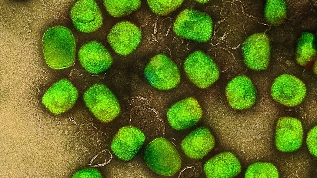
Mpox has the potential to become a significant global health threat if taken too lightly, according to scientists at the University of Surrey. In a letter published in Nature Medicine, researchers highlight how mpox — traditionally spread from animals to humans — is now showing clear signs of sustained human-to-human transmission.
Mpox is a viral infection caused by a virus that belongs to the same family as smallpox.
The virus can cause a painful rash, fever, and swollen glands and, in some cases, lead to more serious illness.
Mpox usually spreads through close contact with an infected person or animal.
Carlos Maluquer de Motes, Reader in Molecular Virology at the University of Surrey, said:
“The most recent outbreaks show that intimate contact is now a significant way the virus spreads. That shift in how it’s transmitted is leading to longer transmission chains and lasting outbreaks.”
The article notes that this change coincided with the rapid spread of clade IIb (a clade is a group of viruses that share a common ancestor) mpox viruses, but different clade I variants are now on the rise too.
Researchers are also concerned because clade I viruses are thought to be more aggressive.
These viruses appear to be accumulating specific genetic mutations — driven by enzymes in the human body — that may be changing viral properties, so the longer these viruses circulate amongst us, the higher the chances these mutations help mpox adapt to humans.
Although mpox was once mainly seen in Central Africa, the virus caused an outbreak worldwide in 2022 and is now causing outbreaks in multiple sub-Saharan countries.
While it currently affects adults the most, the researchers stress that it has the potential to spread among other groups, including children, a group at greater risk of serious illness — although sustained transmission in children has not yet been reported.
Dr Maluquer de Motes added:
“Mpox control has to climb up the global health agenda. We have limited diagnostic tools and even fewer antiviral treatments. We urgently need better surveillance and local or regional capacity to produce what we need — otherwise, we are at risk of future epidemics.”
Unlike smallpox, mpox has an animal reservoir, meaning it can’t be fully eradicated. The authors warn that unless international action is taken now — including investment in point-of-care testing and new treatments — mpox will continue to re-emerge and threaten global health.
Skin cell (keratinocyte)
This normal human skin cell was treated with a growth factor that triggered the formation of specialised protein structures that enable the cell to move. We depend on cell movement for such basic functions as wound healing and launching an immune response. Credit: Torsten Wittmann, University of California, San Francisco
In a study published in Nature, researchers at Karolinska Institutet and SciLifeLab, among others, have identified a new mechanism for how cells deal with stress. This could have implications for treating certain hereditary, neurodegenerative diseases, but may also be relevant for future cancer treatment.
When cells are exposed to stress, such as lack of nutrients or oxygen, a process called the integrated stress response (ISR) is activated. This process helps cells adapt and survive, for example by affecting the production of different proteins.
Researchers have now discovered an alternative stress response, called s-ISR (split ISR), which results in changes in the expression of certain genes that are important for the cell’s energy balance. One of these genes, PCK2, affects the conversion of oxaloacetate to phosphoenolpyruvate, a substance important in the body’s metabolism of sugars and the production of amino acids such as serine and glycine. These amino acids are the building blocks of proteins that are essential for many functions in the body.
“Our discovery challenges the previous understanding of how cells handle stress and opens up new possibilities for understanding and treating diseases where the cells’ stress response is affected,” says Ola Larsson, researcher at the Department of Oncology-Pathology, Karolinska Institutet and the Science for Life Laboratory (SciLifeLab).
May affect cancer cells
One such group of diseases is leukodystrophies – inherited disorders in which the white matter of the brain, the myelin, breaks down. One of these diseases is called VWMD (vanishing white matter disease) and is caused by mutations in a protein involved in the cells’ stress response. Researchers have shown that cells with these mutations activate s-ISR, which can affect their survival under stress.
“Another disease characterised by high stress levels is cancer, and s-ISR may therefore be important for the survival of cancer cells,” says Ola Larsson.
A study from the University of Ottawa’s Human and Environmental Physiology Research Unit (HEPRU) has confirmed that the limits for human thermoregulation – the ability to maintain a stable body temperature in extreme heat – are lower than previously thought.
This research, led by Dr Robert D. Meade, former Senior Postdoctoral Fellow and Dr Glen Kenny, Director of HEPRU and professor of physiology, highlights the urgent need to address the impacts of climate change on human health.
The study, published in the journal PNAS, found that many regions may soon experience heat and humidity levels that exceed the safe limits for human survival. “Our research provided important data supporting recent suggestions that the conditions under which humans can effectively regulate their body temperature are actually much lower than earlier models suggested,” states Kenny. “This is critical information as we face increasing global temperatures.”
Utilizing a widely used technique known as thermal-step protocols, Meade and his team exposed 12 volunteers to various heat and humidity conditions to identify the point at which thermoregulation becomes impossible. What made this study different, was that participants returned to the laboratory for a daylong exposure to conditions just above their estimated limit for thermoregulation. Participants were subjected to extreme conditions, 42°C with 57% humidity, representing a humidex of approximately 62°C. “The results were clear. The participants’ core temperature streamed upwards unabated, and many participants were unable to finish the 9-hour exposure. These data provide the first direct validation of thermal step protocols, which have been used to estimate upper limits for thermoregulation for nearly 50 years”, says Meade.
“Our findings especially timely, given estimated limits for thermoregulation are being increasingly incorporated into large scale climate modelling,” explains Meade. “They also underscore the physiological strain experienced during prolonged exposure to extreme heat, which is becoming more common due to climate change.”
The implications of this research extend beyond academia. As cities prepare for hotter summers, understanding these limits can help guide health policies and public safety measures. “By integrating physiological data with climate models, we hope to better predict and prepare for heat-related health issues,” adds Kenny.
As the world grapples with the realities of climate change, this research aims to spark important conversations about our safety and adaptability in increasingly extreme environments.
April 2nd, 2025, Nairobi, Kenya – Africa stands at the forefront of a revolutionary shift in global health, driven by artificial intelligence (AI) and data science, according to a report released today from the Science for Africa Foundation (SFA Foundation), African institutions and research councils. The report is a first of its kind to comprehensively examine national-level perspectives across Africa on AI and data science for global health. The landscape presents an unprecedented view into the potential to improve AI governance in Africa to reduce the risk and stop the perpetuation of inequity.
Titled “Governance of Artificial Intelligence for Global Health in Africa”, the report is produced through the SFA Foundation’s Science Policy Engagement with Africa’s Research (SPEAR) programme as a culmination of a year-long effort involving convenings across Africa’s five regions, policy analysis and extensive surveys to identify policy gaps and opportunities in AI and data science for global health. Grounded in consultations across 43 African countries, the report incorporates insights from over 300 stakeholders, ensuring a comprehensive and inclusive approach to its findings.
“The global AI governance framework remains ill-suited to Africa’s unique needs and priorities,” said Prof. Tom Kariuki, Chief Executive Officer of the SFA Foundation. “Our report on AI in global health and data sciences champions a shift towards frameworks that reflect Africa’s context, ensuring ethical, equitable, and impactful applications of AI not only for our continent’s health challenges, but also to advance global health.”
Key findings and opportunities
The report identifies key trends, gaps, and opportunities in AI and data science for health across Africa:
Increasing national investments: Countries including Mauritius, Nigeria, Malawi, Ethiopia, Ghana, Rwanda, Senegal, and Tunisia have launched national AI programmes, while at least 39 African countries are actively pursuing AI R&D. Initiatives such as Rwanda’s Seed Investment Fund and Nigeria’s National Centre for AI and Robotics illustrate promising investments in AI startups.
Need for health-specific AI governance: Despite growing interest, there is a critical gap in governance frameworks tailored to health AI across Africa. While health is prioritised in AI discussions, specific frameworks for responsible deployment in health are still underdeveloped.
Inclusive AI policy development: Many existing AI policies lack gender and equity considerations. Closing these gaps is essential to prevent inequalities in access to AI advancements and health outcomes.
“Incorporating AI into healthcare is not just about technology—it is about enhancing our policy frameworks to ensure these advancements lead to better health outcomes for all Africans,” added Dr Uzma Alam, Programme Lead of the Science Policy Engagement with Africa’s Research (SPEAR) programme.
There are existing policy frameworks on which to build and/or consolidate governing of responsible AI and data science: At least 35 African countries have national STI and ICT as well as health research and innovation policy frameworks that contain policies applicable to the development and deployment of AI and data science.
There is a surge in African research on health AI and data science (big data): raising the need for equitable North-South R&D partnerships.
Recommendations and way forward
The report is expected to act as a catalyst for integrating AI into health strategies across the continent, marking a significant step forward in Africa’s journey toward leadership in global health innovation by calling for:
Adaptive and Inclusive AI Governance: The report calls for the integration of diverse perspectives spanning gender, urban-rural dynamics, and indigenous knowledge into AI health governance frameworks. It highlights the need for adaptive policies that balance innovation with equitable access, while leveraging regional collaboration and supporting the informal sector.
Innovative Funding and African Representation: Recognising the potential of local knowledge and practices, the report advocates for creative funding models to bolster AI research and development. It emphasises connecting the informal sector to markets and infrastructure to encourage grassroots innovation.
The Reinforcement of Science Diplomacy: To position Africa as a key player in global AI governance, the report recommends investing in programmes that align AI technologies with Africa’s health priorities. It also stresses the importance of amplifying Africa’s voice in shaping international standards and agreements through robust science-policy collaboration.
The Bridging of Gendered digital divide: To bridge the gendered digital divide in Africa. targeted initiatives are needed to address regional disparities and ensure gender inclusivity in the AI ecosystem. It’s essential to focus on programs that build capacity and improve access to resources.
“The report clearly outlines pathways for leveraging AI to bridge gaps and overcome current capacity constraints, while strengthening Africa’s role as a leader in shaping global health policy,” said Dr Evelyn Gitau, Chief Scientific Officer at the SFA Foundation. “This initiative showcases Africa’s potential to lead, innovate, and influence the global health ecosystem through AI.”
“We envision a world where AI advances health outcomes equitably, benefiting communities around the world. The Science for Africa Foundation’s report brings this vision to life by providing clarity on policy frameworks of AI and data science in global health. This empowers African voices to shape AI policy – not only directing healthcare innovation but setting a precedent for inclusive AI governance across sectors.” – Vilas Dhar, President of the Patrick J. McGovern Foundation.
Post-traumatic stress disorder (PTSD) is common in military veterans but can affect anyone who has suffered or witnessed an extreme physical or emotional event, and it is very hard to treat. More than two-thirds of people fail to respond to treatment with drugs and therapy. Novel treatments are urgently needed.
A recent study from Israel has been showing promise with an unusual treatment: hyperbaric oxygen therapy (HBOT). This involves breathing pure oxygen in a pressurised chamber. HBOT is conventionally used to treat various physical ailments, such as carbon monoxide poisoning and decompression sickness (also known as “the bends”).
The study, published in The Journal of Clinical Psychiatry, included 63 male veterans aged 25 to 60 who had suffered from PTSD for more than five years. Fifty-six subjects completed the study.
Participants in the study were randomly assigned (28 in each group) to either receive the active treatment with 60 sessions of hyperbaric oxygen at a pressure corresponding to diving 10 metres underwater, or a “sham treatment” (the control group), with air just above atmospheric pressure.
Treatments were 90 minutes a day, five days a week for 12 weeks and included air breaks, or simulated air breaks for people in the control group, every 20 minutes. The groups were similar (as expected in a randomised study) and could not correctly guess which treatment they received (this “blinding” helps remove bias from the study).
The group that received hyperbaric oxygen improved much more in self-reported symptoms related to PTSD and depression compared with the control group, immediately after the treatment and three months later.
Interestingly, changes could also be seen in certain areas of the brain associated with PTSD, with magnetic resonance imaging. The study reported some mild side-effects including resurfacing of traumatic memories, which in itself is very interesting and possibly part of the treatment effect.
The study is small but solid, with very interesting results.
More than half a century
Hyperbaric oxygen treatment has been used for more than half a century for its multiple effects on the immune system – in wound healing, infections and chronic inflammation. If we consider PTSD a wound with chronic inflammation, it is not difficult to imagine how it works, even if we do not fully understand the mechanisms involved.
In a wound, the damaged cells release molecules that trigger the immune system and attract stem cells involved in the healing process. This process uses lots of oxygen and the mitochondria that are the power plants of the cell need to work at full capacity.
If the healing is not completed, some cells close to the wound change their behaviour and close down power plants (mitochondria) to survive, instead of choosing to die and be replaced.
This is a natural survival mechanism, important for regeneration after an injury. Cells that are damaged but not lost go into survival mode that can be reversed when the imminent danger is over. But, for unknown reasons, some cells stay in survival mode.
This survival mode is called senescence or ageing. It happens normally as we age but with an injury, many cells can go into survival mode at the same time.
If we look at the body as an ecosystem or society, survival-mode cells are the elderly. The elderly cells are in a way smarter since they carry memories from previous traumas and do not work themselves to death when things get a bit rough. Unfortunately, they are also more sensitive to stress, are not as efficient, and still consume oxygen and energy.
The main reason for functional decline is that we gradually “rust” from the inside due to oxidative stress. We lose anti-oxidative capacity and get an increased number of elderly cells. The elderly cells have fewer mitochondria.
Many effective drugs are developed to reduce the symptoms of oxidative stress. However, since cells are programmed to preserve energy, the normal function may decline further if cells are not challenged. On the other hand, if we cannot reduce oxidative stress naturally, it may reduce our quality of life and lifespan.
Nerve cells in the brain live much longer than any other cell in the body. A brain cell can live 70 years or more. Regions of the brain called the amygdala, hippocampus and prefrontal cortex are important areas that are sensitive to oxidative stress and become dysfunctional in PTSD.
The immune cells patrol the body and their communication with the brain is important in PTSD. In the brain, astrocytes are immune cells with a long lifespan and memory that support and protect the brain cells.
In survival mode, due to mitochondrial dysfunction, this support becomes dysfunctional. It helps the brain cells to survive but is not good for optimal performance and causes chronic inflammation.
Astrocytes form a protective shield of antioxidants for elderly brain cells, which is difficult to overcome under normal circumstances. Hyperbaric oxygen has been shown to stimulate astrocytes to deliver new mitochondria to neurons in a cell model.
By challenging cells with hyperbaric oxygen, they gain enough energy for elderly cells to close down and die, and healthy cells become stronger and more efficient, which leaves room and energy for the next-generation workers (new stem cells). This reconditioning process may be what is at work in healing people with PTSD.
SCP -Using modern CT technology, radiologists can search for narrowed arteries in various parts of the body, including the neck and brain. This process is called CT angiography.
Radiology provides crucial insights into the complications caused by diabetes, allowing for timely diagnosis, effective management and monitoring of disease progression. Early detection of these complications can significantly improve patient outcome and quality of life.
What is diabetes?
Diabetes is known as a ‘silent killer’ because it is quite often asymptomatic at the onset. Diabetes, a major lifestyle disorder, has become one of the most dangerous and common diseases in the world. It is a chronic disease that causes high blood sugar levels and occurs when the body doesn’t produce enough insulin or use insulin properly.
Types of diabetes
Type 1 diabetes: The body’s immune system destroys the cells that produce insulin
Type 2 diabetes: The body doesn’t produce enough insulin or the body’s cells don’t react to insulin as they should
Gestational diabetes: Sometimes occurs during pregnancy when the placenta releases hormones that cause insulin resistance. This tampers with the expectant mom’s blood sugar level, changing the amount of glucose in the blood
Around 4.2 million people in South Africa have diabetes – 90% of whom have type 2 diabetes, a lifestyle disease exacerbated by dietary factors, coupled with too little physical activity and high levels of obesity.
Dr Jean de Villiers, senior partner and radiologist at SCP Radiology, discusses the imaging techniques used to identify and manage complications of diabetes.
Cardiovascular Disease: People with diabetes are at higher risk of developing heart disease and other cardiovascular problems. Imaging techniques such as CT angiography can be used to assess the heart’s blood vessels and detect issues such as atherosclerosis, coronary artery narrowing or blockage of the arteries. CT angiography is also used for the neck, arm and leg arteries, as well as the arteries to the gut.
Stroke: Diabetes increases the risk of stroke by damaging blood vessels through high blood sugar levels, leading to the formation of fatty deposits and clots within the arteries. This can increase the chance of clot formation and block blood flow to the brain and cause a stroke. Imaging techniques such as MRI, CT scans, and ultrasound may be able to detect these fatty deposits in the arteries. The deposits are generally seen as areas of narrowing in the involved arteries or calcification of the walls of the arteries.
Blood vessel damage: Chronic high blood sugar levels can directly damage the lining of blood vessels, making them more susceptible to inflammation and clot formation. Essentially, the excess glucose in the blood weakens and stiffens the blood vessel walls, making them more prone to blockages. CT or MRI scans can be critical in identifying and assessing strokes, transient ischemic attacks (TIAs) or other cerebrovascular issues in diabetic patients.
High blood pressure association: People with diabetes often also have high blood pressure, which can exacerbate the damage to blood vessels and increases stroke risk. A CT of the coronary arteries is used to visualise blockages in the coronary blood vessels and assess the severity of atherosclerosis in diabetic patients. This helps in planning for interventions like stent placement or bypass surgery.
Kidney disease: Diabetes affects your kidneys by potentially damaging the blood vessels within the kidneys due to high blood sugar levels. This can lead to impaired kidney function, causing the kidneys to leak protein into the urine and eventually progressing to chronic kidney disease if left uncontrolled. This condition is often referred to as ‘diabetic nephropathy.’
Diabetic nephropathy can lead to kidney damage and radiology plays a role in assessing kidney size, structure and function. Renal ultrasound can help assess kidney size and detect signs of chronic kidney disease (CKD). In advanced cases, a CT scan or MRI can be used to further evaluate the kidneys for the presence of complications such as renal artery stenosis or renal scarring.
Diabetic neuropathy: Diabetic neuropathy is a complication of diabetes where high blood sugar levels damage nerves throughout the body. Most commonly affected are the nerves in the legs and feet, leading to symptoms like numbness, tingling, pain and sometimes muscle weakness. It can also impact internal organs, depending on which nerves are affected and is considered a serious diabetes complication that can affect up to 50% of diabetics.
While radiology is not typically used for direct diagnosis of diabetic neuropathy, it can help rule out other causes of neuropathy. MRI and CT scans can assess for structural issues, such as spinal problems or other nerve impingements that may be contributing to symptoms.
Infections: Diabetic patients have a higher susceptibility to infections due to impaired immune response.
Diabetic foot ulcers and infections: Over time, high blood sugar levels damage nerves, blood vessels and skin in the feet. Damaged nerves can cause loss of feeling in the feet, while damaged blood vessels slow blood flow to the feet, preventing the healing of injuries.
Imaging techniques like CT, MRI and ultrasound are useful for detecting and monitoring bone and soft tissue infections. These can be critical for determining the appropriate course of antibiotic treatment or surgical intervention. X-rays, CT and MRI can be used to assess for infection in diabetic foot, such as ulcers, osteomyelitis or abscesses that may progress to amputation if left untreated.
Liver disease: Non-alcoholic fatty liver disease (NAFLD) is commonly seen in diabetic patients. Ultrasound is the primary tool for detecting fatty liver, while CT and MRI may offer further details on liver fat content or cirrhosis. Regular monitoring through imaging can help prevent more severe liver damage.
Osteoporosis: Long-term diabetes, especially type 1, can increase the risk of osteoporosis due to lower bone density. A DEXA scan helps assess bone mineral density (BMD), aiding in the early detection of osteoporosis and providing information on fracture risk.
‘As with any lifestyle disease, prevention is best. However, second to this is early detection and timely diagnosis, effective management and monitoring of the disease,’ says Dr de Villiers. ‘In the case of diabetes, we work with physicians and patients to detect possible complications early enough to help improve medical care, monitor treatment response and ultimately, improve quality of life.’